The Earth is home to a wide variety of living beings. It is estimated that about 8.7 million species of living beings are currently on the Earth of which 1.2 million species are known to us. These biotic components have total biomass of about 545.8 gigatons, of which 12.8 % is bacterial biomass, while human accounts for only 0.01%.
Interesting Science Videos
What are Bacteria?
Bacteria are microscopic, unicellular, prokaryotic organisms. They do not have membrane-bound cell organelles and lack a true nucleus, hence are grouped under the domain “Prokaryota” together with Archae.
In a three-domain system, Bacteria is the largest domain. ( Living beings are classified into Archae, Bacteria, and Eukaryota domain in the three-domain system.)
Bacteria, a singular bacterium, is derived from the Ancient Greek word “backērion” meaning “cane”, as the first bacteria observed were bacilli.
The study of ‘Bacteria’ is called ‘Bacteriology’; a branch of ‘Microbiology’.
Evolution of Bacteria
Bacteria are considered as the first life-form to arise on the Earth about 4 billion years ago. All other life-forms are evolved from the bacteria.
A hyperthermophile of about 2.5 – 3.2 billion years ago was the ancestor of bacteria and archaea that are found in the present time.
Endosymbiotic association between different bacteria around 1.6 – 2.0 billion years ago give rise to the first proto-eukaryotic cell, which gradually gives rise to eukaryotes.
Ecology (Habitat) of Bacteria
Bacteria are evolved to adapt and survive in any kind of ecological niches; from normal to extreme environments. Hence, they are ubiquitous.
They are found in every possible habitat on the Earth; soil, air, and water. They are associated with all the biotic and abiotic components of the Earth. They are essential components of every ecosystem.
They are not only in normal ecological habitats but are also found in extreme environments. Such bacteria are called extremophilic bacteria. They are found in extreme cold (Psychrophiles), extremely hot (thermophiles), extreme pH (acidophiles and Alkaliphiles), extreme pressure (barophiles), anoxic environments (anaerobic), desertic area (xerophiles), high radiation area, toxic wastes, barren sand and rocks, deep underground and mountain tip, etc.

Soil is the most heavily habituated place where they constitute about 0.5% W/W of the soil mass. One gram of topsoil may contain as many as one billion bacterial cells.
It is estimated that there are approximately 2×1030 bacteria on the Earth, but only around 2% of them are fully studied to date. Hence, there is a huge research gap on the diversity and ecology of many unknown bacterial species.
Wide varieties of bacteria live in the body of all living beings, including higher plants, animals, and even the human body. In an average human body (normal), there are about 1014 bacterial cells, while the body itself is made up of only 1013 human cells.
Structure of a Bacterial Cell
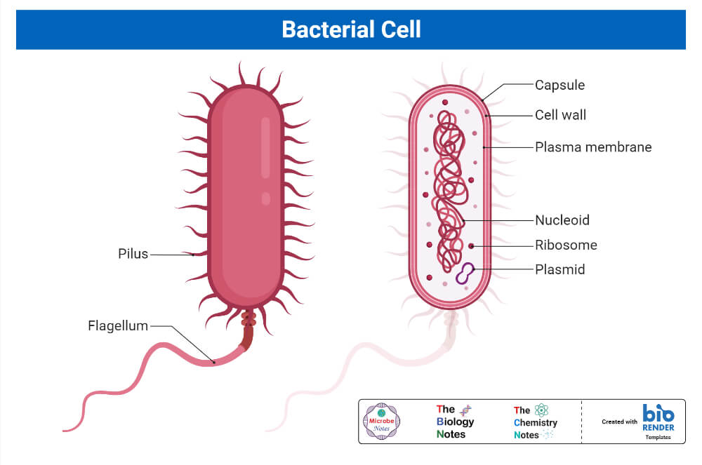
Bacteria are unicellular i.e. made up of a single cell. They are prokaryotes and their cells are different from animal and plant cells. In general, the structure of bacteria can be studied as external and internal structures;
External Structure of a Bacteria
It includes a cell wall and all the structures outside the cell wall.
1. Flagella (sing. Flagellum)
Flagella are long hair-like filamentous structures of about 4 – 5 μm long and 0.01 – 0.03 μm in diameter. They confer motility to the bacteria. Flagella are divided into three parts; filament, hook, and the basal body.
The filament is a threadlike part extending outside the cell wall. It is made up of flagellin protein.
The hook is a short curved structure that joins filament with the basal body. It produces repulsion like the propeller during the revolving of flagella.
The basal body is a set of rings embedded in the cell wall and plasma membrane. It consists of 2 pairs of rings in Gram-Negative bacteria and 1 pair of rings in Gram-Positive bacteria. It synthesizes polymers of the flagellum, produces energy for revolution, and regulates movements of the flagellum.
Functions of Flagella
- Responsible for motility
- Aids in chemotaxis
- Aids in bacterial pathogenicity and survival
2. Pili/Fimbriae
They are the short, hollow, non-helical filamentous structure of about 0.5 μm in length and 0.01 μm in diameter. They are exclusively found in Gram-Negative bacteria.
They are composed of protein ‘pilin’ arranged non-helically. They are short, numerous, and straight than flagella.
Sex pili are a special kind of pili that take part in bacterial conjugation. They are larger than usual pili; 10-20 μm in length. They are few in number, just 1-4 in number. They are further classified into two types; F-pili and I-pili.
Functions of Pili/Fimbriae
- Aids in adherence to host cells
- Sex pili helps in bacterial DNA transfer during bacterial conjugation
3. Capsule
It is a viscous outermost layer surrounding the cell wall. It is composed of either polysaccharides or polypeptides of both (~2%) and water (~98%). They are present only in some species of bacteria. The capsule is of 2 types; macro-capsule (capsule with a thickness of 0.2 μm or more) and micro-capsule (capsule with thickness less than 0.2 μm).

Instead of viscous covering, some bacteria are surrounded by amorphous/paracrystalline colloidal protein materials called the slime layer.
Functions of Capsule
- Aids in adherence
- Prevents from desiccation
- Confer resistance against phagocytosis
- The Slime layer protects from proteolytic enzymes
4. Sheath and Prosthecae
- A sheath is a hollow tube-like structure enclosing chain-forming bacteria, mostly aquatic bacteria. It provides mechanical strength to the chain.
- Prosthecae is a semi-rigid extension of the cell wall and plasma membrane. It increases nutrient absorption and also helps in adhesion.
5. Cell Wall
- The cell wall is a rigid structure made up of peptidoglycan that surrounds the plasma membrane as an external coat. It is 10 -25 μm in thickness.
- Peptidoglycan is a cross-linked polymer of alternately repeating N-Acetylmuramic Acid (NAM) and N-Acetylglucosamine (NAG) polysaccharide sub-units.
- Based on composition, bacterial cell-wall is classified into 2 types; Gram-positive, and Gram-negative cell walls.
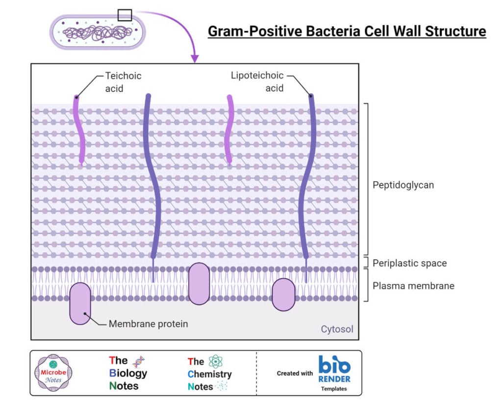
Gram-positive cell wall
The gram-positive cell wall is a thick cell wall containing a large amount of peptidoglycan, about 40 – 90% of the cell wall, arranged in several layers. This type of cell wall also contains acidic sugars like teichoic acids, teichuronic acids, and neutral sugars like mannose, arabinose, rhamnose, and glucosamine as matrix substances.
Teichoic acids are made of polyribitol phosphate or polyglycerol phosphate. They are major surface antigens of gram-positive bacteria. They are of two types; wall teichoic acid and lipoteichoic acid.
Teichuronic acid is a polymer of N-acetylmannuronic acid or D-glucuronic acid.
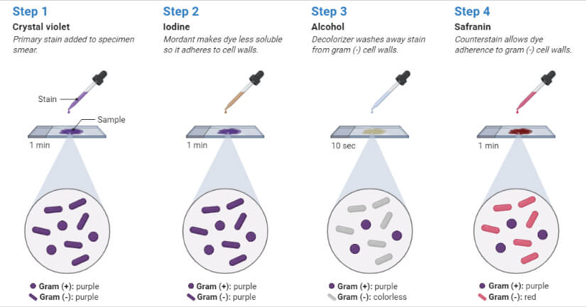
This type of cell wall takes up the crystal violet dye and confer the purple color of the gram-positive bacteria in Gram staining.
Gram-negative cell wall
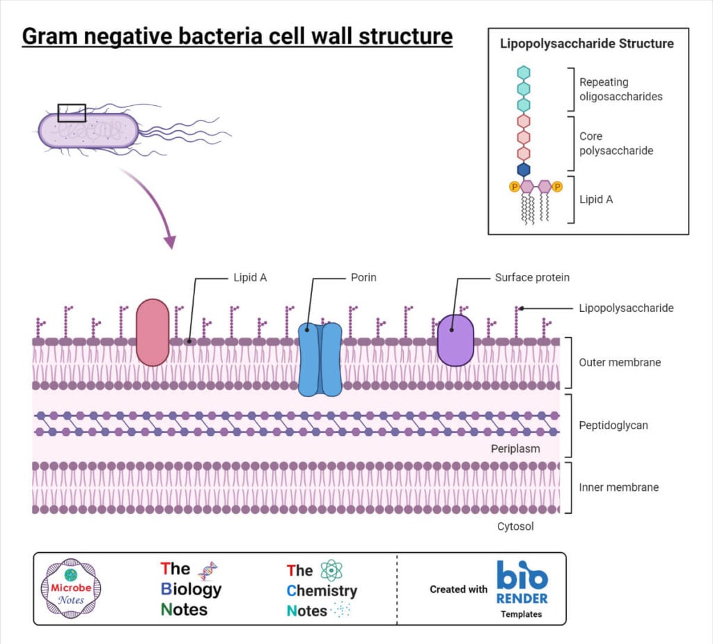
The gram-negative cell wall is a thin cell wall with significantly less amount of peptidoglycan. It is comparatively more complex than the gram-positive cell wall. It contains lipoprotein, lipopolysaccharide, and outer membrane in addition to peptidoglycan.
The lipoprotein layer is composed of Braun’s lipoprotein. It is embedded in the outer membrane and stabilizes the outer membrane.
The outer membrane is a bilayered structure containing an inner layer resembling the plasma membrane in composition, and an outer layer made up of lipopolysaccharide. It is rich in a variety of proteins like ‘porin and outer membrane proteins.
Lipopolysaccharide is a complex molecule consisting of 3 components; Lipid-A, core oligosaccharide, and O-polysaccharide. Lipid-A is composed of phosphorylated glucosamine disaccharides, long-chain fatty acids, and hydrosymyristic acid. Core oligosaccharide is composed of two sugars; keto-deoxy octanoic acid and a heptose sugar bounded together by Lipid A. O-polysaccharide are composed of a wide variety of sugars that differ in between bacterial strains. This confers different antigenic properties to these different bacterial strains.
They lose crystal violet during the Decolorization step and take up safranin during counterstaining, hence providing characteristic pink color to Gram-Negative bacteria.
Cell-wall of Acid-Fast Bacilli
It is unique with a large number of mycolic acids. They resist the Decolorization of acid alcohol or sulfuric acid, hence called acid-fast.
Bacteria without a cell wall
Mycoplasma is a minute (50 -300 nm) bacteria without a cell wall. They do not have a fixed shape. Besides this natural bacteria, there are several other cell walls deficient forms like protoplasts, spheroplasts, and L-forms.
Gram-Positive Cell-Wall vs Gram-Negative Cell-Wall
| Gram-Positive Cell-Wall | Gram-Negative Cell-Wall |
| Thick (20 – 80 nm) | Thin (10 – 15 nm) |
| Higher peptidoglycan content | Lower peptidoglycan content |
| Lower lipid content (2 – 5%) | Higher lipid content (15 – 20%) |
| The main components are peptidoglycan, teichoic acid, and teichuronic acid | The main components are peptidoglycan, lipoprotein, lipopolysaccharide, outer membrane |
| Very few amino acids without any aromatic amino acids | Wide variety of amino acids with different aromatic amino acids |
Internal Structure of Bacteria
It includes the cell membrane and all the structures inside the cell membrane.
1. Cell membrane/Plasma membrane
- It is the innermost phospholipid bilayer, just beneath the cell wall, enclosing cytoplasm. It is a thin (~ 5 -10 nm) semipermeable layer.
- Unlike eukaryotic plasma membrane, they lack sterols (except in Mycoplasma), and comparatively have a higher proportion of proteins. In place of sterols, they have sterol-like compounds, called ‘hapanoids’. They contain a wide variety of fatty acids like usual saturated and unsaturated types and additionally methyl, hydroxyl, or cyclic groups too.
- The plasma membrane is equipped with several porin proteins for the passive transport of nutrients and ions.
Functions of Cell membrane/Plasma membrane
- Selective permeability regulates the inflow and outflow of nutrients, ions, and metabolites
- Electron transport and oxidative phosphorylation
2. Cytoplasm
- It is a colorless, colloidal, viscous fluid with suspended organic and inorganic solutes enclosed within the plasma membrane.
- Unlike eukaryotic cytoplasm, they lack membrane-bound organelles. They have ribosomes, mesosomes, inclusion bodies, nucleic acids floating in them.
2.1 Ribosomes
Bacterial ribosomes are of 70S type and quite smaller than eukaryotic 80S types. They are made up of 2 subunits, the 50S, and 30S. Their main role is to synthesize bacterial proteins and enzymes. They are target sites for different antibiotics like erythromycin, macrolides, aminoglycosides, etc.
2.2 Mesosomes
They are vesicular or branched structures formed by invaginated of the plasma membrane. They represent the eukaryotic mitochondria in function and are the site of action of the bacterial respiration enzymes.
2.3 Inclusion bodies
They are believed to be storage food. They are of two types; (i) organic inclusion bodies, containing glycogen or polyhydroxybutyrate granules, and (ii) inorganic inclusion bodies, containing polyphosphate or sulfur granules.
3. Bacterial Nucleus
They are called nucleoids. Unlike eukaryotic nuclei, they are not enclosed in the nuclear membrane and lack nucleolus and nucleoplasm. It is represented by a dsDNA molecule either in a closed circular form or in coiled form.
Bacterial DNAs are found either in nucleoid as chromosomal DNA or outside nucleoid as a plasmid.
Endospore of a bacteria
Some bacteria under stress form a dormant stage called an endospore. They are produced during unfavorable environmental conditions. They grow to vegetative form when the conditions become favorable.
They have four distinct structural components; (i) core, containing nucleoid and condensed cytoplasm, (ii) spore wall, the innermost wall of peptidoglycan, (iii) cortex, the thickest wall with unusual peptidoglycan, and (iv) protein coat, an outer impermeable layer made of keratin like protein.
Shapes and Arrangement of Bacteria
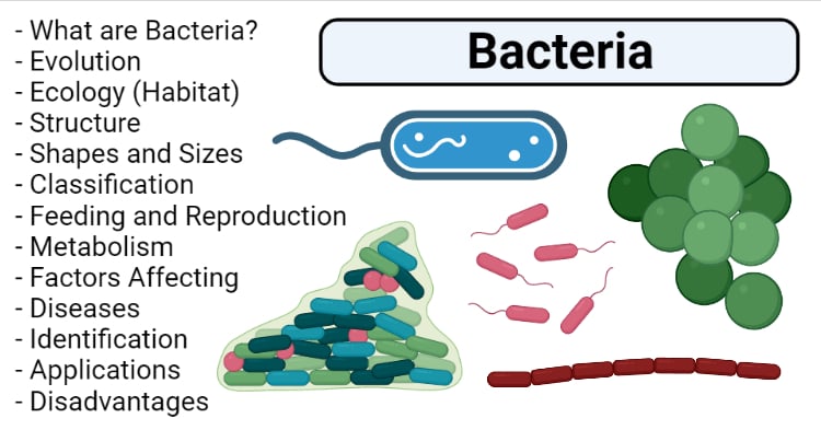
Basically, bacteria are of four distinct shapes, cocci, bacilli, spiral, and comma-shaped.
a. Cocci shape bacteria
They are spherical bacteria. Based on the arrangement of cells they are further sub-grouped as;
- Monococci; singular cocci. Eg. Micrococcus luteus,
- Diplococci; two spherical bacteria are arranged in pairs. Eg. Neisseria spp., Moraxella catarrhalis, Streptococcus pneumoniae, etc.
- Streptococci; spherical bacteria are arranged in a long chain. Eg. Streptococcus pyogenes, S. agalactiae, etc.
- Staphylococci; spherical bacteria arranged in irregular clusters like a bunch of grapes. Eg. Staphylococcus aureus, S. saprophyticus, etc.
- Tetrad; arrangement in a group of 4 cocci. Eg. Aerococcus urinae, Pediococcus spp., etc.
- Sarcinae; arrangement of cocci in a group of 8. Eg. Sarcina spp., Clostridium maximum, etc.
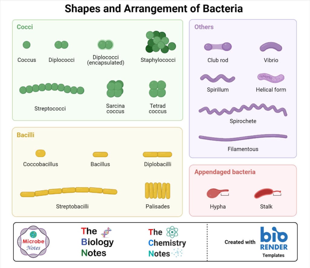
b. Bacilli shape bacteria
They are rod-shaped bacteria. Based on the arrangement of cells they are also sub-grouped as;
- Bacillus /Mono–bacillus; single unattached rod-shaped bacteria. Eg. Salmonella enterica serovars, Bacillus cereus, etc.
- Diplobacilli; bacilli arranged in a pair. Eg. Moraxella bovis, Bacillus licheniformis, etc.
- Streptobacilli; bacilli arranged in a chain. Eg. Streptobacillus moniliform, etc.
- Palisade; bacilli arranged in fence-like form. Eg. Corynebacterium diptheriae, etc.
- Coccobacilli; bacilli with rounded ends or oval-shaped. Eg. Chlamydia spp., H. influenzae, etc.
c. Spiral
They are long helical-shaped or twisted bacteria. Eg. Spirilla spp. , Spirochetes spp. , etc.
d. Comma shaped
They are comma (,) like in structure. Eg. Vibrio spp.
Besides these four basic shapes, several bacteria are found in other shapes like;
- Filamentous (E.g. Actinobacteria, Candidatus savagella, etc. )
- Star shaped (E.g. Stella vacuolata, Stella humosa, etc)
- Appendaged / Budding (E.g. Hypomicrobium, Rhodomicrobium, etc.)
- Pleomorphic (E.g. Mycoplasma spp.)
- Chinese letter like (E.g. Corynebacterium spp.)
- Lobed (E.g. Sulfolobus spp.)
- Stalked (E.g. Caulobacter crescentus )
- Sheathed (E.g. Leptothrix, Clonothrix)
Size of Bacteria
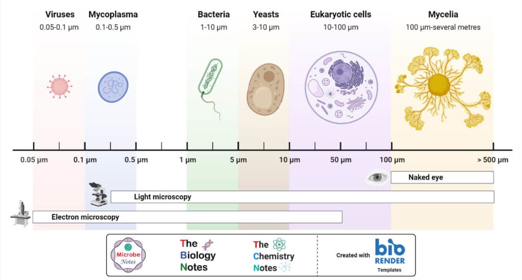
- Bacteria are microscopic with a wide range of sizes from 0.2 μm to 100 μm.
- Cocci are generally of 0.2 to 1.0 μm.
- Bacilli are generally of 1.0 μm 5 μm in length and 0.5 to 1.0 μm in diameter.
- Spirochetes are generally 20 μm in length and 0.1 to 1.0 μm in diameter.
- The smallest bacilli are Pelagibacter ubique ( 370 – 890 nm in length and 120 – 200 nm in diameter).
- The smallest cocci are Mycoplasma genitalium with a diameter of 200 – 300 nm.
- The largest bacteria is Thiomargarita namibiensis with a diameter of 0.75 mm.
Classification of Bacteria
There are different schemes for the classification of bacteria. Some of the most common schemes of classifications are:
a. Classification of Bacteria based on Gram Staining
It is the most common mode of classification used widely in medical and research purposes. Bacteria are grouped into two groups as;
1. Gram-Positive Bacteria
Bacteria having a thick peptidoglycan layer and retaining the purple color of crystal violet during Gram staining are Gram-positive bacteria. E.g. Staphylococcus, Streptococcus, Enterococcus, Corynebacterium, Streptomyces, Bacillus, Haemophilus, Clostridium, Listeria, etc.
2. Gram-Negative Bacteria
Bacteria having a thin peptidoglycan layer and losing crystal violet but retaining pink / red color of counterstain safranine during Gram staining are Gram-negative bacteria. E.g. Escherichia, Salmonella, Shigella, Neisseria, Klebsiella, Proteus, Pseudomonas, Enterobacter, Citrobacter, etc.
| Gram-Positive Bacteria | Gram-Negative Bacteria |
| Stains violet/purple during Gram staining Thick cell wall Thick peptidoglycan layer Higher mucopeptide and very low phospholipid Mesosomes present Fimbriae or pili absent Forms endospores Produce exotoxins Teichoic acid present, Lack an outer layer | Stains red/pink during Gram staining Thin cell wall Thin peptidoglycan layer Lower mucopeptide and very high phospholipid Mesosomes absent (rarely present) Fimbriae or pili present Forms exospores Produce endotoxins Teichoic acid absent, Presence of an outer layer |
b. Classification of Bacteria based on Oxygen Requirements
Bacteria are classified into 3 types as;
1. Aerobic bacteria
They respire aerobically and can’t survive in anoxic environments. E.g. Pseudomonas aeruginosa, Nocardia spp., Mycobacterium tuberculosis, etc.
2. Facultative aerobes
They survive in very low oxygen levels and can survive in both oxygenic and anoxic environments. They are Microaerophiles. E.g. E. coli, Klebsiella pneumoniae, Lactobacillus spp., Staphylococcus spp., etc.
3. Anaerobic bacteria
They respire anaerobically and can’t survive in an oxygen-rich environment. E.g. Clostridium perfinges, Campylobacter, Listeria, Bifidobacterium, Bacteroides, etc.
c. Classification of Bacteria based on Optimum Temperature
Bacteria are classified broadly into 3 types as;
1. Psychrophiles
They have optimum growth temperature at 150C or below. E.g. Chryseobacterium, Psychrobaceter, Polaromonas, Sphingomonas, Alteromonas, Hyphomonas, Listeria monocytogenes, etc.
2. Mesophiles
They have optimum growth temperature at 15 – 450C. Pathogenic bacteria fall in this category. E.g. E. coli, Staphylococcus aureus, Salmonella Typhi, Streptococcus pyogenes, Klebsiella spp., Pseudomonas spp., etc.
3. Thermophiles
They have optimum growth temperature at above 450C. E.g. Bacillus thermophilus, Methanothrix, Archaeglobus, Thermophilus aquaticus, Geogemma barosii (at 1220C), Pyrolobus fumarii (at 1130C), Pyrococcus spp., etc.
d. Classification of Bacteria based on Arrangement of Flagella
Bacteria are classified into 5 types as;
1. Atrichous
They are bacteria without flagella. E.g. Lactobacillus spp., Bacillus anthracis, Staphylococcus spp., Streptococcus spp., etc.
2. Monotrichous
They are bacteria with only one flagellum at one pole. E.g. Campylobacter spp., Vibrio cholerae, etc.

3. Lophotrichus
They are bacteria with multiple flagella at one end. E.g. Spirillum, Helicobacter pylori, Pseudomonas fluorescence, etc.

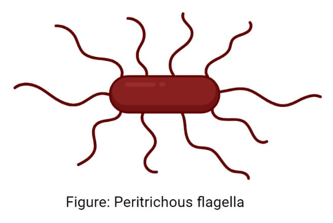
4. Peritrichous
They are bacteria with multiple flagella projecting in all directions. E.g. E. coli, Klebsiella, Proteus, Salmonella Typhi, etc.
5. Amphitrichous
They are bacteria with one flagellum at each pole. E.g. Alcaligenes faecalis, Nitrosomonas, etc.

e. Classification of Bacteria based on mode of nutrition
1. Autotrophic bacteria
They are bacteria capable of assimilating inorganic matters into organic matters i.e. capable of preparing their food like plants. They are of 2 types;
Photoautotrophs; They use energy from sunlight for assimilation. It includes cyanobacteria (Nostoc, Prochlorococcus, etc.), purple sulfur bacteria (Nitrosococcus, Thiococcus, Halochromatium, etc.), purple non-sulfur bacteria (Rhodopseudomonas spp.), green sulfur bacteria (Chlorobium, Chromatium, etc.)
Chemoautotrophs; They use chemical energy for assimilation. It includes sulfur bacteria (Beggiatoa, Thiobacillus, Thiothrix, Sulfolobus, etc.), nitrogen bacteria (Nitrosomonas, Nitrobacter, etc.), hydrogen oxidizing bacteria (H. pylori, Hydrogenbacter, Hydrogenvibrio marinus, etc.), methanotrophs (Methylomonas, Methylococcus, etc), iron bacteria (Thiobacillus ferroxidans, Ferrobacillus, Geobacter metallireducens, etc.)
2. Heterotrophic bacteria
They are bacteria that derive energy by consuming organic compounds, but they do not convert organic compounds to inorganics. They are parasitic or symbiotic types. E.g. E. coli, Rhizobium spp., Staphylococcus spp., Mycobacterium spp., Klebsiella pneumoniae, etc.
3. Saprophytic bacteria
They are bacteria that decompose organic compounds into inorganic and derive energy. They are decomposers and feed on dead plants and animals. E.g. Cellulomonas, Clostridium thermosaccharolyticum, Pseudomonas denitrificans, Acetobacter, etc.
Feeding in Bacteria
Bacteria feed on several organic or inorganic compounds. The food enters the bacterial body either by phagocytosis (active transport) or by osmosis and diffusion or through protein channels (passive transport). They obtain energy by either photo- or chemosynthesis decomposing organic compounds or breaking down inorganic compounds. Based on feeding habits, they are grouped as autotrophs, heterotrophs, and saprophytes.
Reproduction in Bacteria
Bacteria have a very short generation time i.e. they reproduce very quickly. Their reproduction is an asexual type and can be classified into the following types;
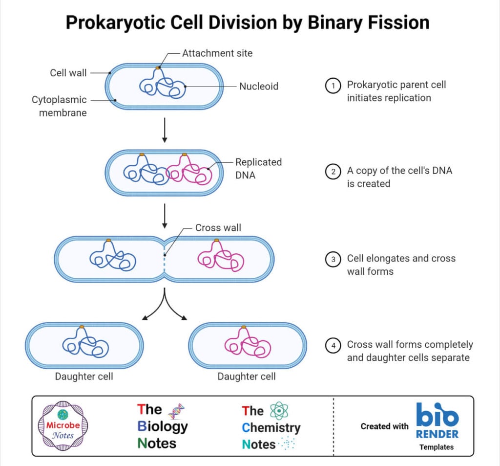
1. Binary fission
It is the most common type. Under favorable conditions, each bacterium divides into two identical bacteria. The bacterial cells first acquire nutrition grow at their maximum size and replicate their DNA. The new replicated DNA called an incipient nucleus, migrates towards opposite poles. A transverse septum begins to develop and separate the two daughter cells.
2. Conidia formation
It is mostly seen in filamentous bacteria like those in actinomycetes, e.g. Streptomyces, Micromonospora, Rhodomicrobium, etc.
3. Budding
The bacterial cells develop small swelling, called protuberance or bud, at one side. Bacterial DNA replicates and one copy enters into the bud. The bud eventually separated and develop into a daughter cell. E.g. Planctomyces spp, Rhodomicrobium vannielia, Hyphomicrobium spp., etc.
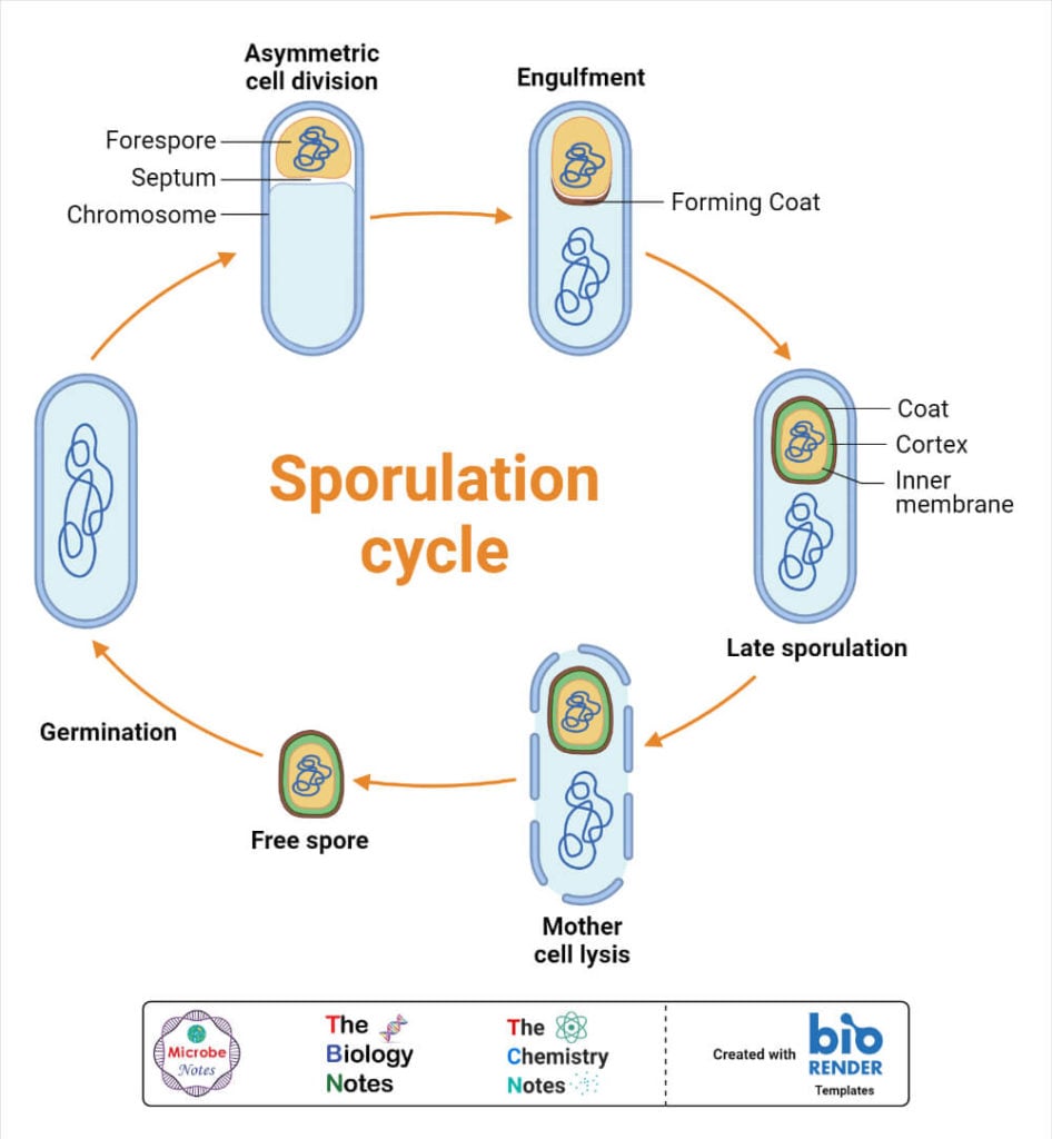
4. Endospore formation
It is seen in some Gram-positive bacteria during unfavorable conditions and environmental stresses. The cytoplasm becomes concentrated around bacterial DNA and a thick, hard, and resistant wall develops around it. E.g. Bacillus spp., Clostridium spp., Sporosarcina spp., etc.
5. Transformation
It is considered a sexual method. In this method, the DNA of one bacterium directly enters into a cell of another bacterium of the same species and forms recombinant DNA. The DNA enters through extracellular environments.

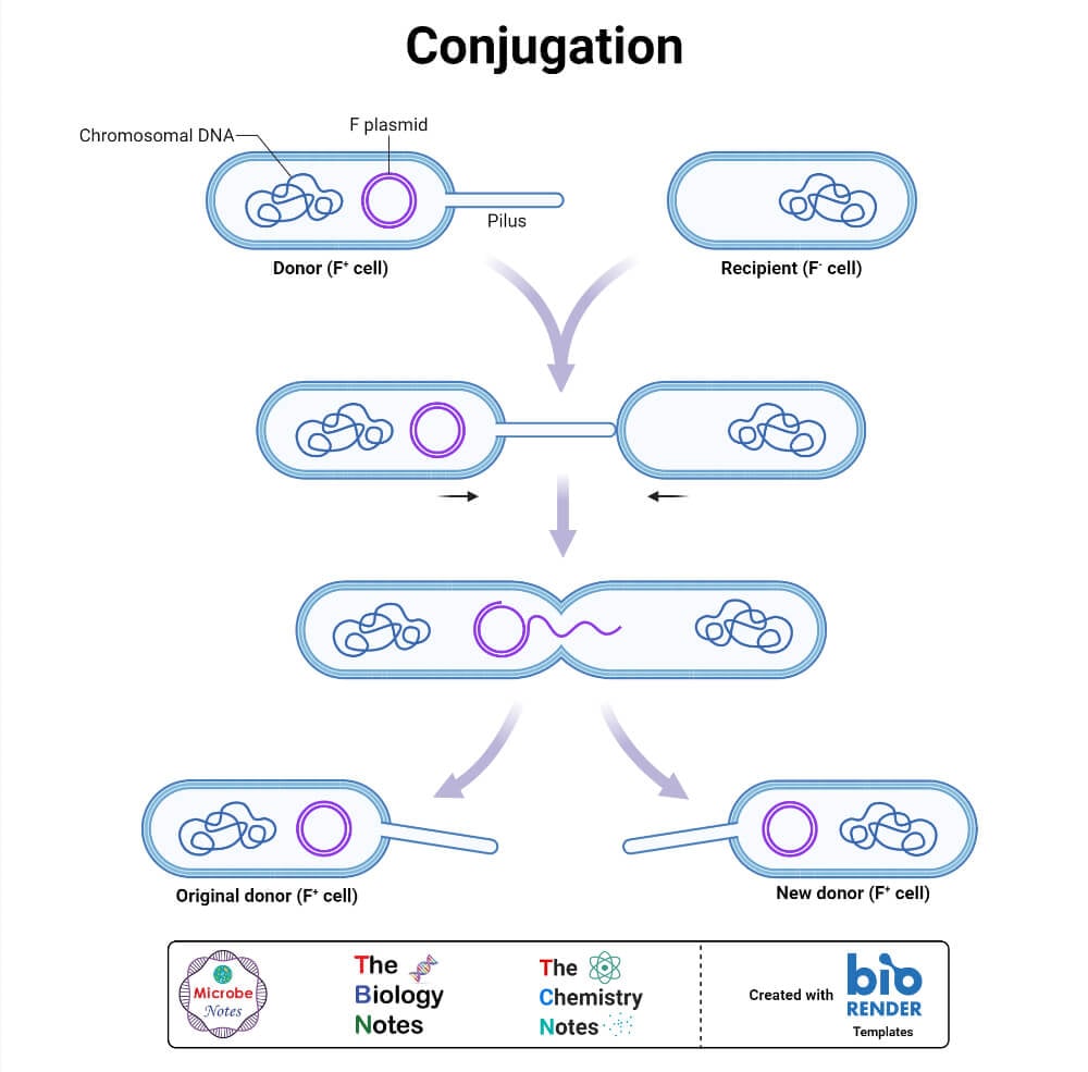
6. Conjugation
It is another sexual method where DNA transformation is by direct contact between donor and recipient bacterium via conjugation tube. Sex pili are responsible for conjugation. Donor cell develops sex pilus and attaches to the recipient cell. A conjugation tube or bridge is formed at the connected point. DNA fragments transform from one bacterium (donor) to another (recipient) through this tube.
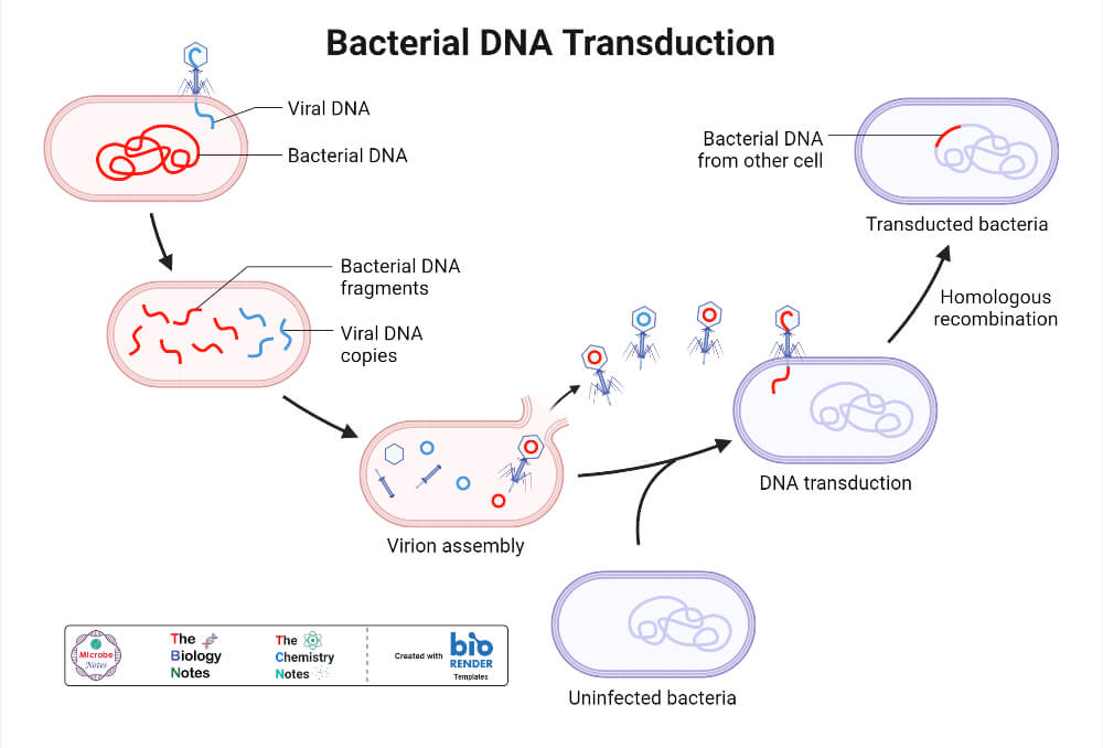
7. Transduction
In this method, DNA fragments are transformed from donor bacterium to recipient bacterium by bacteriophages.
Bacterial Metabolism
It includes all the metabolic/biochemical activities occurring inside bacterial cells. Based on the mode of obtaining carbon, bacterial metabolism can be classified as heterotrophic metabolism and autotrophic metabolism.
1. Heterotrophic Metabolism
In this type, bacteria use organic compounds as carbon and energy source. Carbohydrates, lipids, and proteins are commonly oxidized to form ATP and precursor molecules. There are different processes by which bacteria perform heterotrophic metabolism. Some important types are;
Respiration in Bacteria
- It is the process of obtaining energy (ATP) by complete oxidation of the food (glucose) inside the bacterial cells. Here, glucose completely breaks down into carbon dioxide and water releasing a large amount of energy.
- Respiration can be aerobic or anaerobic respiration.
- In aerobic respiration, bacteria use molecular O2 as a terminal electron acceptor. The general reaction occurring in aerobic respiration can be stated as; C6H12O6 + 6O2 = CO2 + 6H2O + energy (38 ATP)
- In anaerobic respiration, bacteria use nitrate (NO3–), sulfate (SO4-2), CO2, fumarate, etc. as terminal electron acceptors. Along with CO2 and water, H2S, and NH3 are also produced.
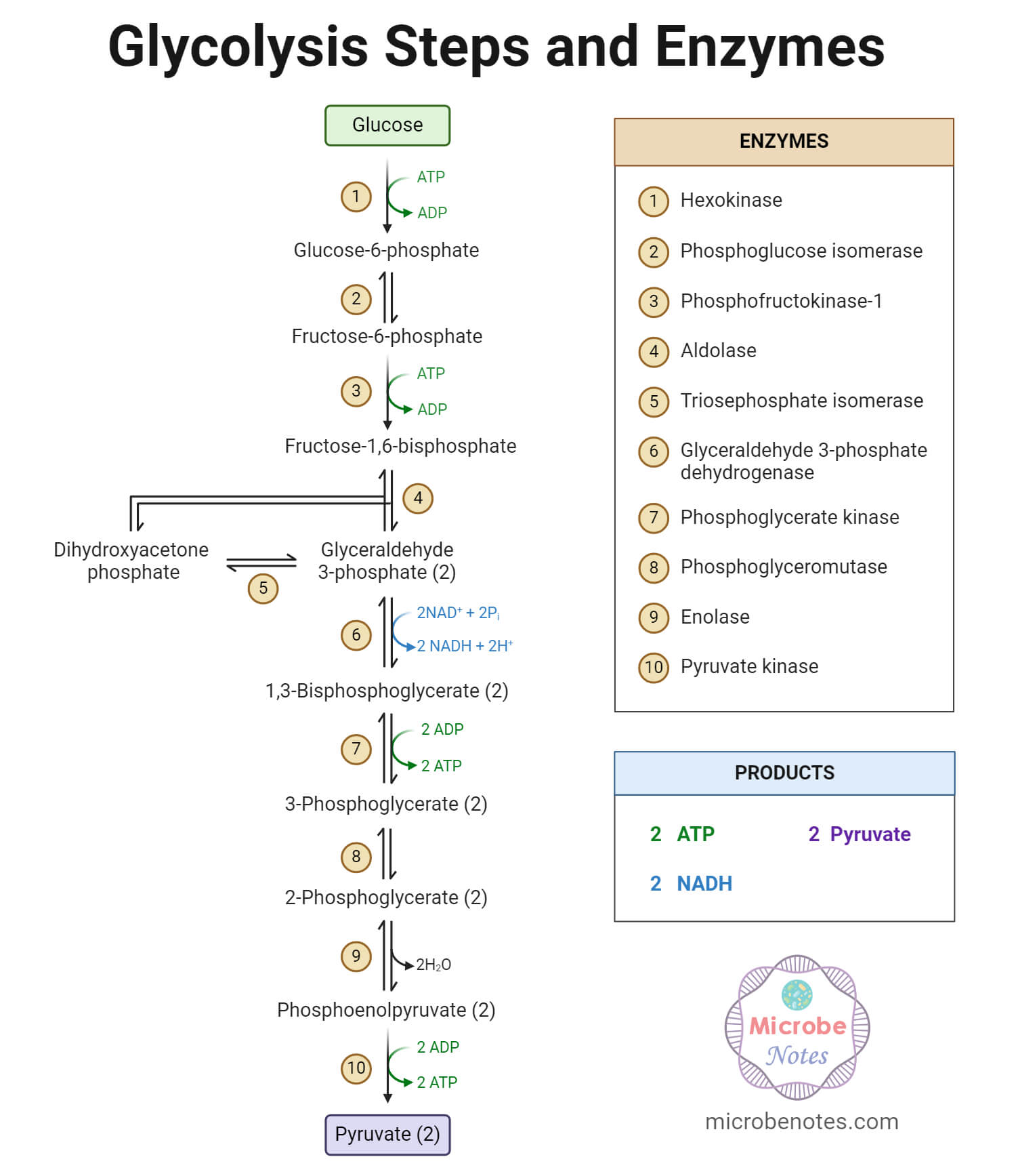
- The complete respiration process includes three basic biochemical pathways; the first is Glycolytic (Embden-Meyerhof-Parnas / EMP pathway), the second is Tricarboxylic acid (TCA) cycle (Kreb’s cycle), and the final one is oxidative phosphorylation (electron transport chain / ETC).
- Besides these, there are other minor pathways like the phosphoketolase pathway, oxidative pentose phosphate pathway (HMP shunt), and the Entner-Doudoroff pathway (ED pathway).
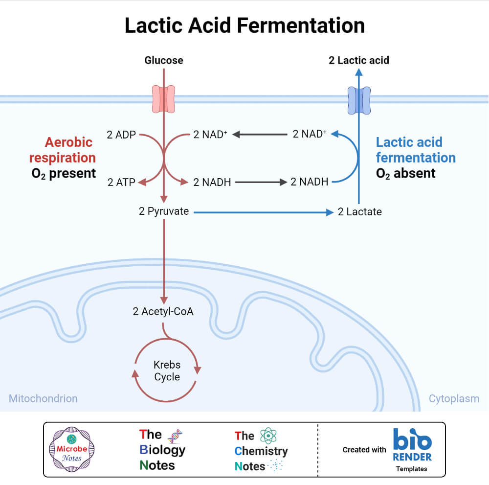
Fermentation in Bacteria
- It is the process where glucose is enzymatically broken down into simpler organic end products like alcohols or acids. Dehydrogenation of glucose releases energy (ATP) along with end products.
- In the homo-fermentation process, bacteria ferment glucose into a single end product. Mostly glycolytic pathway is followed to produce pyruvic acid, which can be further reduced to acetaldehydes, -acetolactate, acetyl-SCoA, and lactyl – SCoA. Some can produce lactic acid, butyric acid, ethanol, etc.
- In the hetero-fermentation process, bacteria ferment glucose into a mixture of multiple end products like ethanol, acetic acid, formic acid, lactic acid, H2, and CO2. This type of fermentation is more common in natural bacterial flora.
There are other types of minor heterotrophic metabolisms like:
(a) methylotrophy, where bacteria use C1-carbon compounds like methanol, formaldehyde, methylamines, etc. as an energy source. This is seen in methanotrophs like Methylobacter spp., Methylomonas spp., Methylococcus spp., etc. Methane is produced during methanogenesis.
(b) syntrophy, where one species of bacteria use the metabolic end product of another species of bacteria. Here different bacteria pair to achieve a chemical reaction, which they can’t perform individually.
2. Autotrophic Metabolism
In this system bacteria directly oxidize inorganic compounds (without using solar energy) to generate energy. It is also called chemolithotrophy or chemoautotrophy or chemotrophic. The most common metabolic pathways include Calvin (reductive pentose phosphate) pathway, reductive TCA cycle, and the acetyl-CoA pathway.
Based on different types of inorganic compounds used as the substrate, there occur different oxidative reactions. Common reactions are; hydrogen oxidation, sulfur oxidation, ferrous oxidation, nitrification, anammox, manganese oxidation, etc.
3. Phototrophic Metabolism
In this system, bacteria use light energy to oxidize inorganic compounds and produce energy (ATP). There are two types of phototropism in bacteria; oxygenic and anoxygenic phototropism.
In Oxygenic phototropism, H2O is oxidized to O2 to obtain electrons by using light energy. It is seen in Cyanobacteria (BGA) containing chloroplast pigment. Two photosystems(PS), PS-I and PS-II are involved in the process.
In Anoxygenic phototropism, H2S or S2 or H2 or other organic compounds are used as electron donors. It is seen in Green Sulfur Bacteria, Green non-sulfur bacteria, purple sulfur bacteria, and purple non-sulfur bacteria. Only one photosystem is involved; PS-I in green bacteria and PS-II in purple bacteria.
Factors Affecting Bacterial Growth
- Water Availability / Water Activity; bacteria have a normal water activity requirement of 0.91 and above. Water is required for maintaining osmotic pressure, conducting metabolisms, regulating physiology, regulating pH, etc.
- Nutrition Level; different bacteria have different nutritional requirements. Bacteria that require very high nutritional requirements are called fastidious bacteria. The bacteria that survive at very low nutrient levels are called non-fastidious bacteria. Along with the increase in nutrition concentration, bacterial growth increases up to a certain limit, but further increment can’t increase the growth rate.
- Temperature; different bacteria have a different optimum temperatures for growth. Based on temperature requirements, bacteria are classified as Mesophiles, Thermophiles, and Psychrophiles. The most common bacteria, including pathogens, are Mesophiles with an optimum temperature of about 370C.
- Gaseous concentration; mostly O2 and CO2 influence bacterial growth. Strict Aerobes require high O2 content. Facultative aerobes can grow at very low O2 content. Anaerobes can’t survive in an environment with O2.
- pH / Hydrogen Ion Concentration; bacteria mostly grow in pH around neutrality (6.5 -7.5). Acidophiles have an optimum pH requirement of pH below 5. Alkaliphiles have an optimum pH requirement of pH above 9. pH affect the enzyme system, proteins, and membrane integrity of bacteria.
- Salinity; salt concentration also affect bacterial growth by influencing homeostasis and enzymatic actions. Halophiles are organisms that have very high optimum ion concentration needed for growth.
- Light intensity; phototrophic bacteria require light for preparing food.
Bacterial Diseases
The bacteria that can cause infection (disease) are called pathogenic bacteria, and such diseases are called bacterial diseases. Most of the bacteria known to us are non-pathogenic. Only <5% are pathogenic. To be pathogenic bacteria, the bacteria must fulfill Koch’s Postulates.
Some common bacterial diseases with their causative species are listed in the table below.
| Disease | Causative Bacteria |
| Tuberculosis Pneumonia Urinary Tract Infection (UTI) Meningitis Gastroenteritis Typhus, Rocky Mountain Spotted Fever Typhoid Cholera Tetanus Syphilis Influenza Cellulitis or wounds | Mycobacterium tuberculosis Klebsiella pneumoniae, Streptococcus pneumoniae, etc. E. coli, Klebsiella, Proteus, Staphylococcus, etc. Neisseria meningitidis, Streptococcus pneumoniae, etc. E. coli, Clostridium, Salmonella, Shigella, Rickettsia spp. Salmonella Typhi and Salmonella Paratyphi Vibrio cholerae Clostridium tetani Treponema pallidum Haemophilus influenzae Staphylococcus aureus, Streptococcus pyogenes, etc. |
Bacterial Identification
This is the method of identifying genera and species of isolated bacteria i.e. to identify which bacteria are isolated. There are several methods designed and used for bacterial identification.
a. Cultural Methods for Bacterial Identification
It is the method of identifying bacteria by studying their cultural characters in a specific culture media. Several selective and indicator media are used for bacterial identification. In this method, we study colonial characters like;
- The shape of colonies (circular, irregular, rhizoid, etc.)
- Size of colonies (micro, small, medium, large, etc.)
- Pigmentation
- Elevation of colonies (concave, convex, flat)
- The margin of colonies (smooth, rough, dented, wavy, etc.)
b. Staining and Microscopy for Bacterial Identification
It is another useful and commonly used method for bacterial identification.
- Gram staining is the most important type of staining method used in microbiology for bacterial identification. It is a differential staining technique used to differentiate bacteria into two groups; Gram Positive and Gram Negative, and to study bacterial morphology. Crystal Violet is used as the primary stain, Gram’s Iodine is used to fix the CV stain, Acetone/Ethanol is used as a decolorizer and Safranine is used as a counter stain.
- Besides there are other staining techniques like simple staining, negative staining, Acid Fast Staining (ZN staining), Giemsa staining, Flagella staining, Endospore staining, etc.
- Light microscopy, Fluorescent microscopy, Dark Field microscopy, and Electron microscopy are used.
c. Biochemical Tests for Bacterial Identification
These tests are the methods of identifying bacteria based on their biochemical activities. Here, we study the ability of bacteria to utilize substrate or to produce certain metabolites and chemicals. This is a traditional method and is still widely used for the phenotypic identification of bacteria.
Visual detection of bacterial growth and color change of media is key to identifying bacteria. The principle of biochemical tests is that different bacteria have different physiology and metabolism, hence showing different biochemical reactions.
Biochemical tests are easy, simple, inexpensive, and the most widely used methods. However, they have the disadvantage of a high probability of false-positive and false-negative results.
The most common biochemical reactions used are;
- Indole test; is a qualitative test that detects the ability of bacteria to produce ‘indole’ by deamination and hydrolysis of ‘tryptophan’ by producing ‘tryptophanase’ enzymes. It is used to differentiate members of the Enterobacteriaceae family. Tryptophan-containing media like Tryptophan broth, Tryptic soy broth, Sulfide Indole Motility (SIM) medium, etc. are commonly used in this method. Bacteria are grown in a medium containing tryptophan and incubated for 24 hours. Following incubation, an indole reagent is added and the color change is noted. Development of red or pink color denoted indole production.
- Methyl-Red (MR) test; is used to detect the production of acid by fermentation of glucose in the medium. MR-VP broth is used in this test. Bacteria are inoculated in MR-VP broth and incubated overnight. Following incubation, a methyl red indicator is added. If bacteria ferment glucose in the medium-producing acid, then the medium will turn red.
- Voges Proskauer (VP) test; is used to detect the ability of bacteria to produce neutral products like acetoin or 2,3-butanediol. Bacteria are inoculated in MR-VP broth and incubated overnight. Following incubation, VP reagents I and II are added and the color change is observed. Development of cherry red / pink color indicates a positive reaction.
- Citrate test; is used to detect the ability of bacteria to utilize citrate as a source of carbon. Simmon’s citrate agar is mostly used.
- Urease test; is used to detect the ability of bacteria to ferment urea to ammonia by producing urease enzyme. Urea containing medium like Christensen Urea Agar is used.
- Triple Sugar Iron (TSI) test; is used to detect the ability of bacteria to ferment glucose and lactose or sucrose and release H2S gas. TSI agar is used for this test. Color change in slant and butt of TSI agar slant is studied. Change in color from red to yellow denoted sugar fermentation. If the color of the slant is changed to yellow, it denotes the fermentation of glucose alone. If the color of the butt is also changed to yellow, it denotes fermentation of either sucrose, lactose, or both. If black coloration is developed, it denoted the production of H2S.
- Catalase test; is used to detect the ability of bacteria to produce catalase enzymes.
- Oxidase test; is used to detect the ability of bacteria to produce the cytochrome oxidase enzyme.
- Sugar Fermentation test; is used to study the ability of bacteria to ferment different types of sugars (glucose, lactose, sucrose, mannitol, sorbitol, arabinose, etc.)
- There are several other tests used like; DNAse test, Nitrate reduction test, Esculin hydrolysis test, Microdase test, Gelatin hydrolysis test, PYR test, ONPG test, Decarboxylase test, Coagulase test, Sulfur reduction test, Starch hydrolysis test, Phenylalanine deaminase test, CAMP test, Bile solubility test, etc.
d. Molecular Methods for Bacterial Identification
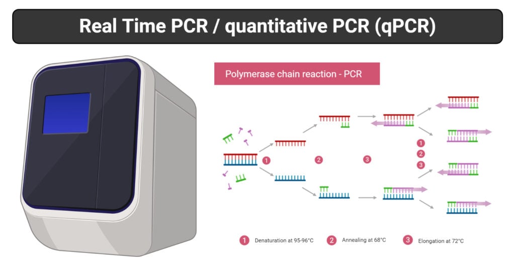
This test includes the study of a bacterial genome and genomic sequences. This is the most advanced and accurate method used when very precise identification is required. We can classify bacteria into sub-species, strains, serotypes, or pathovar levels using molecular methods. This method includes Polymerase Chain Reactions (PCR), DNA / RNA probe tests, Microarray, Electrophoresis, Proteomics, etc.
e. Immunological Methods for Bacterial Identification
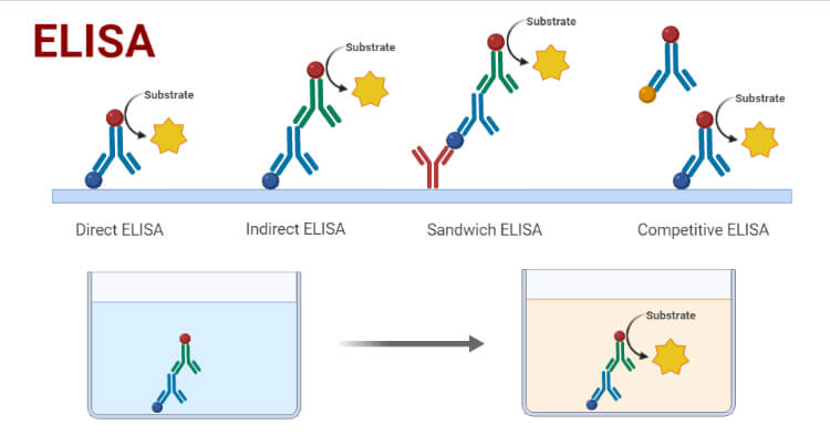
This method is limited to the identification of pathogenic bacteria only. In this method, we identify bacteria-specific antibodies or antigens in the body of an infected person. The identified antibody or antigen is correlated with the identification of the infecting bacteria.
Enzyme-Linked Immunosorbent Assay (ELISA), Radio-immunoassay (RIA), Fluoro Immuno Assay (FIA), Immuno chromatography tests, etc are commonly used tests.
Importance, Uses and Applications of Bacteria
- They are responsible for recycling several nutrients like nitrogen, carbon, sulfur, phosphorus, oxygen, etc. They play the most important role in assimilation and dissimilation of the organic compounds during any biogeochemical cycle.
- They play a very important role in regulating atmospheric oxygen levels. Photosynthetic bacteria (Cyanobacteria, Green Sulfur bacteria) play a very important role in the production of oxygen during photosynthesis.
- They are responsible for biodegradation, composting, decomposition, and bioremediation. They play a very important role in the management of organic wastes and dead organisms and their parts.
- Several bacteria are used industrially for the production of several enzymes. These enzymes are used in industrial processes, medical purposes, food processing, etc. Amylase, lipase, cellulases, proteases, hemicellulases, zymase, penicillinases, polymerases, etc. are produced by bacteria.
- Bacteria are genetically modified and used in biotechnological applications to produce hormones like insulin and enzymes.
- They are used in an anaerobic fermentation process to produce biogas (methane) which is used as fuel.
- Different genera of Actinomycetes and other bacteria are the source of antibiotics used for pharmaceutical purposes.
- Several bacterial species like Bifidobacterium, E.coli, Lactobacillus, etc. are used as probiotics.
- Bacteria are used in producing fermented food products like fermented dairy products, sausages, fermented fruit juices, etc.
- They are used in the bioremediation of oil spillage, xenobiotic, radiation wastes, heavy metal wastes, bio-hazardous wastes, toxic wastes, and other organic and inorganic wastes.
- Bacteria are used in genetic engineering and molecular research. Their genes are being used in producing different Genetically Modified Organisms (GMOs).
- The bacterial fuel cell is new technology to convert chemical energy into electric energy. They can be used as an alternative source of energy.
- In agriculture, they are used as bio-pesticides, bio-fertilizers, and bio-insecticides.
- Bacteria are the pioneer of life forms in barren lands like deserts, rocks, etc. Every living organism living today are evolved from some eukaryotes which were developed from bacteria some 2.0 billion years ago.
- Bacteria are present as normal flora in our body. They help fight against invading pathogens, boost immune response, and help in the digestion process.
Disadvantages and Limitations of Bacteria
- Different pathogenic bacteria are responsible for a wide variety of human diseases from simple to life-threatening. Bacterial diseases are responsible for thousands of death each year.
- Bacterial spoilage of foods feeds and pharmaceutical products is another disadvantage. The food and pharma industries have to bear huge losses due to bacterial spoilage.
- Several bacteria like denitrifying bacteria, sulfur-oxidizing bacteria, etc. are responsible for decreasing the fertility of the soil, ultimately reducing crop yields.
- Bacteria can cause disease to crop plants and domestic animals. This will reduce agricultural production.
- Bacteria cause deterioration and degradation of useful organic products like furniture, textiles, etc.
References
- Krasner, Robert (2014). The Microbial Challenge: a public health perspective. Burlington, Mass: Jones & Bartlett Learning. ISBN 978-1-4496-7375-8
- Woese CR, Fox GE (November 1977). “Phylogenetic structure of the prokaryotic domain: the primary kingdoms”. Proceedings of the National Academy of Sciences of the United States of America. 74 (11): 5088–90. doi:10.1073/pnas.74.11.5088
- Parija, S.C.(2013).Textbook of Microbiology and Immunology (2nd Ed). Elsevier Health Sciences. ISBN: 978-81-312-2810-4
- Baron S, editor. Medical Microbiology. 4th edition. Galveston (TX): University of Texas Medical Branch at Galveston; 1996. Introduction to Bacteriology.
- Jurtshuk P Jr. Bacterial Metabolism. In: Baron S, editor. Medical Microbiology. 4th edition. Galveston (TX): University of Texas Medical Branch at Galveston; 1996. Chapter 4.
- Jurtshuk, P., Jr. (1996). Bacterial Metabolism. In S. Baron (Ed.), Medical Microbiology. (4th ed.). University of Texas Medical Branch at Galveston.
- Bacteria – Definition, Shapes, Characteristics, Types & Examples (biologydictionary.net)
- Bacteria | What is microbiology? | Microbiology Society
- Bacteria – Diversity of structure of bacteria | Britannica
- Bacterial cell structure and function – Online Biology Notes
- Structure of Bacterial Cell (With Diagram) (biologydiscussion.com)
- Morphology, Different shapes of the bacterial cell (byjus.com)
- Shapes of Bacteria | Bacteria Shapes and Arrangements | Biology Explorer (bioexplorer.net)
- Images created with biorender.com

Amazing 🥰,well summarised notes!
This is very nice content.
Nice coverage
I had a wonderful experience am grateful
Green sulfur bacteria don’t produce oxygen during photosynthesis