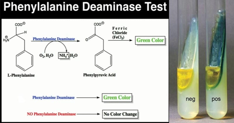
Phenylalanine Deaminase Test (PDA) is the biochemical test used to determine the ability of bacteria to synthesize the phenylalanine deaminase enzyme.
Bacteria capable of producing phenylalanine deaminase enzyme can deaminate phenylalanine resulting in phenyl pyruvic acid and ammonia. This ability of bacteria was first studied and demonstrated by Hendriksen in 1950 on Proteus spp. Later in 1957, Ewing EH and his team formulated a medium containing phenylalanine to test the bacterial capacity to deaminate the phenylalanine oxidatively.
Similarly, in 1971, Ederer GM and his associates developed a disc to rapidly detect the bacteria’s ability to produce urease and phenylalanine deaminase enzymes. These methods are modified over the course of time and are now rapidly used in the laboratory, mostly to differentiate Proteus spp. from other Enterobacterales, as a PDA test. As phenyl pyruvic acid is formed during the course of the reaction, this test is also called the ‘Phenylpyruvic Acid (PPA) Test.’
Interesting Science Videos
Objectives
- To determine the ability of bacteria to synthesize the phenylalanine deaminase enzyme.
- To differentiate Enterobacterales and identify the Proteus spp.
Principle of Phenylalanine Deaminase Test
Some bacteria have the capacity to synthesize phenylalanine deaminase enzyme, which oxidatively deaminates (removes NH2) from the amino acid phenylalanine. Upon deamination of phenylalanine by the deaminase enzyme, phenyl pyruvic acid and ammonia are released. Thus released phenyl pyruvic acid reacts with the chelating agent ferric chloride in the reagent and results in the formation of light to deep-green colored complex, indicating a positive reaction.
Requirements for Phenylalanine Deaminase Test
Culture Media
Phenylalanine agar medium (also called the phenylalanine deaminase medium) is used for the PDA test.
Composition of Phenylalanine Medium per 1000 mL
Yeast extract- 3.00 grams
DL-Phenylalanine- 2.00 grams
Sodium Chloride- 5.00 grams
Disodium hydrogen phosphate- 1.00 grams
Agar- 15.0 grams
Final pH 7.3±0.2 at 25°C
(Reference: Phenylalanine Agar (himedialabs.com))
Preparation of Phenylalanine Medium
- Measure the appropriate amount of phenylalanine powder (or the media components) and mix in the water of the required volume in a conical flask (or glass bottle) according to the instruction of the manufacturing company.
- Stir well using a magnetic stirrer or manually and heat to boiling so that all the components dissolve completely in water.
- Dispense the media in a test tube (about 5 mL in each or appropriate volume as per requirement) and loosely put on the screw cap (or use a cotton plug to cover the opening).
- Autoclave the tubes with the medium at 121°C and 15 lbs pressure for 15 minutes.
- Let it cool and solidify in a slanting position at room temperature.
Reagents
Acidic 10% Ferric Chloride Solution
Preparation Method
- Dissolve 12 grams of ferric chloride in 97.5 mL of distilled water
- Slowly add 2.5 mL of concentrated Hydrochloric acid (HCl) and mix well
(Alternatively, 10% aqueous ferric chloride solution; 10 grams of ferric chloride in 100 mL distilled water can be used in place of acidic ferric chloride solution.)
1 N HCl and Urea-PDA disks (for a rapid test)
Equipment
| Test-tubes Dropper | Weighing Machine Autoclave | Bunsen burner Incubator | Inoculating loop |
PPE and other general laboratory materials
Test organism (sample bacteria)
Positive Control: Proteus mirabilis ATCC 12453
Negative Control: Escherichia coli ATCC 25922
Procedure of Phenylalanine Deaminase Test
Agar Method
It is the most common and sensitive method used to detect the phenylalanine deaminase enzyme.
- Using a sterile loop, pick up a heavy load of test bacteria from a well-isolated colony (or pure culture) or fresh culture (18 to 24 hours old culture) and heavily inoculate the slant by streaking method.
- Incubate the inoculated tube aerobically at 35±2°C for 18 to 24 hours. (If heavy inoculum is used for streaking, incubation for about 6 hours can be enough to detect the enzyme production.)
- After incubation, drop 4 to 5 drops of acidic 10% ferric chloride solution directly over the slant.
- Roll the tube gently so that the reagent reaches all over the slant surface.
- Observe for color change and development of green color within 5 minutes.
Rapid Method
- In a test tube (preferably a plastic test tube), add 0.25 mL of sterile saline and make a heavy suspension of the test bacteria from a fresh, actively growing bacterial culture.
- Add a urea-PDA disk.
- Incubate the tube aerobically at 37°C for up to 2 hours.
- Observe for the development of pink color over the disk, indicating a positive urease test.
- Add 2 drops of 1 N HCl.
- Add 2 drops of acidic 10% ferric chloride solution and shake well and observe for the development of green color.
Result and Interpretation of Phenylalanine Deaminase Test
- Development of light-green to dark-green over the slant (or in suspension in rapid method) within 1 to 5 minutes indicates a positive reaction.
- The absence of green color development (remaining yellow color due to ferric chloride) indicates a negative reaction.

PDA Test Positive Bacteria
Proteus spp., Morganella spp., Providencia spp.
PDA Test Negative Bacteria
E. coli, Klebsiella spp., (most of Enterobacteriaceae)
Quality Control
Positive Control: Proteus mirabilis ATCC 12453 cause the development of green color over agar slant (or saline suspension) after the addition of acidic ferric chloride solution.
Negative Control: Escherichia coli ATCC 25922 doesn’t form green color on the slant (or saline suspension) after the addition of an acidic ferric chloride solution.
Precautions
- Use heavy inoculum of fresh culture for inoculation.
- Store ferric chloride solution in a dark bottle and dark space away from light.
- Always check the urea-PDA disk for discoloration before use.
Applications of Phenylalanine Deaminase Test
- To differentiate Proteus spp., Morganella spp., and Providencia spp. among other Enterobacterales.
- Identification of Proteus spp. in clinical isolates.
- Used in biochemical identification of unknown bacteria.
Limitations of Phenylalanine Deaminase Test
- It is not a confirmatory test; hence, it needs other tests for the complete identification of unknown bacteria.
- The developed green color fades rapidly, so the result must be read within 5 minutes of the addition of ferric chloride solution.
References
- Leber, Amy L., editor in chief. (2016). Clinical microbiology procedures handbook (Fourth edition). Washington, DC : ASM Press 1752 N St., N.W., [2016]
- Tille, P. M., & Forbes, B. A. (2014). Bailey & Scott’s diagnostic microbiology (Thirteenth edition.). St. Louis, Missouri: Elsevier.
- Phenylalanine Deaminase Test – Procedure, Principle and Uses – Laboratoryinfo.com
- Phenylalanine Deaminase Test: Principle, Procedure, Results • Microbe Online
- Phenylalanine Agar Test – Principle, Procedure, Uses and Interpretation (microbiologyinfo.com)
- Phenylalanine deaminase test: Principle, Requirements, Procedure and Results interpretations – Online Biology Notes
- Phenylalanine Deaminize Test: Principle, Procedure, Results and Uses – BIOCHEMINSIDER
- Phenylalanine Deaminase (PDA) Test: Introduction, Principle, (medicallabnotes.com)
- Phenylalanine Deaminase Test: Result, Principle, & Reagents (researchtweet.com)
- Phenylalanine Deaminase test: Principle, procedure, result interpretation (universe84a.com)
- Welcome to Microbugz – Phenylalanine Deaminase Test (austincc.edu)
- Phenylalanine Agar (himedialabs.com)
