Note: Each image source is given below in this post of respective subheadings.
Interesting Science Videos
1. Amoeba under the microscope
- Amoeba is a unicellular organism in the Kingdom Protozoa.
- It is a eukaryote and thus has membrane-bound cell organelles and protein-bound genetic material with a nuclear membrane.
- Amoeba moves with their pseudopodia, which is a specialized form of the plasma membrane that results in a crawling motion of the organism.
- Because these are unicellular organisms, they cannot be seen through the naked eyes and thus are microscopic organisms.
- Amoeba can be observed under the microscope either directly without staining or after staining and fixing with a particular dye.
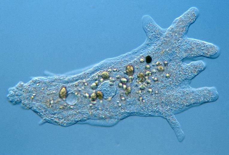
Figure: Amoeba under the microscope. Image Source: Onview.net Ltd.
Direct observation
- Direct observation allows the viewing of living organisms as they move around.
- For direct observation, a sample of water can be directly observed under a microscope, or the organisms can be cultured to increase the number before the inspection.
- Under direct observation, Amoeba appears like a transparent jelly-like structure that shows the crawling movement of the organism through the field.
- Pseudopodia can be observed as the cell membrane protrudes, forming long finger-like projections.
- Inside the organism vacuoles are seen as large empty spaces and food particles are seen as tiny black dots.
Observation after staining
- In order to observe the internal cell organelles of the organism, fixing and staining procedures are performed.
- Fixing kills the organisms and thus isn’t useful to observe the motility of the organism.
- However, fixing and staining provide a better understanding of the structure and morphology of the organism.
- After staining the organism, it can be observed that Amoeba’s cell organelles and cytoplasm are enclosed inside the cell membrane.
- The cytoplasm is stained, which allows the observation of food vacuoles, nucleus, and other essential cell organelles.
- Amoeba usually has one or more nucleus, which is present inside the nuclear membrane.
- Food particles can be seen present inside vacuoles where they are stored and digested.
- Similarly, to maintain the osmotic balance, contractile vacuole can also be seen throughout the cytoplasm.
2. Algae under the microscope
- Algae are photosynthetic organisms that are mostly found in either freshwater or marine sources.
- Most algae are provided with pigments that assist the organisms in producing food or oxygen.
- The structure of algae is quite different from other organisms like plants and animals.
- Some algae are microscopic whereas some are large extending up to 200 feet in length.
- Based on the complexity of the algae, they can either be collected along with the water sample or by cutting the large kelps. Algae in the soil are difficult to obtain, so it is better to culture them before observation.
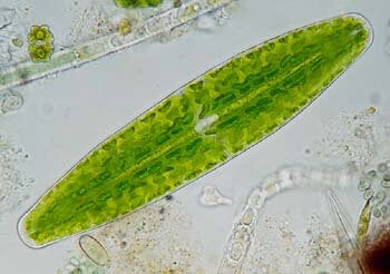
Figure: The desmid Netrium digitus has a beautifully folded chloroplast. Image Source: Onview.net Ltd.
Based on their morphology, algae are divided into separate groups:
Chlorophyta
- Under the microscope, Chlorophytas are seen as green structures enclosed into compartments arranged in the form of chains.
- Inside each of such compartments, a large vacuole is observed and two layers of the cell wall can also be seen.
- The shape and size of the algae vary depending on the genus.
- Some algae in this category are motile while some are non-motile.
Chromophyta
- This group of algae contains species that are barely chained and instead appear as drum-shaped, amoeboid or pear-shaped in structure.
- Some species are provided with hair-like appendages or flagella that sometimes, extend longer than the body of the organism.
- The shape and size of some algae might change throughout their life, depending on the stage of life and habitat.
Cryptophyta
- The algae in this group appear comma-shaped with red or similar pigments.
- Some species might have a groove in their cell membrane while others don’t.
- The pigments are usually positioned towards the side while the nucleus is present in the center near the vacuole.
Rhodophyta
- These are filamentous where the body is characterized by thallus with calcareous deposits resulting in a solid structure.
- The color of the organism ranges from pink to purple, red, yellow, green, or even white.
- Some species are photosynthetic and thus have green pigments deposited in the interior of the cell wall.
Dinoflagellata
- These are unicellular organisms that appear golden-brown due to the presence of golden-brown plastids.
- They have a dented cell membrane and show distinct swimming patterns.
- The nucleus is rather large with visible chromosomes.
- They have two dissimilar flagella protruding from the cell membrane.
- Some dinoflagellates are macroscopic and can be seen even without any microscope.
Euglenophyta
- Under the microscope, they have a large elongated green structure. The shape might change from one species to another.
- They have two to four flagella with chloroplast deposits throughout the cytoplasm.
- In Euglena, an orange spot is seen towards the periphery that is called the eyespot of the organism.
3. Animal cell under the microscope
- A typical animal cell is 10–20 μm in diameter, which is about one-fifth the size of the smallest particle visible to the naked eye.
- Under the microscope, animal cells appear different based on the type of the cell. However, the internal structure and organelles are more or less similar.
- Animal cells usually are transparent and colorless, and the thickness of the cell differs throughout the cytoplasm.
- Compared to the plant cell, animal cells have a more pleomorphic shape as they don’t have a cell wall and thus can change their shape throughout their life.
Direct observation
- Because animal cells are transparent and colorless, it is challenging to observe them directly without staining.
- However, under a phase-contrast microscope, the nucleus is visible as a solid structure because it is denser than other parts of the cell.
- The cell membrane appears as a border enclosing all the components inside the cells.
- Direct observation, however, allows the observation of living cells without any components being lost or distorted during specimen preparation.
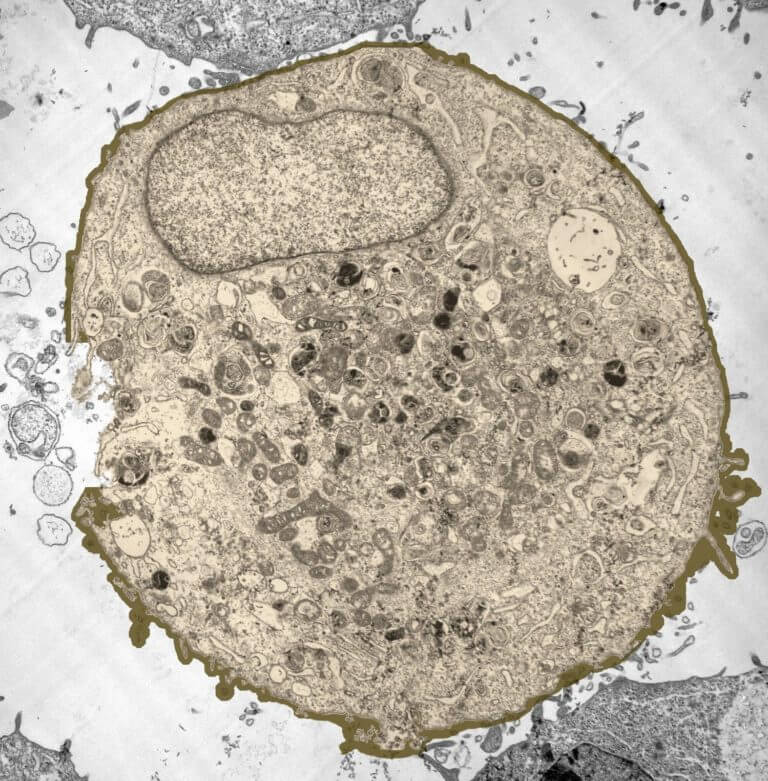
Figure: Animal cell under the microscope. Image Source: The Greatest Garden.
Observation after staining
- Staining allows the viewing of the cellular organelles present in the cytoplasm.
- Separate stains are available for the staining of a distinct part of the cell which allows a more detailed study of different components of a cell.
- Under a low-power microscope, the cell membrane is observed as a thin line, while the cytoplasm is completely stained.
- The cell organelles are seen as tiny dots throughout the cytoplasm, whereas the nucleus is seen as a thick drop.
- Under a higher power microscope, the organelles like mitochondria and ribosomes can also be seen.
- In some cells, the chromosomes present inside the nucleus can also be seen.
- In the case of tissues, other structures like microvilli and cilia can also be observed.
4. Ant under the microscope
- Ants are one of the most common terrestrial insects found in various ecosystems.
- These are macroscopic organisms and can be easily viewed without a microscope. However, under a microscope, different parts of the ant can be seen in more detail.
- The size of the ants differs depending on the stage of life as well as in different species.
- The color of ants ranges from black to brown to rusty red in color.
- Ants are social animals and therefore are usually found as colonies, and each colony has one or more egg-laying queens and an army of female worker ants.
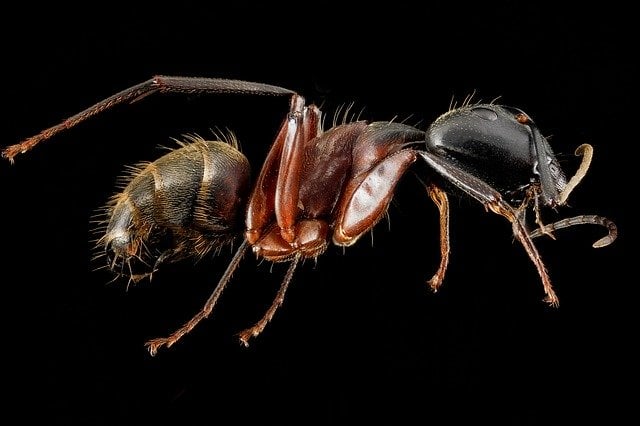
Figure: Ant under the microscope. Image Source: Microscope Master.
Ants under the magnifying glass
- For the observation, one can take either alive or dead ants.
- Under the microscope, ants appear to have three main body parts; head, thorax, and abdomen.
- The head is more movable than other parts while the thorax is the middle part, and the body consists of six pairs of appendages.
- If viewed closely, the head has a pair of antenna and a couple of compound eyes.
- Similarly, on the sides of the head are two mandibles that are the mouthparts of the insect.
- In the thorax region, male ants have two pairs of wings as sterile female ants don’t have wings.
- The queen ants, however, do have wings and are sometimes even more significant in size than the male ants.
- The entire body of the ant is covered with an exoskeleton made up of chitin that protects the internal organs of the insect.
Ants under a light microscope
- As under a magnifying glass, three body parts of the ants can also be seen under a light microscope.
- Additionally, the thorax can further be seen divided into three segments, where the second two segments carry the wings.
- Each wing appears to have a network of irregular veins that strengthen the wing. The wings are usually colorless.
- Under the microscope, a small structure called petiolus can be seen between the thorax and the abdomen, which provides a range of motion to the abdomen.
- The antenna on the head is bent which is divided into segments towards the end.
- In the compound eyes, numerous units called ommatidia can be seen. Three smaller eyes can further be seen in the head arranged in a triangle.
- The light microscope also provides a better view of the mouthparts of the ant. The mouth is made up of two large upper mandibles, two lower mandibles known as maxilla, the upper lip (labrum), as well as the lower lip (labium.
5. Atom under the microscope
- Atoms are the smallest unit of an element in that the particles within atom-like electrons and neutrons do not show the properties of the element.
- The size of an atom is about 1× 10⁻¹⁰ m which means that the atoms are not visible with a light microscope.
- However, a number of other microscopes are available through which the structure of an atom can be observed.
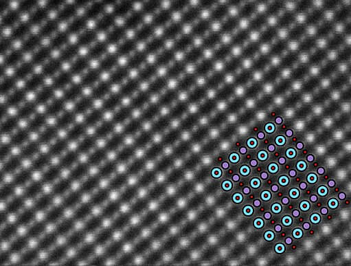
Figure: Atom under the microscope. Image Source: Microscope Master.
Electron microscope
- The images from the transmission electron microscope show a razor-thin layer, just two atoms thick, of two atoms bonded together.
- The microscope can not only distinguish between individual atoms but even see them when they were about only 0.4 angstroms apart, half the length of a chemical bond.
- Some variation of this microscope can also penetrate down to the subatomic particles like electrons.
- Energy-filtered transmission electron microscopes can even observe individual electrons orbiting around the nucleus.
- A scanning transmission electron microscope (STEM) is often used to observe crystals or compounds that reveal the atoms present inside the compounds with some electrons being used to identify atoms of a particular element through the microscope.
- The structure of an atom is visible with these microscopes. However, the internal components like neutrons, protons, and electrons are only observed as waves. The detailed arrangements of these components are yet to be seen.
6. Bacteria under the microscope
- Bacteria are unicellular prokaryotes in which the genetic material is not enclosed inside a nuclear membrane.
- These are simpler organisms consisting of membrane-less cell organelles.
- The size of bacteria ranges from 0.5 to 5 µm, and therefore the bacteria are microscopic.
- The bacteria are varying in shape and size and their components.
- It is hard to observe bacteria directly from their source and thus need to be cultured to increase their number.
- Bacteria are very hard to observe without staining as they are colorless and transparent and tiny in size.
- Because of the varying shape and size of the bacteria, it is also challenging to distinguish bacteria from other dust particles without staining.
- A number of different staining processes can be done to obtain a more detailed structure of these bacteria.
- Some of these procedures even allow the differentiation of bacteria into separate groups based on their staining results.
Observation under simple staining
- This method is usually performed to detect and observe bacteria simply.
- In this case, all the bacteria are stained with a distinct colored stain which causes the entire surface of the bacteria to be stained with that color.
- Based on the shape of the bacteria, they are classified into cocci, bacilli, spirilla, and other groups.
- Some bacteria might be seen in chains while some are observed in groups in a grape-like structure.
- However, some bacteria exist alone as a singular unit.
- Under a higher power microscope, it is possible to observe the internal cellular components in the bacteria.
- For a more detailed structure of the cellular organelles, however, separate staining of the internal organelles is to be performed.
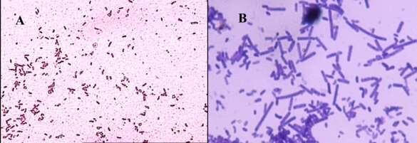
Figure: Bacterial cell under microscope A; Gram-negative B; Gram-positive bacteria. Image Source: https://doi.org/10.24897/acn.64.68.503
Observation under Gram staining
- Gram staining is usually performed to distinguish bacteria into groups.
- Gram-positive bacteria appear purple whereas Gram-negative bacteria appear red under the microscope.
- Based on the result of the staining, the thickness of the cell wall of the bacteria can be assumed.
- Under a high-power microscope like the scanning transmission electron microscope, it is possible even to stain and observe the detailed structure of the cellular organelles.
- The nucleus appears as a large black spot in the center where they are not necessarily surrounded by any membrane.
- The cytoplasm is also stained, which reveals other structures as tiny dots or long filamentous structures.
7. Blood under the microscope
- Blood is the liquid connective tissue in animals that transfers nutrition, water, oxygen, and carbon dioxide to different parts of the body.
- Blood consists of a liquid portion called plasma and other blood cells.
- Blood transfers throughout the body through the blood vessels.
- The blood also consists of other particles like dissolved glucose, other nutrients, and proteins that assist in the functions of the blood.
- Blood appears as a red-colored liquid due to the presence of hemoglobin.
- Under the microscope at 40X, a colorless liquid is seen called plasma that occupies about half of the volume of the blood.
- Other components, like blood cells, are seen suspended in the plasma.
- As the power of the microscope increases (under 100X), red blood cells and white blood cells can be distinguished.
- The volume of red blood cells is higher than that of white blood cells.
- Red blood cells are smaller and don’t have any nucleus whereas white blood cells are larger in size with the nucleus that appears as a dark stain.
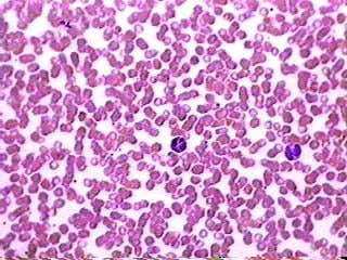
Figure: Blood under the microscope. Image Source: MicroscopeMaster.
- Under a higher power (400X), red blood cells are seen stacked on top of each other, and some granules can be seen inside the white blood cells.
- At this point, platelets can also be seen between the red and white blood cells as tiny dots.
- In an electron microscope, it is even possible to see other proteins and elements present in the blood other than plasma and blood cells.
8. Blood cells under the microscope
- Blood cells are cellular structures found suspended in the plasma of the blood.
- Human blood contains a number of blood cells on the basis of their purpose and structure.
- The red blood cells occupy most of the blood cells in the blood, followed by white blood cells and then the platelets.
- The red blood cells are red in color due to the presence of hemoglobin. These cells are formed in the bone marrow through erythropoiesis.
- The red blood cell is responsible for the transfer of oxygen to different parts of the body.
- The white blood cells, on the other hand, do not have hemoglobin and are involved in providing protection against foreign invaders.
- Other cells termed platelets are also present in the blood, which helps in the clotting of the blood.
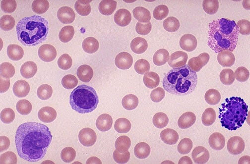
Figure: Blood cells under the microscope. Image Source: Quizlet.
Red blood cells
- Under the microscope, red blood cells appear as red-colored circular cells that are thick at the periphery and thin in the center.
- The red blood cells do not have a nucleus or any other cellular organelles.
- They appear as biconcave discs that are empty on the inside under a microscope.
- In the case of a fresh blood sample, the red blood cells appear yellow-green in color with pale centers containing no visible internal structures.
- The structure of the cells, however, might not be uniform as they get distorted while traveling through the blood capillaries.
White blood cells
- White blood cells or leukocytes are comparatively fewer in blood and thus are difficult to find under the microscope.
- Because they are colorless, it is also difficult to observe them without staining.
- After staining, however, different types of leukocytes can be seen in the microscopic field.
Neutrophils
- They appear spherical in shape with a darkly stained nucleus which is usually segmented into 2-5 lobes.
- Further, tiny granules can be seen in the cytoplasm along with small threads connecting different lobes of the nucleus.
Eosinophils
- These cells also appear spherical in shape under the microscope.
- The cytoplasm contains granules along with a darkly stained nucleus with just two lobes. The nucleus is horse-shoe-shaped.
Basophils
- Basophils are larger in size than other leukocytes and have irregular nuclei inside the spherical cell.
- The nucleus of the basophil is seen bluish in color which is not as defined as in other leukocytes.
Mast cells
- Mast cells are very few and thus difficult to detect; however, they appear enormous compared to other cells and have more granules in their cytoplasm than other cells.
Lymphocytes
- Lymphocytes are the cells that are comparatively smaller in size and under the microscope appear spherical in shape with minimal cytoplasm.
- The nucleus is large and round, occupying most of the volume inside the cell.
- They don’t have any granules in the cytoplasm.
Monocytes
- Monocytes appear larger than lymphocytes and have a kidney or bean-shaped nucleus.
- These cells, like lymphocytes, don’t have granules in their cytoplasm.
- They have more cytoplasm than lymphocytes.
9. Cheek cells under the microscope
- The cells in the cheeks are eukaryotic cells with a defined nucleus enclosed inside a nuclear membrane along with other cell organelles.
- These cells line the buccal cavity in humans and are usually shed during mastication and even talking.
- Under direct observation, only the shape and size of the cell are visible because the cells are transparent and colorless.
- After staining, however, other components like the nucleus are visible under the microscope.
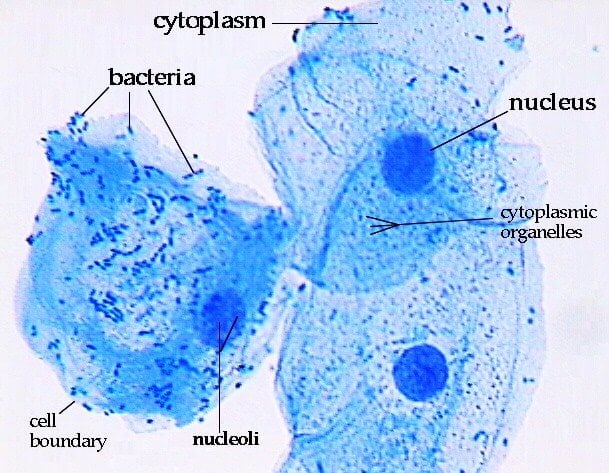
Figure: Cheek cells under the microscope. Image Source: Paul Anderson (John Abbott College).
Observation after staining
- The cells in the cheek are not uniform in shape but are more or less circular in shape.
- The cell membrane is visible as a dark stained border, and the nucleus is seen as a dark spot in the center.
- Similarly, the cytoplasm is also stained, which allows the differentiation of the nucleus and the cytoplasm.
- The cytoplasm is granulated with tiny dots all over.
- Under a high-power microscope, the cell organelles are more differentiated and allow the observation of individual structures.
- Because of the affinity of the stain with the DNA and RNA of the cell, the components inside the nucleus might also be visible.
10. DNA under the microscope
- DNA (Deoxyribonucleic acid) is a molecule present inside the nucleus consisting of two polynucleotide chains coiled around each other to form a helical structure.
- DNA is present in the chromosomes inside the nucleus, which is responsible for controlling all activities of a cell.
- The structure of the DNA was first discovered through X-ray crystallography.
- Even though the overall length of a DNA molecule is about 2 inches, it is not possible to see DNA through light microscopy as the DNA is present inside the nucleus inside the cell.
- DNA that has been extracted might be seen through naked eyes as a long thread-like structure.
- It is, however, possible to observe DNA through a high-resolution microscope like an electron microscope.
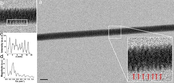
Figure: TEM image with intensity profile and corresponding FFT pitch calculation of λ-DNA fibers. Image Source: Nano Lett.
Observation under Scanning Transmission Electron Microscope
- Under STEM, DNA can be distinguished from other biological molecules as it operates in a dark field.
- This method allows the stained visualization of DNA strands inside the cell.
- Without staining, the DNA appears corkscrew thread of the DNA double helix.
- Usually, through this method, rather small segments of DNA are visible as the electron breaks up the entire DNA into shorter strands.
- Under Cryo-electron tomography, DNA strands are visible in a 3-D structure that allows the visualization of DNA from different angles.
- Through this technique, it is even possible to measure the length of the DNA strands.
11. E. coli under the microscope
- Escherichia coli (E. coli) is a bacterium commonly found in various ecosystems like land and water.
- Most of the strains of E. coli are harmless, but some strains are known to cause diarrhea and even UTIs.
- E. coli is commonly studied as they are considered as a standard for the study of different bacteria.
- Because E.coli is a motile organism, it is beneficial to observe E. coli directly without staining.
- E. coli being a prokaryote, doesn’t have a membrane-bound nucleus and has primitive cell organelles.
- E. coli is a bacillus and has an elongated structure with round edges.
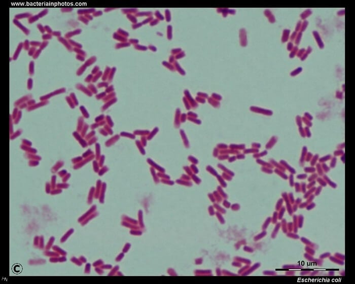
Figure: E. coli under the microscope. Image Source: bacteriainphotos.
Hanging drop method
- This technique is performed to observe the motility of the organism.
- Under this method, the living organisms are observed, which allows a more life-like observation of the organism.
- The structure of the organism can be observed with this technique in which E. coli is seen as a bacillus arranged in chains.
- The internal structure and organelles are not visible through this method as the organism itself is colorless.
- The motility of the organism is, however, possible to observe where the organism moves in a different direction while changing position rather than showing a Brownian movement.
Simple staining
- This method is usually performed to detect and observe E. coli simply.
- Here, the organism is stained with a distinct colored stain which causes the entire surface of the bacteria to be stained with that color.
- E. coli is seen as a rod-shaped organism ranging in size from 1-2 µm. The bacteria are seen mostly in chains.
- However, some bacteria exist alone as a singular unit.
- Under a higher power microscope, it is possible to observe the internal cellular components in the organism.
- For a more detailed structure of the cellular organelles, however, separate staining of the internal organelles is to be performed.
Gram Staining
- After Gram staining, E. coli appears pink in color under the compound microscope.
- This indicates that the bacteria are Gram-negative and have an additional layer in the cell membrane made up of phospholipids and lipopolysaccharides.
- Under a high-power microscope like the scanning transmission electron microscope, it is possible even to stain and observe the detailed structure of the cellular organelles.
- The nucleus appears as a large black spot in the center where they are not surrounded by any membrane.
- The cytoplasm is also stained, which reveals other structures as tiny dots or long filamentous structures.
- On the surface of the cell membrane, a long filamentous structure called flagellum is seen.
12. Euglena under the microscope
- Euglena is a single-celled organism that belongs in the kingdom Protista.
- These are usually found in pond water or marshy places.
- These are algae and thus are capable of producing their own food. They carry the pigment chlorophyll.
- As they are easily found in water and other areas, they are easy to collect and observe.
- Because they are unicellular organisms, they cannot be viewed through the naked eyes but can be easily seen through a compound microscope.
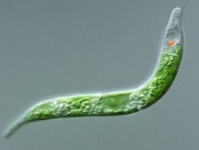
Figure: Euglena mutabilis under the microscope. Image Source: djpmapfer.
Observation under the compound microscope
- As the sample is usually collected from pond water, it might be contaminated with Amoeba and other such organisms.
- It is possible to distinguish between Amoeba and Euglena as the latter is an elongated organism while Amoeba has a more irregular shape.
- Under 40X magnification, Euglena is seen as tiny particles making sudden movement in the field as they are motile.
- As the resolution increases, green spots are seen in the organisms indicating the presence of chloroplast.
- Inside the organisms, dark spots are also observed which refer to the nuclear material of the organism along with a whip-like flagellum at the end. The flagellum is colorless and transparent and thus might be difficult to detect.
- As the resolution increases, the orange-colored spot is seen at the periphery of the organism, which indicates the eyespot of Euglena known to detect light.
13. Hair under the microscope
- Hair is a keratinized structure that is characteristic of mammals.
- A hair filament grows from the follicles present underneath the skin.
- The entire skin surface of humans except some glabrous skin is covered with hair.
- The hair has two parts; root present inside the skin and shaft present above the surface.
- New cells are formed at the root when then add up and reach the outside of the skin, where they become keratinized and convert into dead cells.
- Over time, the microscopic examination of hair has become very important as it allows the distinction of color, shape, structure, and texture of the hair.
- Through observation under microscopic, it is possible to examine the condition of the scalp, its pigmentation, and its condition.
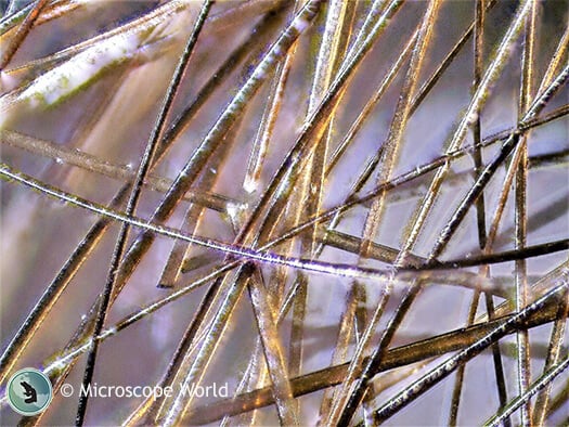
Figure: Hair under the microscope. Image Source: Microscope World.
Observation under the stereo microscope
- Stereo microscopes allow up to 90X magnification for the observation of the general structure and condition of the hair.
- The external characteristics like color, shape, texture, and length of hair can be seen easily through a stereomicroscope.
- Under this microscope, the hair will appear to have tiny fragments or fiber on its surface.
- It allows the observation of how uniform the thickness and pigmentation of the hair are.
- When placed on the scalp, the microscope also provides information on the condition and composition of the scalp.
- The outer scales on the hair can be observed to some extent through this microscope.
Observation under the compound microscope
- The compound microscope provides a more detailed visualization of the hair fragment.
- Through the compound microscope, it is possible to distinguish hairs on the basis of their thickness and also allows the differentiation of different scales present on the hair.
- The scales are seen to be present in an annular pattern which is usually different in different animals.
- Through a compound microscope, it is possible to distinguish the three layers of hair; cuticle, medulla, and cortex.
- The cuticle consists of scales made up of keratinized structures in the form of rings followed by the cortex that provides moisture and pigmentation to the hair.
- The medulla, in turn, is seen either as a long continuous thread or is fragmented or even absent in some hair.
14. Paramecium under the microscope
- Paramecium is a single-celled organism resembling in shape to that of the sole of a shoe.
- It is a eukaryote that has developed cellular organelles with a nucleus enclosed inside a nuclear membrane.
- It is a ciliated organism with cilia present throughout the body of the organism.
- Paramecium is a freshwater protist that can be easily collected along with the water sample.
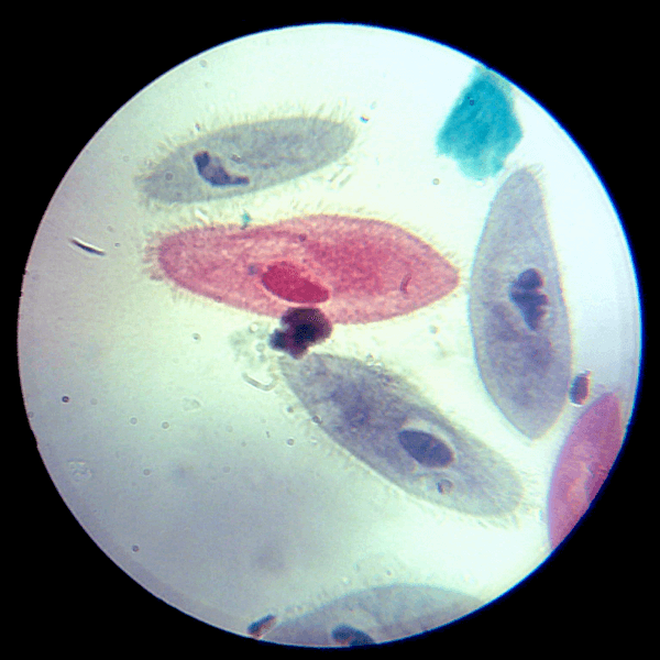
Figure: Paramecium under the microscope. Image Source: Office for Science and Society, McGill University.
Direct observation
- When observed directly under the microscope, this organism appears like the sole of a shoe and thus is named “slipper animalcules”.
- The cilia move in a coordinated way to propel the organism forward. The movement can be seen under the microscope if observed directly.
- The body of the organism is transparent and thus is very difficult to observe without staining.
- The cytoplasm of the organisms is seen as a transparent jelly that moves throughout the microscopic field.
Observation after staining
- After staining, it is easier to distinguish the organism from other particles.
- The cytoplasm of the organism is stained, revealing the contents of the cytoplasm as tiny colored dots.
- The nucleus is seen as a dark stained elongated structure at the center of the cytoplasm.
- On the surface of the cell membrane of the organism, tiny hair-like projections are seen throughout the body.
- A folded structure is observed on the side of the cell membrane, which is the oral groove.
15. Plant cell under the microscope
- Plants cells are larger than animal cells ranging in size from 10-100 µm in length.
- The structure and shape of the cell are more rigid when compared to animal cells as plant cells have a rigid cell wall that provides a more solid structure to the plant cell.
- The shape of the plant cell is usually rectangular in shape even though some plant cells have a triangular shape.
- A plant cell is also turgid than animal cells as the cell membrane can withstand more pressure than animal cells.
- Green plants have pigment deposits on their cell, which might provide some color to the cell.
- It is difficult to differentiate a single plant cell from others, and thus these are usually observed in the form of tissues.
- Because the structure of living and dead plant cells is not much different, plant cells are mostly observed after staining.
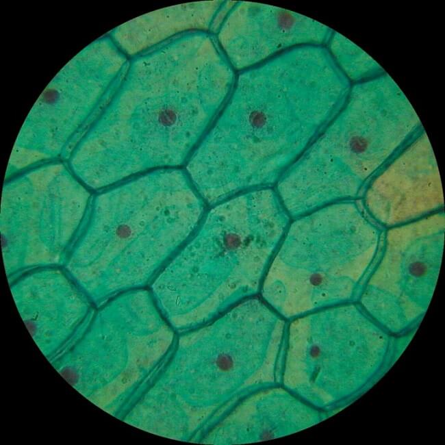
Figure: Plant cell under the microscope. Image Source: Glenda Stovall (Puplbits).
Observation after staining
- Under the microscope, plant cells are seen as large rectangular interlocking blocks.
- The cell wall is distinctly visible around each cell. The cell wall is somewhat thick and is seen rightly when stained.
- The cytoplasm is also lightly stained containing a darkly stained nucleus at the periphery of the cell.
- Similarly, a large empty vacuole occupies most of the cell.
- Throughout the cytoplasm, tiny dots or granules are seen indicating the presence of starch granules.
- The plant cells from the green parts of the plant might even have some green pigments deposited on some parts of the cytoplasm.
16. Pollen under the microscope
- Pollens are the male gametes in sexually reproducing plants.
- Pollen is a small grain consisting of few cells.
- These are macroscopic structures that can be observed with naked eyes.
- Macroscopically, they appear as yellow dust-like particles that can be easily moved by wind or water.
- Pollen is produced in the anther of the male reproductive part of the plant.
- These are haploid having half the number of chromosomes as in regular plant cells.
- Because these are macroscopic structures, they can be observed easily even through a stereomicroscope.
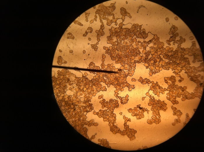
Figure: Pollen under the microscope. Image Source: Hanny van Arkel.
Observation under the stereo microscope
- In the stereo microscope, pollen appears irregularly shaped with random structures.
- They are yellow in color, and each pollen is different from the other in structure and shape.
Observation under the compound microscope
- Different structures within the pollen appear better under staining as it provides contrast.
- Under a compound microscope, pollen appears ovoid and is provided on the surface with scales or similar structures.
- The structure of the pollen also depends on the type of plant.
Observation under the electron microscope
- Under the electron microscope, pollens appear as inflated or deflated ovoid structures.
- The surface of the pollen is provided with cleavages and marks which are different in different pollen.
- The scales on the surface are irregularly placed with some pollen having scales throughout the surface and some having them only at the polar region.
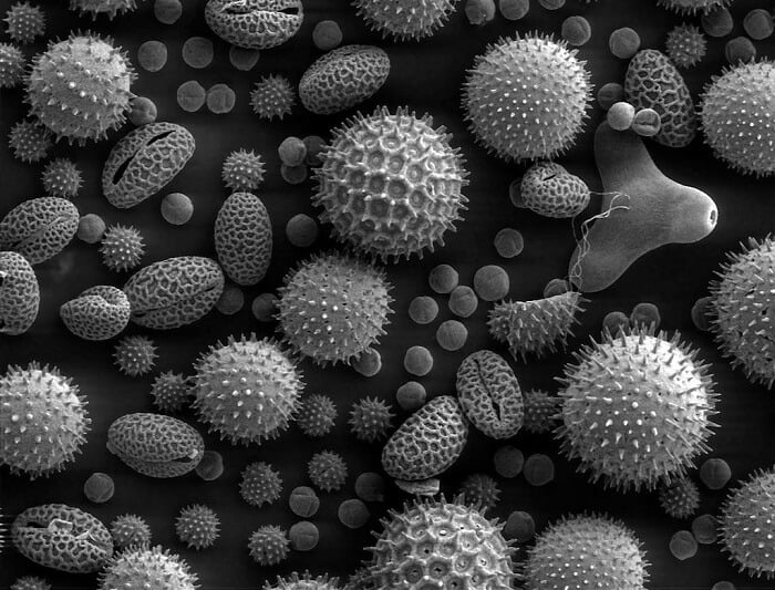
Figure: Pollen under the microscope (SEM). Image Source: Dartmouth College.
17. Salt under the microscope
- Salts are mineral compounds that are usually found in nature and can also be made through acid-base reactions.
- Salt is essential for the living being as it provides the necessary minerals to the body.
- Salt exists in the form of a crystal and is made up of two or more electrons.
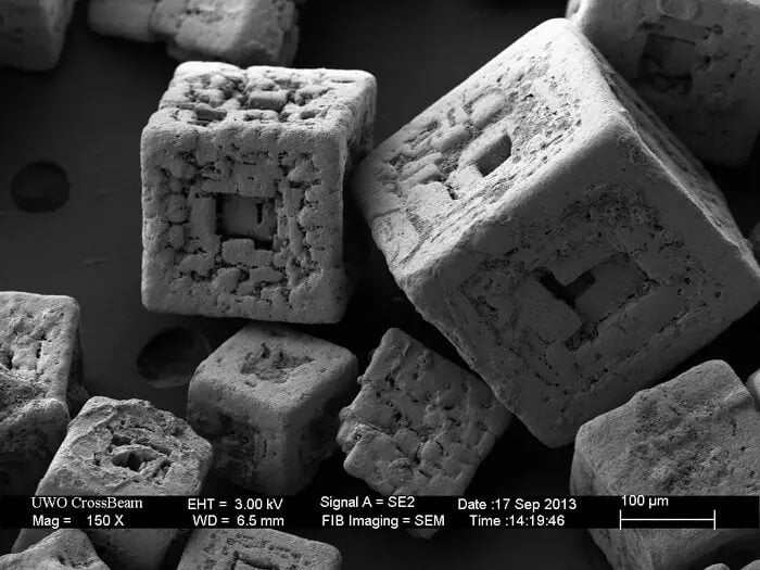
Figure: Salt under the microscope (SEM). Image Source: ZEISS (Flickr).
- The shape of different salt crystals may not be the same as they go through wear and tear.
- The internal structure or chemical makeup, however, is the same in all salt crystals.
- Salt crystals are macroscopic structures and thus can easily be viewed through a compound microscope.
- Under the microscope, salt crystals appear cubical in shape.
- The elements are arranged in the form of lattices arranged in separate planes.
- Since they are 3-Dimensional, with a compound microscope, you will see a fuzzy outline on the edge where there is an out-of-focus section.
- The arrangement of the lattices in the salt crystal results in nice, shiny crystal faces.
- The structure of the crystals might differ in different salts with some salt crystals having rectangular or hexagonal structures.
18. Sand under the microscope
- Sand is a loose granular material consisting of finely divided rocks and other mineral particles.
- The composition of sand and the ratio of its components vary from one location to another.
- Sand is made up of fine particles called sand grains having a diameter ranging from 0.06 mm to 2 mm.
- Sand and all other types of soil are formed by breaking soil by the process of weathering.
- The properties of sand can be used to determine the place of their origin.
- Sand particles are microscopic particles that can be seen with our naked eyes.
- Macroscopically, the color of the sand particles and their size can be determined.
- However, in order to determine other physical properties of sand particles, we can observe these particles either with a magnifying glass or with a compound microscope.
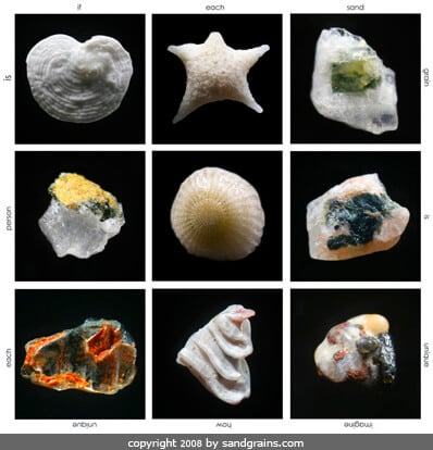
Figure: Nine Sand grains under the microscope. Image Source: Gary Greenberg (Sand Grains).
Observation under a magnifying glass
- Under a magnifying glass, it is possible to observe individual grains of sand particles and distinguish the color of these particles.
- Based on the color and size of these particles, their place of origin can be determined.
- While observing sand particles under a magnifying glass, we can see that the size and color of the particles are not always uniform which might be because the sand particles are moved around because of wind and other environmental factors.
Observation under the compound microscope
- Under a compound microscope, the differences between the sand particles become more apparent.
- It is visible that the shape, size color, and texture of individual particles vary within the sand collected from the same place.
- Some grains might appear smooth, while others appear irregular and sharp. The softer grains indicate that they were formed earlier in time than the sharp and irregular ones.
- The color of the sand particles and their opacity determine the composition of the sand particles.
- Particles that are translucent and shiny usually have a higher ratio of quartz. In contrast, other particles that are dull and black often have iron and other metals as their main component.
- Pink, peach, or such light-colored sand particles tends to have granite as their main component.
- Sand particles with holes or some texture on the surface indicate the remains of some marine life forms.
19. Skeletal muscle under the microscope
- Skeletal muscles are the muscles that are attached to the bones of the skeleton system that are connected by the bundle of collagen termed tendons.
- These are striated muscles that are voluntary and move in the direction of the somatic nervous system.
- The shape, size, and arrangement of fibers in skeletal muscle vary according to the position of the muscle in the body.
- These muscles are essential for the movement of the bones and also provide the elasticity required for contraction and relaxation.
- An individual cell of the skeletal muscle is a unicellular unit; however, the muscle formed by the bundle of these cells is multicellular and can be seen with naked eyes.
- The skeletal muscles are red in color because of the presence of myoglobin and a large number of mitochondria.
- When observed under a microscope, however, they might be confused with other connective tissue, which is why microscopic observation after staining is recommended.
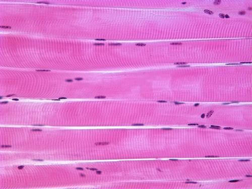
Figure: Skeletal muscle under the microscope. Image Source: School of Biomedical Sciences, Newcastle University.
40X magnification
- Under the microscope at the magnification of 40X, bundles of muscle fibers termed fascicles are seen where each of such bundles is separated by connective tissue, perimysium.
- Similarly, nuclei of the cells might also be visible, which appear like tiny dots.
100X magnification
- Under a higher magnification of 100X, nuclei of the cells appear towards the periphery because of the proteins present in the cytoplasm of the muscle cells.
- With the increased magnification, we can observe individual muscle cells connected to each other through another connective tissue, the endomysium.
- The nuclei of the cells of the connective tissue might also be seen that are smaller and more rounded than that of the muscle cells.
400X magnification
- In this case, the nucleus appears more flat and oval if the muscle sample taken is sectioned transversely.
- Faint lines are seen across each of the muscle cells, which are termed striations. This is why the skeletal muscles are included in the striated muscles category.
- These striations, however, are not actual structures inside the cell but are the reflection of light caused by the proteins present inside the cells.
20. Skin under the microscope
- Skin is the largest and one of the most important organs of our body.
- The skin is constituted by three layers: epidermis, papillary dermis, and reticular dermis, composed respectively by squamous stratified epithelium, loose connective, and connective containing compact collagen fibers.
- According to the location, the thickness of the epidermis varies from 0.06 to 1 mm.
- The most predominant cell type in the epidermis is the keratinocyte and several morphologically distinct epidermis layers are formed as the keratinocytes move from the basement membrane to the skin surface.
- Skin, as an organ, is a multicellular structure; however, individual skin cells are microscopic and can only be viewed under a microscope.
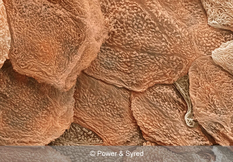
Figure: Skin under the microscope. Image Source: PS micrographs.
- The general surface of the sin can be viewed through a hand-held stereo microscope.
- This reveals the outer surface of the skin arranged in the form of scales and pores are seen throughout the skin.
- These pores are the openings of the sweat and sebaceous glands distributed throughout the skin.
- In addition, individual hair strands are also visible, which are present close to the pores.
- Under a high-power microscope, the different layers of the skin are seen.
- The outermost epidermis and inner dermis are visible through the compound microscope.
- The cells on the epidermis appear more irregular and are formed of fewer layers, whereas the cells in the dermis are more uniform and have more layers.
- The nucleus of the cells is visible towards the base of the cells.
- The projection of hair strands can also be seen origination from the roots present inside the skin.
- Besides, ducts of the different glands can be seen passing through the cell and opening on the surface of the skin.
21. Snowflake under the microscope
- Snowflake is the term used to describe individual ice/ snow crystals that together larger crystal balls of snow.
- Snowflakes are interchangeably also termed snow crystals.
- These flakes are formed from water vapor as they freeze under lower temperatures and the snowflakes take shapes as more water molecule freezes on the surface of the seed crystal.
- Each snowflake might have an individual shape and structure as well as patterns on its surface.
- Snowflakes are macroscopic and can be seen with naked eyes; however, the structure and pattern present cannot be viewed without a microscope.
- Because of their macroscopic structure, they can be viewed merely under a stereomicroscope.
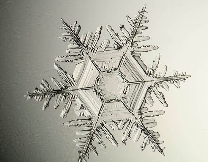
Figure: Snowflake under the microscope. Image Source: Michael Peres.
- Under a magnifying glass or a stereo microscope, the shape and structure of the snowflake can be determined. For the pattern present on the surface, however, a compound microscope is to be used.
- Under a compound microscope, all snowflakes have a geometric crystalline shape.
- These flakes are symmetric and usually have a six-sided hexagonal shape.
- In addition, different patterns can be seen on the surface, which is different in different flakes.
- The difference in the pattern of the flakes is due to the differences in the way the molecules of water are joined.
22. Sperm under the microscope
- Sperms are male gametes that are formed in the testes of the male reproductive system in humans and other animals.
- Sperms are haploid and carry only 23 chromosomes in humans.
- The general morphology of a sperm cell is composed of a clear head, midpiece, and tail.
- Sperms are highly motile and thus require a large amount of energy which is provided by a large number of mitochondria present in the cell.
Direct observation
- One of the most distinctive characters of sperms is their motility, and thus, direct observation of sperms is usually done before staining to ensure the presence of sperms.
- Through direct observation, it is possible to detect the motility of sperm, which is rapid and random.
- Similarly, the basic structure of sperm can also be identified through the microscope.
- The head and body of the sperm appear as one under direct observation whereas the tail is distinguishable as a long flagella-like structure.
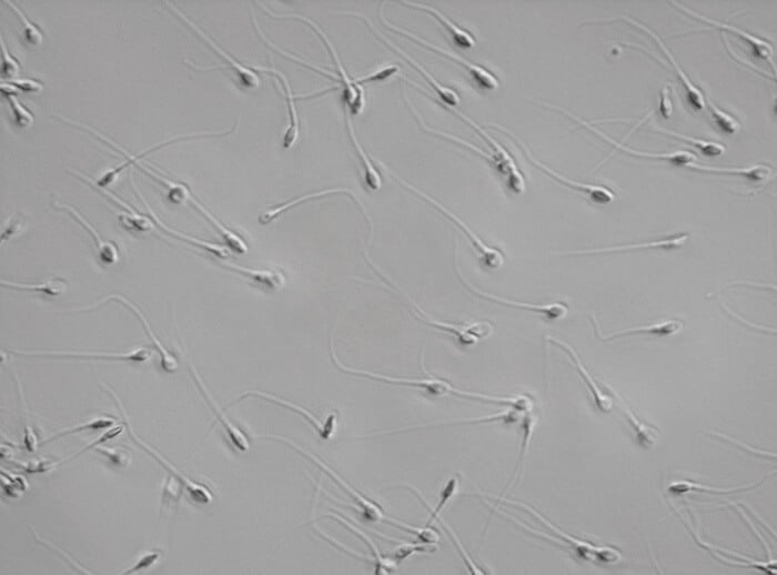
Figure: Sperm under the microscope. Image Source: Zeiss.
Observation after staining
- After staining the sperm with appropriate dye, the body of the sperm appears red while the acrosome and the tail appear green.
- The head appears as a smooth oval structure that resembles an egg.
- The head is important as it carried the chromosomes and also has the acrosome on the anterior part.
- The acrosome and acrosome cap are present together at the top of the head that appear conical in shape.
- The nucleus is seen as a stained dot and also has a nucleus vacuole.
- After the head is a short piece that carries all the mitochondria necessary to generate the energy required for the motility of the sperm.
- Similarly, a centriole is also present between the head and the midpiece.
- At last, the tail appears a long elongated structure that occupies about 80%of the entire sperm.
- The tail is transparent and thus is difficult to detect under a low-power microscope.
23. Spirogyra under the microscope
- Spirogyra is a green alga found mostly in freshwater in the form of green clumps.
- Spirogyra is unicellular, but because it clumps together, it can be seen in the pond even with our naked eyes.
- These organisms have green pigments that are arranged in the form of ribbons in the cytoplasm.
- Spirogyra exists in chains where individual cells are stacked on top of another.
- The name spirogyra is given because of the spiral/ helical structure of chloroplasts present in the cytoplasm.
- Because they are pigmented, they can be easily viewed directly without any staining.
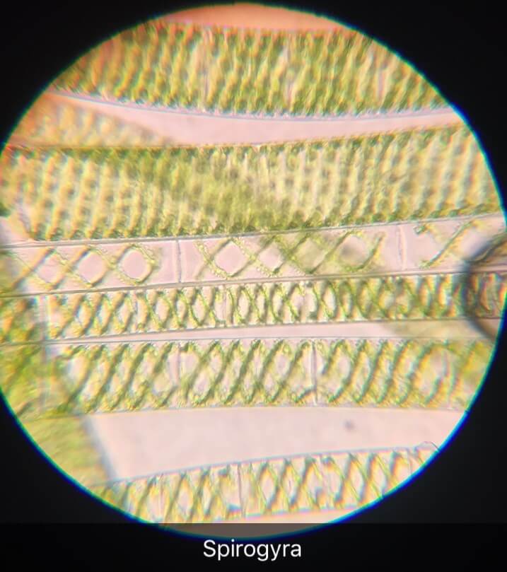
Figure: Spirogyra under the microscope. Image Source: Motherdragonair.
- Under the microscope, spirogyras appear surrounded by a slimy jelly-like substance which is the outer wall of the organism dissolved in water.
- The next layer of the cell wall is present on the outside of the cell that appears transparent.
- The cytoplasm is also transparent except the chloroplast arranged in the form of ribbons.
- These ribbons are observed as helical structures in the cytoplasm.
- For the differentiation of the nucleus and other cell organelles, staining has to be performed.
- After staining, the nucleus is visible as a stained spot at the side of the cytoplasm beside the ribbons of chloroplasts.
24. Virus under the microscope
- Viruses are particles that are considered obligatory parasites as they don’t grow or survive outside a living organism.
- The size of viruses ranges from 20 nm to 200-450 nm in diameter.
- Because viruses are tiny as compared to bacteria, they cannot be viewed with a compound microscope.
- Instead, high-power microscopes like fluorescence microscopes or transmission electron microscopes are to be used.
Fluorescence microscope
- Under fluorescence microscopes, the viruses appear the color of the fluorescent particle used.
- It is difficult to distinguish the structure of the virus, but this technique is useful for the quantitative estimation of the virus.
- The fluorescent dyes are specific for certain proteins which allows them to detect the desired particles.
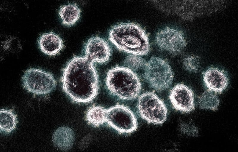
Figure: Virus (SARS-CoV-2) under the microscope (TEM). Image Source: NIAID (Flickr).
Transmission electron microscope
- Transmission electron microscopes are better for the observation of viruses as they provide up to 1000X magnification of particles.
- Through this type of microscope, it is possible to observe viruses inside the cells of living beings.
- Like in fluorescent microscopy, this technique also utilizes dyes that are specific for the proteins in the viruses which allow the visualization of the viruses.
- When the structure of a virus is viewed under a powerful microscope, it may be icosahedral or helical.
- The shape and structure of each virus are different from the other, but the composition is similar.
- All viruses have genetic material which can be either DNA or RNA enclosed inside a protein coat.
- If glycoprotein spikes are present like in the influenza virus, those can also be visible.
- In the case of bacteriophage viruses, the tail and tail fibers are also visible and are found attached on the surface of bacterial cells.
- The protein head can be seen as a hexagonal capsid inside which the genetic material is present in the form of coiled strands.
25. Volvox under the microscope
- Volvox is an alga usually found in ponds, ditches, and shallow puddles.
- These are unicellular organisms and thus cannot be seen through naked eyes.
- These organisms like spirogyra have chloroplasts deposited in the cytoplasm of the organism.
- Volvox exists in colonies and thus appears larger than their cells.
- Their size ranges from 350-500 µm but appears larger as they exist in the form of colonies.
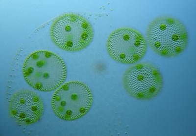
Figure: Volvox under the microscope. Image Source: Wim van Egmond.
- Under the microscope, about 200-50,000 individual cells are arranged in the form of a hollow sphere.
- Within the parent colony, further many daughter colonies can be seen.
- Once the parent colony bursts, the daughter colonies are released which then develops into new parent colonies.
- Each volvox cell appears to have two flagella that beat together to move around in the water.
- Individual volvox cell is spherical and occupies cytoplasm, a transparent nucleus, and green colored granules.
- Towards the periphery, a red eyespot can be seen that receives sunlight for the preparation of food.
26. Worm under the microscope
- Worms are invertebrates that are further divided into three groups; roundworms, flatworms, and segmented worms.
- Although the shape and structure of worms vary, worms are generally characterized by an elongated, legless body where the organisms move by crawling movement.
- Worms are found throughout the world in different habitats, but most of them are terrestrial and are found in soil.
- Some worms might even be parasitic.
- Worms are macroscopic organisms; however, the internal structure and components are not visible with the naked eyes.
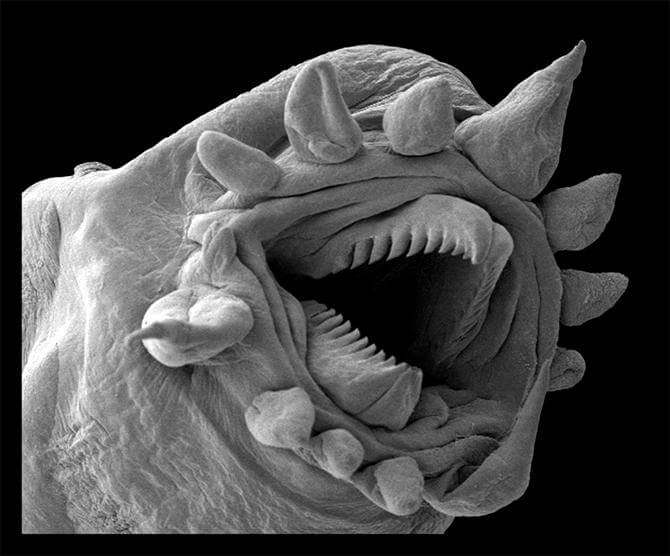
Figure: Worm under the microscope. Image Source: Philippe Crassous.
Observation under the magnifying glass
- Under the magnifying glass, segmented worms like earthworms appear visible.
- The uppermost segment is the head that is smaller than other segments.
- The dorsal part of the body might appear dark due to the epidermis whereas the ventral surface is lighter in color and thus more clearly visible.
- A more distinct and thick segment is present in the upper part of the body called the clitellum.
- In addition, fine hair-like projections called setae are also visible in each segment.
- The flatworms, in turn, are smaller than segmented worms and have a flattened leaf-like body.
- The anterior part of the body appears broader than the posterior end.
- A closer look may also reveal eyespots at the head region as well as a pharynx located near the middle (central part of the body).
Observation under the compound microscope
- Under a high-power microscope, a muscular flap might be visible at the anterior end of the body, which is the prostomium.
- Prostomium surrounds the mouthparts of the worm.
- A septum is also visible, separating each segment on the body of the worm.
- After a closer look, the ventral surface of the worm appears flatter than the dorsal surface.
- The setae or hairs will be more visible than with the magnifying glass.
- Apart from the hair, pores are also visible on the surface of the worm. Some pores appear more significant than others.
- Additionally, to observe the internal organs of the worm, worms can be dissected.
27. Yeast under microscope
- Yeasts are unicellular eukaryotic organisms that are mostly found in plants and soil.
- Some yeasts are also found on the surface of the skin and even inside the body of some animals.
- Yeasts mostly exist in a budding form with few cells found as single or pairs.
- These are microscopic organisms but are visible with naked eyes when present in large numbers.
- Some yeast cells are visible without staining under bright field microscopes.
- In a bright field microscope, yeast appears as oval-shaped cells with tiny buds visible in some cells.
- These are colorless but under a bright-field microscope might appear creamy to off-white in color.
- For the observation of cellular organelles, yeast cells have to be stained.
- In fluorescent microscopes, different dyes can be used for different organelles to obtain a more detailed structure of the organelles.
- Using separate dyes for separate organelles increases the contrast and allows a better distinction between them.
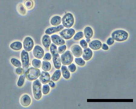
Figure: Yeast under the microscope. Image Source: microbiological garden.
References and Sources
- Alberts B, Johnson A, Lewis J, et al. Molecular Biology of the Cell. 4th edition. New York: Garland Science; 2002. Looking at the Structure of Cells in the Microscope. Available from: https://www.ncbi.nlm.nih.gov/books/NBK26880/
- <1% – https://www.worldatlas.com/feature/what-are-the-differences-between-plant-cells-and-animal-cells.html
- <1% – https://www.wisegeek.com/what-is-blood-tissue.htm
- <1% – https://www.wired.com/story/new-microscope-shows-the-quantum-world-in-crazy-detail/
- <1% – https://www.wikihow.com/Identify-Ants
- <1% – https://www.thoughtco.com/gram-stain-procedure-4147683
- <1% – https://www.thoughtco.com/cytoplasm-defined-373301
- <1% – https://www.studymode.com/essays/Lab-Report-1-Microscopy-And-Staining-922932.html
- <1% – https://www.slideshare.net/diojoeyrichard/male-reproductive-system-15216719
- <1% – https://www.sciencedirect.com/topics/agricultural-and-biological-sciences/cytoplasm
- <1% – https://www.sciencedirect.com/topics/agricultural-and-biological-sciences/yeasts
- <1% – https://www.sciencedaily.com/terms/ant.htm
- <1% – https://www.reddit.com/r/explainlikeimfive/comments/2jji7r/eli5why_is_the_atom_considered_the_smallest_piece/
- <1% – https://www.quora.com/What-is-the-dark-spot-at-the-center-of-the-nucleus-called
- <1% – https://www.quora.com/What-does-glass-and-other-transparent-matter-look-like-under-a-microscope
- <1% – https://www.quora.com/Do-white-blood-cells-have-a-nucleus-If-so-why
- <1% – https://www.qsstudy.com/biology/describe-with-labelled-diagram-the-structure-of-spirogyra
- <1% – https://www.ncbi.nlm.nih.gov/pmc/articles/PMC6133778/
- <1% – https://www.ncbi.nlm.nih.gov/pmc/articles/PMC3925548/
- <1% – https://www.ncbi.nlm.nih.gov/pmc/articles/PMC3224570/
- <1% – https://www.ncbi.nlm.nih.gov/books/NBK26880/
- <1% – https://www.microscopemaster.com/worm-under-a-microscope.html
- <1% – https://www.microscopemaster.com/red-blood-cells.html
- <1% – https://www.microscopemaster.com/e-coli-under-microscope.html
- <1% – https://www.microscopemaster.com/ants-under-a-microscope.html
- <1% – https://www.microscopemaster.com/amoeba-under-the-microscope.html
- <1% – https://www.microscopeinternational.com/what-is-a-compound-microscope/
- <1% – https://www.manoa.hawaii.edu/exploringourfluidearth/biological/invertebrates/worms-phyla-platyhelmintes-nematoda-and-annelida
- <1% – https://www.leica-microsystems.com/science-lab/factors-to-consider-when-selecting-a-stereo-microscope/
- <1% – https://www.lab.anhb.uwa.edu.au/mb140/CorePages/Blood/Blood.htm
- <1% – https://www.ivis.org/library/concise-review-of-veterinary-virology/general-characteristics-structure-and-taxonomy-of
- <1% – https://www.invitra.com/en/sperm-cell/structure-and-parts-of-a-sperm-cell/
- <1% – https://www.infovisual.info/en/biology-animal/insect
- <1% – https://www.hematology.org/education/patients/blood-basics
- <1% – https://www.dummies.com/education/science/anatomy/the-anatomy-of-skin/
- <1% – https://www.colgate.com/en-us/oral-health/basics/mouth-and-teeth-anatomy/parts-of-the-mouth-and-their-functions-0415
- <1% – https://www.britannica.com/science/thorax
- <1% – https://www.britannica.com/science/skeletal-muscle
- <1% – https://www.britannica.com/science/prokaryote
- <1% – https://www.britannica.com/science/cilium
- <1% – https://www.biologyonline.com/dictionary/algae
- <1% – https://www.answers.com/Q/Why_are_red_blood_cells_shaped_as_biconcave_disk
- <1% – https://www.answers.com/Q/What_is_The_most_superior_segment_of_the_upper_limb_is_called
- <1% – https://theydiffer.com/difference-between-red-and-white-blood-cells/
- <1% – https://study.com/academy/lesson/functions-of-red-blood-cells-white-blood-cells-platelets.html
- <1% – https://sciencing.com/what-crystal-how-does-form-4925052.html
- <1% – https://sciencestruck.com/dissecting-microscope-vs-compound-microscope
- <1% – https://sciencespot.net/Media/FrnsScience/hairfibercard.pdf
- <1% – https://quizlet.com/87558617/salts-flash-cards/
- <1% – https://quizlet.com/80154963/plants-vocab-grade-7-flash-cards/
- <1% – https://quizlet.com/36858773/12-striated-smooth-and-cardiac-muscle-flash-cards/
- <1% – https://quizlet.com/3369374/algae-and-lower-plants-flash-cards/
- <1% – https://quizlet.com/123172441/annelids-chapter-12-flash-cards/
- <1% – https://quizlet.com/109401870/chapter-5-hair-flash-cards/
- <1% – https://quizlet.com/106882058/chapter-9-muscular-system-flash-cards/
- <1% – https://pediaa.com/difference-between-dna-and-rna-viruses/
- <1% – https://microscope-microscope.org/microscope-info/phase-contrast-microscope/
- <1% – https://microscope-microscope.org/microscope-applications/microscopic-sand/
- <1% – https://microbiologynotes.org/deoxyribonucleic-acid-dna/
- <1% – https://medical-dictionary.thefreedictionary.com/basophilic+leukocyte
- <1% – https://hypertextbook.com/facts/2001/JenniferShloming.shtml
- <1% – https://encyclopedia2.thefreedictionary.com/Phylum+Chlorophyta
- <1% – https://en.wikipedia.org/wiki/Reptile_scale
- <1% – https://en.wikipedia.org/wiki/Prokaryotic_cells
- <1% – https://en.wikipedia.org/wiki/Pathogenic_Escherichia_coli
- <1% – https://en.wikipedia.org/wiki/Electron_microscope
- <1% – https://en.wikipedia.org/wiki/Coccus
- <1% – https://employees.csbsju.edu/ssaupe/biol327/Lecture/cell-wall.htm
- <1% – https://educheer.com/reports/cheek-cell/
- <1% – https://differencecamp.com/plant-cell-vs-animal-cell/
- <1% – https://courses.lumenlearning.com/boundless-chemistry/chapter/the-structure-of-the-atom/
- <1% – https://courses.lumenlearning.com/boundless-biology/chapter/osmoregulation-and-osmotic-balance/
- <1% – https://courses.lumenlearning.com/boundless-ap/chapter/the-skin/
- <1% – https://civiltoday.com/civil-engineering-materials/sand/233-sand-composition-types
- <1% – https://chem.libretexts.org/Bookshelves/Physical_and_Theoretical_Chemistry_Textbook_Maps/Supplemental_Modules_(Physical_and_Theoretical_Chemistry)/Physical_Properties_of_Matter/States_of_Matter/Properties_of_Liquids/Unusual_Properties_of_Water
- <1% – https://byjus.com/biology/plant-cell/
- <1% – https://bodytomy.com/function-of-red-blood-cells
- <1% – https://biologywise.com/what-is-spirogyra
- <1% – https://biologyreader.com/difference-between-light-and-electron-microscope.html
- <1% – https://bio.libretexts.org/Bookshelves/Microbiology/Book%3A_Microbiology_(Bruslind)/06%3A_Bacteria_-_Surface_Structures
- <1% – https://answers.yahoo.com/question/index?qid=20070628145438AAJN52E
- <1% – https://activilong.com/en/content/95-structure-composition-of-the-hair
- <1% – http://www2.centralcatholichs.com/Bio1site/Cells/plasma%20membrane2.pdf
- <1% – http://www1.biologie.uni-hamburg.de/b-online/e03/03e.htm
- <1% – http://www.webexhibits.org/pigments/intro/greens.html
- <1% – http://www.mriquestions.com/why-are-veins-blue.html
- <1% – http://www.microbehunter.com/phase-contrast-vs-bright-field-microscopy/
- <1% – http://www.funscience.in/study-zone/Biology/Respiration/RespirationInAmoeba.php
- <1% – http://www.crossroadsacademy.org/crossroads/wp-content/uploads/2016//05/Tissues-and-Organs.pdf
- <1% – http://medcell.med.yale.edu/systems_cell_biology/blood_lab.php
- <1% – http://downloads.lww.com/wolterskluwer_vitalstream_com/sample-content/9780781797597_Cui/samples/Chapter_04.pdf

The worm was cool and scary at the same time.
The worm was so weird because i didn’t know that worms have teeth
the worm is disgusting
I think it was cool but the worm was scary.
Would you please advise how I may identify microscopic, I will suggest mites coloured tan, white and a black with the later having a vicious nature bites, white and the tan noticeably boring under skin. Medically they see nothing but I can see with my hobby microscope but they are not defined. Large numbers, had a contaminated water tank so bacteria. Fungi, mould, yeast as the mites are attracted to yeast and multiply in and out of body continuously.
This is so very interesting. I admire your ability to collect these images and go forth with accurate observations and analysis. Contact me dirving1927@gmail.com