Interesting Science Videos
What are Eosinophils?
Definition of Eosinophils
Eosinophils are motile phagocytic cells that play an important homeostatic role in providing defense against parasitic infections.
- Eosinophils are bone marrow-derived granulocytes that remain in the bloodstream for a shorter period of time and mostly reside in tissues.
- The functions of eosinophils are multifaceted, including antigen presentation, the release of peptides, lipids, and cytokine mediators.
- However, these can also be a part of malignant neoplasms and autoimmune conditions, and connective tissue disorders.
- Eosinophils represent about 6% of the total circulating leukocytes and can be found in tissues of the lung, thymus, mammary glands, spleen, and uterus.
- It has been long believed that eosinophils that leave the bone marrow are mature cells; however, recent studies have indicated that multiple tissue-specific subtypes of the cell that are differentiated on the basis of the cell surface marker expression exist.
- Eosinophils present in the circulation are recruited into the physiological locations and inflammatory sites in response to eosinophil-specific chemokines and a variety of other stimuli.
- These cells remain localized around invading worms where their membranes are damaged by the activity of proteins released from the eosinophils.
- Besides, eosinophils are known to important roles in asthma and allergy symptoms in areas where parasitic infections are less prevalent.
- Eosinophils are also associated with certain medical conditions like eosinophilia, where the number of eosinophils ranges between 450-550 cells/µL in the bloodstream.

Structure of Eosinophils
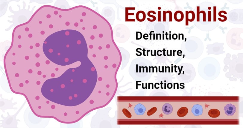
- Eosinophils are granulocytes, measuring in size between 10-16 µm in diameter. The cells contain a segmented or bi-lobed nucleus where the nuclei: cytoplasm ratio is about 30%.
- An important morphological feature of eosinophils is the content of their cytoplasmic granules, which contain specific cationic proteins.
- The specific granules of eosinophils are the principal distinctive feature of the cells. These granules contain a unique crystalloid core, a dense matrix surrounded by a membrane. The core is responsible for the cardinal properties of eosinophils.
- The granules also contain different mediators like proteins, cytokines, chemokine, and enzymes that can induce inflammation and tissue damage.
- The cytoplasm also contains Golgi bodies, endoplasmic reticulum, and mitochondria that function to produce proteins and ATPs in the cell.
- The histological structure of eosinophils depends on the level of activation of the cells as the number of vesicles in the cells is only observed while the cell undergoes piecemeal degranulation.
- The plasma membrane of eosinophils contains numerous receptors that are specific for certain cytokines that regulate the maturation and other physiological functions of the cell.
How Eosinophils work against pathogens? (Immunity)
- Eosinophils are formed from the multipotent hematopoietic stem cells under the influence of soluble mediators and transcription factors.
- The mature eosinophils contain a molecular system that allows the docking and fusion of vesicles to the membrane of the cell.
- Degranulation or the release of proteins and other components inside the vesicles are released via regulated exocytosis. The docking complex is composed of soluble N-ethylmaleimide-sensitive factor attachment protein receptors present on the vesicles and the target membrane.
- Besides degranulation, eosinophils have also been associated with antigen presentation to T cells.
- The process usually occurs post allergen exposure, where the cell expresses machinery for antigen presentation as well as a full set of co-stimulatory molecules like MHC II.
- The eosinophils present in tissues migrate to draining lymph nodes, ultimately reaching the proliferation zone by a process that is independent of the eotaxin receptor CCR3.
- The antigen-presenting eosinophils also promote antigen-specific T-cell proliferation.
- The process of accumulation of eosinophils at a particular side of inflammation involves numerous sequential interactions that allow the cells to adhere and transmigrate through the endothelium layer.
- The adherence of eosinophils to the endothelium layer involves different pathways, including CD18-dependent pathways, adherence to E-selection, and P-selection.
- In the case of allergen-induced recruitment of eosinophils, the movement of cells is dependent on CD4+ T cells and interferon-γ.
Functions of Eosinophils
The following are some of the functions of eosinophils:
- The most obvious function of eosinophils is their role in host defense against parasitic infections.
- Besides, eosinophils are also known to release lipid, peptide, and cytokines that mediate inflammation and host defense.
- Some eosinophils also function as antigen-presenting cells as these can be induced to express MHC class II proteins.
- Eosinophils are effector cells that release lipid mediators like leukotriene C4, lipoxins, and PAF that contribute to the acute manifestations of allergic or immunological responses.
- Eosinophils also have the potential to regulate mast cell functions through the release of granule proteins and cytokines.
- A panel of cytokines like IL-2, IL-4, IL-6, IL-12 are released by eosinophils which are capable of promoting T cell proliferation and activation of Th1 or Th2 polarization.
- Eosinophils in the lungs has been recognized as a significant contributor to airway hyperreactivity and asthma.
References
- Peter J. Delves, Seamus J. Martin, Dennis R. Burton, and Ivan M. Roitt(2017). Roitt’s Essential Immunology, Thirteenth Edition. John Wiley & Sons, Ltd.
- Judith A. Owen, Jenni Punt, Sharon A. Stranford (2013). Kuby Immunology. Seventh Edition. W. H. Freeman and Company.
- Ramirez, Giuseppe A et al. “Eosinophils from Physiology to Disease: A Comprehensive Review.” BioMed research international vol. 2018 9095275. 28 Jan. 2018, doi:10.1155/2018/9095275
- Wen, Ting, and Marc E Rothenberg. “The Regulatory Function of Eosinophils.” Microbiology spectrum vol. 4,5 (2016): 10.1128/microbiolspec.MCHD-0020-2015. doi:10.1128/microbiolspec.MCHD-0020-2015
- Kovalszki, Anna, and Peter F Weller. “Eosinophilia.” Primary care vol. 43,4 (2016): 607-617. doi:10.1016/j.pop.2016.07.010
- Blanchard, Carine, and Marc E Rothenberg. “Biology of the eosinophil.” Advances in immunology vol. 101 (2009): 81-121. doi:10.1016/S0065-2776(08)01003-1
- Akuthota, Praveen, and Peter F Weller. “Eosinophils and disease pathogenesis.” Seminars in hematology vol. 49,2 (2012): 113-9. doi:10.1053/j.seminhematol.2012.01.005
- Peter F Weller. Eosinophils: structure and functions. Current Opinion in Immunology. Volume 6, Issue 1. 1994. Pages 85-90. https://doi.org/10.1016/0952-7915(94)90038-8.
- McBrien Claire N., Menzies-Gow Andrew. The Biology of Eosinophils and Their Role in Asthma. Frontiers in Medicine. VOLUME=4. 2017.93. DOI=10.3389/fmed.2017.00093.
Sources
- https://www.researchgate.net/publication/319625167_Functions_of_tissue-resident_eosinophils – 20%
- https://www.frontiersin.org/articles/10.3389/fmed.2017.00093/full – 6%
- https://cancerimmunolres.aacrjournals.org/content/canimm/2/1/1.full.pdf – 2%
- https://www.sciencedirect.com/science/article/pii/S0065277608010031 – 1%
- http://europepmc.org/articles/PMC5088784 – 1%
- https://www.sciencedirect.com/topics/medicine-and-dentistry/eosinophil-in-asthma – 1%
