Interesting Science Videos
What are Basophils?
Definition of Basophils
Basophils are nonphagocytic granulocytes that are the least common leukocytes occurring in the blood circulation and are critical to the immune response against parasites.
- Basophils are similar to eosinophils in that these are produced in response to parasitic infections; however, in the absence of parasitic infections, basophils are involved in allergy symptoms.
- Even though basophils account for a small percentage (0.5%) of the total circulating blood cells, the number can increase rapidly in response to inflammatory signals.
- Basophils are formed from the granulocyte-monocyte progenitors present in the bone marrow, which then populate the peripheral blood as mature cells.
- Studies related to the functions, roles, and their responses have been difficult due to their short lifespan and scarcity.
- Basophils are developmentally related to mast cells as both the cells express high-affinity receptors for IgE and can release a similar spectrum of mediators in response to IgE binding.
- Some of the common markers that can be used to distinguish basophils include ckit–FcεRI+, CD11b+, IL-3Rhi, etc.
- As a result of their involvement in allergic reactions, a basophil activation test has been prepared that detects allergic reactions to drugs, food, and venom in patients.
- Basophils are considered circulating innate cells that not only elicit microvascular permeability to enable the transmigration of leukocytes into the inflamed tissues but also contribute to the Th2 immune response.
- The paucity of basophils in circulating blood is a regulatory mechanism in innate and acquired immunity rather than hypersensitivity and atopic response.

Structure of Basophils
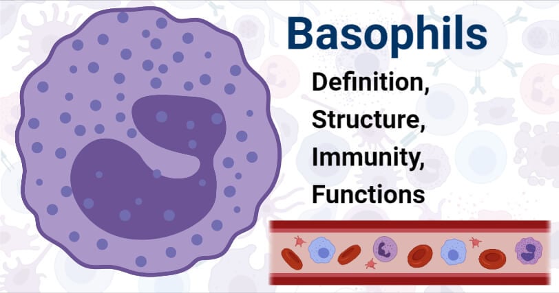
- Basophils are the smallest granulocytes with a diameter ranging between 10-14 µm. These polymorphonuclear cells with polylobed nucleus and prominent, brightly metachromatic cytoplasmic granules.
- Even though most basophils are rounded cells, some elongated, narrow uropods or tails have been described in humans.
- The nucleus contains condensed nuclear chromatin, but the nucleolus is absent.
- The cytoplasm contains organelles like mitochondria, vesicles, glycogen, and granules. The mature granules usually contain dense particles.
- Usually, human basophil cytoplasmic granules are round to angular, membrane-bound structures with sizes ranging up to 1.2 µm.
- The substructure of these granules contains dense particles in a less dense matrix occasional complex membrane.
- The cytoplasm has a complex vesicular system that is involved in basophil degranulation in the presence of an appropriate stimulus.
- The structure of basophils is similar to mast cells, and the differentiation can be made on the basis of glycogen content and the plasmalemma ridges.
- Basophils have a higher glycogen content in the cytoplasm, whereas these lack the plasmalemmal ridges and folds that are present in mast cells.
How do Basophils work against pathogens? (Immunity)
- Basophils are formed from the granulocyte-monocyte progenitor cells of the bone marrow, which are then released into the peripheral blood as fully differentiated cells.
- The differentiation of precursor cells into the basophil lineage is dependent on the transcription factor, C/EBPα.
- The level of basophils in the blood remains maintained under normal conditions, but the number increases as a result of parasitic infections or allergies.
- The exact nature of stimuli responsible for basophil development during parasitic infections is not clear yet, but the hematopoietic cytokine IL-3 has been suggested to play an essential role.
- Other parasite-associated molecules like proteases, glycoproteins, or structural components like chitin can also act as stimuli for the differentiation of basophils.
- Basophils, like mast cells, are activated by the cross-linkage between the surface IgE receptor, FcεRI, and the IgE antibodies present in the blood.
- The binding of IgE to basophils results in an activation signal which causes a rapid release of intracellular mediators like histamine and leukotrienes. The increased level of these compounds causes increased secretion of cytokines like IL-4 by basophils.
- Besides, IL-4-independent pathways involving IL-3 have also been described, which result in basophil activation.
- Activated basophils produce a large amount of IL-4, which might even be greater than that produced by T cells.
- The IL-4 produced by basophils induces the activation of parasite-specific naïve T cells to Th2 type effector T cells.
Functions of Basophils
The following are some of the functions of basophils:
- The most important function of basophils is the induction of immune response against parasitic infections, especially infections involving helminths.
- Studies have also indicated that basophils can function as professional antigen-presenting cells required for T cell differentiation. Basophils, once bound to IgE, lead to the differentiation of antigen-specific CD4 T cells into Th2 cells.
- Basophils work with other immune cells via cross-talk to maintain an orchestrated mechanism of allergy.
- Basophils also decrease hypersensitive response by the modulation of releasibility of either histamine or lipid mediators.
- Basophils activation test is a test developed to test allergic reactions to different foods, venoms, and drugs.
- One of the important physiological roles of basophils is the B cell maturation, resulting in IgE production.
References
- Peter J. Delves, Seamus J. Martin, Dennis R. Burton, and Ivan M. Roitt(2017). Roitt’s Essential Immunology, Thirteenth Edition. John Wiley & Sons, Ltd.
- Judith A. Owen, Jenni Punt, Sharon A. Stranford (2013). Kuby Immunology. Seventh Edition. W. H. Freeman and Company.
- Chirumbolo, Salvatore et al. “The role of basophils as innate immune regulatory cells in allergy and immunotherapy.” Human vaccines & immunotherapeutics vol. 14,4 (2018): 815-831. doi:10.1080/21645515.2017.1417711
- Cromheecke, Jessica L et al. “Emerging role of human basophil biology in health and disease.” Current allergy and asthma reports vol. 14,1 (2014): 408. doi:10.1007/s11882-013-0408-2
- Voehringer, David. “Recent advances in understanding basophil functions in vivo.” F1000Research vol. 6 1464. 15 Aug. 2017, doi:10.12688/f1000research.11697.1
- Siracusa, Mark C et al. “Basophils and allergic inflammation.” The Journal of allergy and clinical immunology vol. 132,4 (2013): 789-801; quiz 788. doi:10.1016/j.jaci.2013.07.046
- Min, Booki et al. “Understanding the roles of basophils: breaking dawn.” Immunology vol. 135,3 (2012): 192-7. doi:10.1111/j.1365-2567.2011.03530.x
- Min, Booki, and William E Paul. “Basophils and type 2 immunity.” Current opinion in hematology vol. 15,1 (2008): 59-63. doi:10.1097/MOH.0b013e3282f13ce8
- Dvorak A.M. (1988) The Fine Structure of Human Basophils and Mast Cells. In: Holgate S.T. (eds) Mast Cells, Mediators and Disease. Immunology and Medicine Series, vol 7. Springer, Dordrecht. https://doi.org/10.1007/978-94-009-1287-8_2
- Galli S.J., Dvorak H.F. (1979) Basophils and Mast Cells: Structure, Function, and Role in Hypersensitivity. In: Gupta S., Good R.A. (eds) Cellular, Molecular, and Clinical Aspects of Allergic Disorders. Comprehensive Immunology, vol 6. Springer, Boston, MA. https://doi.org/10.1007/978-1-4684-0988-8_1
Sources
- https://oncohemakey.com/basophils-mast-cells-and-related-disorders/ – 8%
- https://www.slideshare.net/vartika1428/cells-of-immune-system-37288603 – 5%
- https://europepmc.org/article/MED/29257936 – 4%
- https://www.medicalnewstoday.com/articles/324188 – 1%
- https://www.ncbi.nlm.nih.gov/pubmed/29017820 – 1%
- https://www.researchgate.net/publication/230796941_Recent_Developments_in_Basophil_Research_Do_Basophils_Initiate_and_Perpetuate_Type_2_T-Helper_Cell_Responses – 1%
- https://pediaa.com/difference-between-leukocytes-and-lymphocytes/ – 1%

Good the sir and mrs thanks the structure of the diagram agranulocyte