Interesting Science Videos
What are Monocytes?
Definition of Monocytes
Monocytes are cells of the immune system that represent immune effector cells with chemokine receptors and pathogen recognition receptors that circulate through blood or remain localized in lymphoid organs.
- These cells account for about 5% of the total circulating nucleated cells in human blood and have a half-life of three days.
- Monocytes are essential for both the innate immune system and the adaptive immune system as these differentiate into macrophages and dendritic cells.
- Monocytes are one of the master cells of the immune system as these can perform a group of different functions performed by different immune cells.
- These can recognize danger signals or stimuli via their pattern recognition receptors, resulting in phagocytosis. These can also present antigens to other cells and secrete chemokine and cytokines.
- Monocytes can be defined as circulating blood cells that develop in the bone marrow from the common myeloid progenitor cells.
- However, monocytes are known for their developmental plasticity as these can differentiate into osteoclasts or other immune cells depending on the inflammatory response.
- Monocytes occur in all vertebrates, but the concentration of monocytes in the bloodstream differs in different species.
- Monocytes are considered cells of the innate immune system as these are involved in responses against viral, bacterial, fungal, or parasitic infections.
- These are mononuclear phagocytic cells of the immune system, which are described by different morphological and physiological characteristics depending on the stage of differentiation of the cells.

Structure of Monocytes
- Monocytes are the largest cells in the peripheral blood, with the diameter ranging between 14-20 µm in diameter.
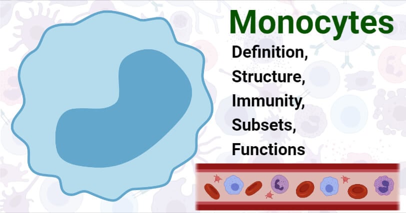
- The morphological features of the cells include an irregular cell shape, an oval or kidney-shaped nucleus, cytoplasmic vesicles, and a high nucleus to cytoplasm ratio (3:1).
- The nucleus is prominent and remains folded rather than multilobed. The cytoplasm, in turn, contains a large number of cytoplasmic granules, which are usually numerous towards the cell membrane.
- The nucleus contains a characteristic chromatin net with strands bridging tiny chromatin clumps. The chromatin clumps are arranged on the inner side of the nuclear membrane.
- The surface of a monocyte contains ruffles and surface blebs which are of functional significance.
- Monocytes are motile and phagocytic; thus, the reduction of curvature of the cell by the formation of ruffles reduces repulsive forces with negative charge groups approach the cell.
- The cytoplasm contains mitochondria that are numerous, small in size, and elongated. The Golgi complex is also present together with the centrosome within the nucleus.
- The cell membrane is also characterized by numerous microvilli, which help in locomotion and adherence to other cells.
- The cytoplasm also has cytoplasmic granules that are small (0.05 to 0.2 µm in diameter) that are dense and homogenous with a limiting membrane.
Subsets of Monocytes
- Monocytes found in humans can be divided into two subsets on the basis of the expression of CD14 and CD16. CD14 is a component of the lipopolysaccharide receptor complex, whereas CD16 is the TcγRIII immunoglobulin receptor.
- Both of these monocytes express distinct chemokines, immunoglobulins, adhesion proteins, and scavenger receptors.
- The first subset, CD14highCD16– monocytes, also called CD14+ monocytes are larger with a diameter of 18 µm and represent about 80-90% of the total circulating monocytes.
- The next subset, CD16highCD14–, also called CD16+ monocytes are smaller with a diameter of 16 µm and account for about 10% of the total circulating monocytes in humans.
- The CD16+ subset of monocytes produces a high level of tumor necrosis factor and low levels of IL-10 as a response to stimulation by Toll-Like Receptors. These are also called proinflammatory monocytes.
- Besides, human blood also contains another smaller subset of monocytes that are distinguished by surface molecule expression.
- CD14+CD16+CD64+ monocytes are a subset of highly phagocytic monocytes that express high levels of MHC class II. This subset is also called the transitional monocytes that can activate T cells.
How do Monocytes work against pathogens? (Immunity)
- Monocytes have toll-like receptors on the cell membrane, which can interact with pathogen-associated molecular patterns (PAMPs) that occur in invading pathogens.
- The binding produces a signal which causes monocytes to migrate from the bone marrow to the peripheral blood circulation within 12 to 24 hours.
- In order to enter the affected areas, monocytes should first bind to the endothelium and then move through the vascular surface.
- Finally, the monocytes adhere to the endothelium and finally make their way through the endothelial cells by the process of diapedesis. The monocytes can then penetrate the endothelial basement membrane and migrate to the area of the inflammation.
- The differentiation of monocytes occurs at the site of inflammation, and the differentiation depends on the growth factors and cytokines produced during the process.
- Monocytes in the area of inflammation can also act as phagocytic cells that engulf microorganisms, foreign materials, and dead and damaged cells.
- Some monocytes release cytokines, which helps in the recruitment of other cells and compounds into the affected area and induce further inflammation.
Functions of Monocytes
The following are some of the functions of monocytes:
- Monocytes are one of the most important components of the innate immune system as these differentiate into populations of dendritic cells and macrophages, which are involved in the regulation of cellular homeostasis.
- Monocytes also regularly patrol the body for pathogens and regulate an immune response during infection and inflammation.
- Monocytes function as phagocytic cells and antigen-presenting cells in the peripheral blood to remove microorganisms, antigens, and dead or damaged cells.
- Different subsets of monocytes produce different cytokines that recruit additional cells and proteins to affected areas to generate an effective immune response.
- Monocytes are highly plastic and heterogenous as these can change their functional phenotypes depending on the environmental stimuli.
- A particular subset of monocytes, called transitional monocytes, is involved in the activation of T cells.
References
- Peter J. Delves, Seamus J. Martin, Dennis R. Burton, and Ivan M. Roitt(2017). Roitt’s Essential Immunology, Thirteenth Edition. John Wiley & Sons, Ltd.
- Judith A. Owen, Jenni Punt, Sharon A. Stranford (2013). Kuby Immunology. Seventh Edition. W. H. Freeman and Company.
- Espinoza VE, Emmady PD. Histology, Monocytes. [Updated 2020 May 4]. In: StatPearls [Internet]. Treasure Island (FL): StatPearls Publishing; 2021 Jan-. Available from: https://www.ncbi.nlm.nih.gov/books/NBK557618/
- Serbina, Natalya V et al. “Monocyte-mediated defense against microbial pathogens.” Annual review of immunology vol. 26 (2008): 421-52. doi:10.1146/annurev.immunol.26.021607.090326
- Yona S, Jung S. Monocytes: subsets, origins, fates and functions. Curr Opin Hematol. 2010 Jan;17(1):53-9. doi: 10.1097/MOH.0b013e3283324f80. PMID: 19770654.
- Geissmann, Frederic et al. “Development of monocytes, macrophages, and dendritic cells.” Science (New York, N.Y.) vol. 327,5966 (2010): 656-61. doi:10.1126/science.1178331
- Karlmark, K R et al. “Monocytes in health and disease – Minireview.” European journal of microbiology & immunology vol. 2,2 (2012): 97-102. doi:10.1556/EuJMI.2.2012.2.1
- Chiu, Stephen, and Ankit Bharat. “Role of monocytes and macrophages in regulating immune response following lung transplantation.” Current opinion in organ transplantation vol. 21,3 (2016): 239-45. doi:10.1097/MOT.0000000000000313
- Yang, Jiyeon et al. “Monocyte and macrophage differentiation: circulation inflammatory monocyte as biomarker for inflammatory diseases.” Biomarker research vol. 2,1 1. 7 Jan. 2014, doi:10.1186/2050-7771-2-1
- Wacleche, Vanessa Sue et al. “The Biology of Monocytes and Dendritic Cells: Contribution to HIV Pathogenesis.” Viruses vol. 10,2 65. 6 Feb. 2018, doi:10.3390/v10020065
- Zanvil A. Cohn. The Structure and Function of Monocytes and Macrophages. Advances in Immunology. Academic Press. Volume 9. 1968. Pages 163-214. https://doi.org/10.1016/S0065-2776(08)60443-5.
Sources
- https://oncohemakey.com/structure-receptors-and-functions-of-monocytes-and-macrophages/- 11%
- https://www.sciencedirect.com/topics/neuroscience/monocyte – 9%
- http://edoc.mdc-berlin.de/17822/1/17822oa.pdf – 1%
- https://ecampusontario.pressbooks.pub/medicalterminology/chapter/cardiovascular-system/ – 1%
- https://study.com/academy/lesson/what-are-monocytes-definition-function-blood-test.html – 1%
