Interesting Science Videos
What is a Dissecting microscope (Stereo microscope)?
These are also known as stereoscopic microscopes. This is a type of digital optical microscope designed with a low magnification power (5x-250x), by use of light reflected from the surface of the specimen, and not the light reflected the specimen. Its primary role is for dissection of specimens and viewing and qualitatively analyzing the dissected samples.
It was first designed by Cherudin d’Orleans in 1677 by making a small microscope with two separate eyepieces and objective lenses. Later in 1852, an inventor named Charles Wheatstone describe the principle of stereoscopic visualization and published it with the title ‘On Some Remarkable, and Hitherto Unobserved, Phenomena of Binocular Vision’. It was later advanced by John Leornld Riddell who equally published it on the Journal of Microscopical Science as ‘ On Binocular Microscope’.
Horatio S. Greenough, an American Biologist developed the stereo Microscope, with two separate but identical optical paths and manufactured it with the Carl Zeiss Company, to which the scope of the Dissecting Microscope is built to date.
Principle of Dissecting microscope (Stereo microscope)
The working principle of the dissecting Microscope depends on the two types of light paths used by the microscope’s objectives and eyepiece. Each light path provides a different angle of viewing. They have the top light which is used while dissecting and the bottom light that is used to view the images. This lighting is enabled by the construction of two eyepieces (binocular stereoscope) each showing a different type of light pathway, each providing a viewing comfort area. Being a digital microscope, the images are viewed live on a computer monitor screen in 3-dimensional visuals. They also offer a very close observation of small specimens such as insects where the image produced is normally larger than the sample size, an effect known as macro-photography. The image is recorded and in complex samples, the topography (surface) is analyzed in 3D. The dissecting microscope works with two magnification systems: Fixed (primary) magnification where two objective lenses provide a degree of magnification and the Zoom (pancratic) magnification which offers a continuous magnification at variant ranges, using the auxiliary objectives whose function is to increase total magnification depending on some factors. The variance between the zoom and the fixed magnification can be achieved by changing the eyepiece lenses. Between the fixed and the zoom magnification is an optical system known as the Galilean optical system which has fixed-focus lenses that confer fixed magnification for different sets of magnification such as two sets of magnifications offers four-magnifications, three sets offer six-magnifications, etc.
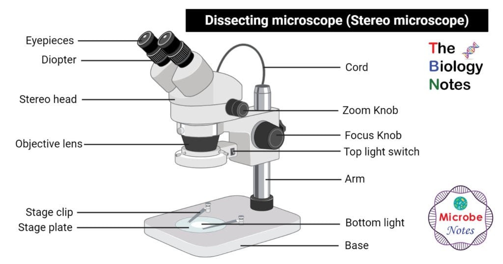
Parts of Dissecting microscope (Stereo microscope)

Figure: Labeled Dissecting microscope (Stereo or stereoscopic microscope). Image created using biorender.com
- LED illuminators- For some of the dissecting Microscopes, they have an inbuilt LED illuminator as a source of light.
- Eyepieces- They have two eyepieces each focusing different pathways of the light into and out of the specimen, each with its own magnification power. To increase the magnification, the use of auxiliary eyepieces can be used.
- Objective lenses- They also have different magnifications, focusing the image on the digital camera, and to improve magnification, the addition of auxiliary objectives is used.
- Stage- This is the apparatus for placing the specimen. they are large hence they can hold large specimen apparatus.
- Optical system- This is the system that is found between the fixed and the zoom magnification which offers fixed-focus lensing.
- Digital camera- It is temporarily fixed for most dissecting microscopes for capturing images and recording the outcome of the images. It captures 2D and 3D images depending on the type of digital camera that is installed.
Types of Dissecting microscopes (Stereo microscope)
- Stereo Zoom Dissecting Microscope- They are trinocular or binocular dissecting microscopes with a zooming range of 6.7x-45x. They can be attached to the digital camera which takes photos of the viewing images. They have a dual-LED illuminator and they can rotate at 360°. Magnification can be changed by adding auxiliary objectives or different eyepieces.
- Digital Tablet Dissection Microscope- These are high-end dissecting microscopes. They have a touch screen LCD tablet camera with a continuous magnification of 6.7x-45x. They have auxiliary eyepieces whose magnification ranges can be increased or decreased. Auxiliary lenses can also be added to the objectives for modifying the magnification. They have a 5.0-megapixel digital camera for capturing images and recording videos directly into the tablet or a USB cable. They have an in-built LED illuminators both at the top and bottom of the microscope, each operating separately.
- Stereo zoom boom stand microscopes- The have a large base and the largest stage for viewing large samples. They have LED lighting or optional dual-pipe lighting. They have a zooming range of 6x-45x which can be altered upward by adding auxiliary lenses or eyepieces.
- Stereo Zoom Dissecting Microscope- this is a compact stereo zoom dissecting Microscope with a zooming range of 10x-30x producing sharp parfocalled images. They have a rotating head hence the eyepiece can be positioned away and toward the specimen. they have a halogen lamp of 10 watts and fluorescent lighting of 5 watts.
- Dual Power Dissecting Microscope- It has a dual-powered dissecting microscope of 10x and 30x, with a 360° rotation ability, for focussing and viewing. They have a dual objective pair, parfocalled, parcentered, and achromatic. Rotating the lenses allows a change of image magnification. It also uses a high LED intensity light ring which fills the surface with light. With a flexible stand, it can be lifted head high for viewing larger specimens.
- Single Power Stereo Dissection Microscope- they have very low magnification power ranging from 10x-40x with inclined eyepieces at 45°C. they also have diopter adjustments of 50mm to 70mm.
- Single Magnification Handheld Pocket Microscope- This is a single powered handheld dissecting microscope with two magnification powers and it does not require light. Manufactured in Japan, it has high optical quality glass with a lot of ease in useability and its compact size makes it portable.
Applications of Dissecting microscope (Stereo microscope)
Like most microscopes, it is used in a wide range of fields including manufacturing, medical, quality control, inspection and biomedical studies like the entomological study of insects and some of its functions include:
- Studying the topography of solid samples
- For dissection
- For microsurgical procedures
- For the manufacturing of watches, circuit boards, and their inspections
- Used for inspection of fractures (fractography)
- Used in forensic engineering
Advantages of Dissecting microscope (Stereo microscope)
- It can be used in a wide range of fields making it one of the most important microscopic techniques.
- The use of two light pathways offers great magnification differences for visualizing the image.
- The attachment of a digital camera allows recording and imaging the image produced.
- It is easily portable and easy to use.
- They are used to view whole specimens and not in pieces.
Disadvantages of Dissecting microscope (Stereo microscope)
- The Galilean Optical Systems which are an essential part of the microscope are quite expensive to purchase.
- Generally, the microscope is costly to purchase.
- They have a low magnification power hence they are not able to view images of high magnification, above 100x hence they cant be used to view tissue structures and other structures.
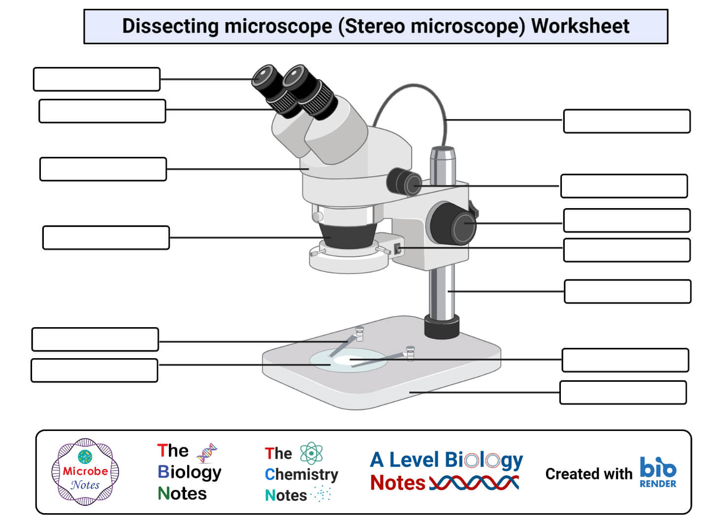
Dissecting microscope (Stereo microscope) Free Worksheet

References and Sources
- https://www.merriam-webster.com/dictionary/dissecting%20microscope
- https://www.britannica.com/technology/microscope/Stereoscopic-microscopes
- https://prezi.com/piodcrdu3r08/stereo-dissecting-microscope/
- 1% – https://www.microscopeworld.com/t-dissecting_microscopes.aspx
- 1% – https://www.microscope.com/stereo-microscopes/
- 1% – https://quizlet.com/218886987/chapter-25-digital-imaging-flash-cards/
- 1% – https://archive.org/details/philtrans09004566
- 1% – http://www.agarscientific.net/microscope-configurations-a-brief-history-of-the-compound-inverted-and-stereo-microscopes/
- <1% – https://sciencing.com/function-microscope-6575328.html
- <1% – https://microbenotes.com/light-microscope/
- <1% – https://classroom.synonym.com/parts-dissecting-microscope-2577.html
- <1% – http://www.microscope-depot.com/p1.asp?pn=s-01522

This is very helpful…Tye information provided in this site is very clear and concise. Thank you for your hard work Ms Faith
It’s very impressive to me I have learnt alot from the stereo microscope