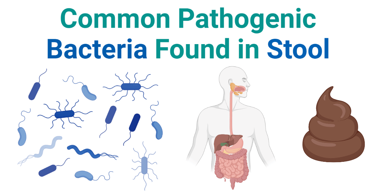
The stool is undigested food material and other wastes produced in the digestion process and eliminated from the digestive tract through the anus/cloaca. Stool culture is a microbiological test done to detect microorganisms causing gastrointestinal infections. Bacteria, viruses, protozoans, and parasites (helminths) are responsible for infection in the GI tract and associated organs. These organisms can be detected in stool. Bacteria are a common cause of gastroenteritis including diarrhea, ulcer, vomiting, food poisoning, etc. Bacteria causing gastrointestinal infections are detected by growing them in culture media using a stool as a sample. The stool is full of the normal flora of the GI tract, so it will be tricky to isolate the specific pathogens among the normal flora contamination.

Interesting Science Videos
List of Bacteria Isolated From Stool
| Gram-positive Bacteria in Stool | Gram-negative Bacteria in Stool |
| Clostridium spp. Staphylococcus spp. Enterococcus spp. Bacillus spp. Listeria monocytogenes | E. coli Klebsiella spp. Salmonella serovars Shigella spp. Helicobacter pylori Yersinia spp. Proteus spp. Campylobacter spp. Vibrio spp. Bacteroides spp. Fusobacterium nucleatum |
Gram -ve and Gram +ve Bacteria Found in Stool
E. coli in Stool
E. coli is a Gram-negative, rod-shaped, lactose fermenting, facultatively anaerobic, mesophilic coliform bacteria of the genus Escherichia in the Enterobacteriaceae family. E. coli are normal flora of the human GI tract, so their presence in stool is considered normal. But, several pathogenic strains of E. coli are responsible for dangerous diarrheal diseases.
Enteropathogenic E. coli (EPEC), Enteroaggregative E. coli (EAEC), Enterohemorrhagic E. coli ( EHEC), Enterotoxigenic E. coli (ETEC) {or Shiga toxin-producing E. coli (STEC) or Verotoxin producing E. coli (VTEC)}, Enteroinvasive E. coli (EIEC), Diffusely adherent E. coli (DAEC) are the pathogenic strains of E. coli associated with several gastrointestinal infections including diarrhea.
E. coli O157:H7 (STEC) is one of the common causes of a severe form of hemorrhagic diarrhea and epidemic diarrheal outbreaks worldwide.
Klebsiella spp. in Stool
Klebsiella is a genus of Gram-negative, rod-shaped, and facultative anaerobic and capsule-forming coliform bacteria in the family Enterobacteriaceae. Klebsiella spp. is common in the GI tract of vertebrates. They are however responsible for gastroenteritis in case of weakened immune condition or case of any physical damage to the GI tract.
K. pneumoniae is common species isolated in the stool of patients with gastrointestinal diseases such as Crohn’s disease, ulcerative colitis, and colorectal cancer.
K. oxytoca is another species reported for hemorrhagic enteritis. Toxin-producing K. oxytoca is reported in necrotizing enterocolitis, bloody diarrhea, and other gastroenteritis.
K. quasipneumoniae is another species responsible for opportunistic gastroenteritis. It is mainly reported in patients whose natural gut microflora is disturbed due to long-term administration of antibiotics.
Salmonella spp. in Stool
Salmonella is a genus of Gram-negative, rod-shaped, flagellated, facultative anaerobic Gammaproteobacteria in the family Enterobacteriaceae. It is commonly found in the human GI tract and fecal contaminated environmental aspects. Salmonella spp. is responsible for food poisoning, diarrhea, and enteric fever (Typhoidal fever).
S. enterica serovars are responsible for several forms of gastroenteritis in humans. S. enterica subspecies enterica serovar Typhi and Paratyphi are responsible for enteric fever (Typhoid and Paratyphoid). They can be isolated in the stool of patients with enteric fever before the administration of antibiotics.
Non-typhoidal Salmonella (NTS) is the major cause of gastroenteritis worldwide. They usually cause self-limiting gastroenteritis, but in infants and immunosuppressed patients, the case may be fatal. S. enterica serovar Typhimurium, S. enterica serovar Enteritidis are most common serovars associated with Salmonella gastroenteritis.
Shigella spp. in Stool
Shigella is a genus of Gram-negative, rod-shaped, non-motile, facultative anaerobic Gammaproteobacteria in the family Enterobacteriaceae genetically related to E. coli. It is responsible for a gastrointestinal infection called Shigellosis in humans and is the leading cause of diarrhea (bacillary dysentery) worldwide.
S. dysenteriae is the main species causing shigellosis. S. dysenteriae subtype 1 (SD1) produces the Shiga toxin. It is responsible for the most severe form of Shigellosis like severe dysentery with systemic complications such as electrolyte imbalance, seizures, and hemolytic uremic syndrome (HUS) and has the highest mortality.
S. flexneri is another common species causing shigellosis. It is responsible for most of the shigellosis in South East Asia. It mostly causes a milder form of diarrhea but in some cases, they are reported for severe forms including HUS and seizures.
S. sonnei and S. boydii are responsible for a milder form of Shigellosis.
Vibrio spp. in Stool
Vibrio is a genus of Gram-negative, rod-shaped (comma-shaped), motile, non-sporing, facultative anaerobic Gammaproteobacteria in the family Vibrionaceae with characteristic 2 chromosomes. It is responsible for waterborne gastroenteritis.
V. cholerae is the most important pathogenic species responsible for a cholera outbreak. It is responsible for endemic cholera in about 69 countries. Strains capable of producing Cholera enterotoxin cause a severe form of cholera with mortality of >50% if untreated. V. cholerae O1 and O139 are the most common serotypes causing epidemic cholera outbreaks.
V. parahaemolyticus is the second most common Vibrio species causing bacterial gastroenteritis, mostly in the Asian region. However, the severity is very less than V. cholerae infection.
V. vulnificus is another species responsible for a milder form of gastroenteritis with diarrhea, vomiting, and fever.
Helicobacter pylori in Stool
Helicobacter is a genus of Gram-negative, helical, microaerophilic Epsilonproteobacteria in the family Helicobaceteriaceae. They are adapted to tolerate the acidity of a human stomach.
H. pylori is normally present in the duodenum of most humans. But in some cases, they can cause gastrointestinal inflammation, gastritis, ulcer, diarrhea, gastric cancer, and other gastrointestinal problems.
Yersinia spp. in Stool
Yersinia is a genus of Gram-negative, coccobacilli, facultative anaerobic Gammaproteobacteria in the family Yersiniaceae.
Y. enterocolitica is associated with about 2% of gastroenteritis. It is usually underestimated as most of the cases are self-limiting, but cases of Y. enetrocolitica gastroenteritis are high usually in children and younger adults.
Y. pestis-related gastroenteritis incidences are comparatively low.
Y. pseudotuberculosis is another species that very rarely cause food-borne infection in human, mostly in cold places.
Proteus spp. in Stool
Proteus is a genus of Gram-negative, rod-shaped, aerobic, and facultative anaerobic motile bacteria in the family Enterobacteriaceae best known for their swarming colonies. Proteus spp. are responsible for gastroenteritis, appendicitis, Crohn’s disease, colitis, and colonization of nasogastric tubes.
P. mirabilis is the most prevalent Proteus species in gastroenteritis. P. vulgaris and P. penneri are rarely involved in gastrointestinal infections.
Clostridium spp. in Stool
Clostridium is a genus of Gram-positive, rod-shaped, strictly anaerobic, spore-forming bacteria in the family Clostridiaceae.
C. difficile is one of the most important causes of nosocomial diarrhea. Toxin-producing strains are responsible for several forms of gastroenteritis, mostly diarrhea, and colitis.
C. perfringens is another species associated with acute gastrointestinal infections. It is responsible for food poisoning cases. It results in mild diarrhea to severe necrotizing enterocolitis. They produce Type A and Type C toxins that are responsible for diarrheal diseases.
Staphylococcus spp. in Stool
Staphylococcus is a genus of Gram-positive, catalase-positive, cocci bacteria belonging to the family Staphylococcaceae, typically known for producing grape-like clusters under a microscope. They are normal flora of the skin.
S. aureus is a major human pathogen producing Staphylococcal enterotoxins (SEs) resulting Staphylococcal food poisoning. It is one of the major causes of food poisoning.
Enterococcus spp in Stool
Enterococcus is a genus of Gram-positive, facultatively anaerobic, lactose fermenting cocci (diplococci) bacteria in the family Enterococcaceae having the capacity to tolerate bile salt concentrations up to 40%. They are commensal to the GI tract and are rarely associated with gastrointestinal infections. They are associated with intra-abdominal infections and gastritis. E. hirae and E. durans are detected as the cause of gastritis in some. Vancomycin-resistant E. faecalis is another species responsible for some cases of gastroenteritis.
Campylobacter spp. in Stool
Campylobacter is a genus of Gram-negative, rod-shaped (comma-shaped), microaerophilic motile bacteria in the family Campylobacteraceae. It is responsible for a bacterial gastrointestinal infection called Campylobacteriosis.
C. jejuni is one of the most common causes of acute bacterial gastroenteritis in children and adults. It causes diarrhea, vomiting, and abdominal pain.
Listeria spp. in Stool
Listeria is a genus of Gram-positive, rod-shaped, non-sporing, facultatively anaerobic bacteria in the family Listeriaceae. It is responsible for a disease called listeriosis.
L. monocytogenes can cause acute, self-limiting, febrile gastroenteritis and invasive gastroenteritis. Listeria gastroenteritis has a very high mortality rate making it the food-borne illness with the third highest mortality rate.
Bacteroides spp. in Stool
Bacteroides is a genus of Gram-negative, rod-shaped, bile resistant, non-sporing, motile or non-motile, obligate anaerobic bacteria in the family Bacteroidaceae. They are part of human gastrointestinal microflora.
B. fragilis is the only species reported to form abscesses in the intestine. They have a capsule and produce a toxin that can cause tissue destruction. If left untreated they disrupt the intestinal wall and cause intra-abdominal sepsis and appendicitis. It has a mortality rate of 19%, and when untreated it raises to 60%.
Fusobacterium nucleatum in Stool
Fusobacterium is a genus of Gram-negative, non-sporing, rod-shaped anaerobic bacteria in the family Fusobacteriaceae. F. nucleatum is found to be associated with colorectal cancer.
Bacillus spp. in Stool
Bacillus is a genus of Gram-positive, rod-shaped, endospore-forming, aerobic or facultative anaerobic bacteria in the family Bacillaceae.
B. cereus is one of the common pathogens responsible for food poisoning. It results in emetic syndrome or diarrheal syndrome.
References
- Bacterial Infections of the Gastrointestinal Tract | Microbiology (lumenlearning.com)
- 19.3 Bacterial Infections of the Gastrointestinal Tract – Allied Health Microbiology (oregonstate.education)
- Graves N. S. (2013). Acute gastroenteritis. Primary care, 40(3), 727–741. https://doi.org/10.1016/j.pop.2013.05.006
- Kim, Y. J., Park, K. H., Park, D. A., Park, J., Bang, B. W., Lee, S. S., Lee, E. J., Lee, H. J., Hong, S. K., & Kim, Y. R. (2019). Guideline for the Antibiotic Use in Acute Gastroenteritis. Infection & chemotherapy, 51(2), 217–243. https://doi.org/10.3947/ic.2019.51.2.217
- Sell, Jarrett; Dolan, Bevin (2018). Common Gastrointestinal Infections. Primary Care: Clinics in Office Practice, (), S0095454318300459–. doi:10.1016/j.pop.2018.05.008
- Kaur, C. P., Vadivelu, J., & Chandramathi, S. (2018). Impact of Klebsiella pneumoniae in lower gastrointestinal tract diseases. Journal of digestive diseases, 19(5), 262–271. https://doi.org/10.1111/1751-2980.12595
- Karaliute, I., Ramonaite, R., Bernatoniene, J. et al. Reduction of gastrointestinal tract colonization by Klebsiella quasipneumoniae using antimicrobial protein KvarIa. Gut Pathog 14, 17 (2022). https://doi.org/10.1186/s13099-022-00492-2
- Totani T. (1978). Nihon rinsho. Japanese journal of clinical medicine, Suppl, 1308–1309.
- Giannella RA. Salmonella. In: Baron S, editor. Medical Microbiology. 4th edition. Galveston (TX): University of Texas Medical Branch at Galveston; 1996. Chapter 21. Available from: https://www.ncbi.nlm.nih.gov/books/NBK8435/
- Ajmera A, Shabbir N. Salmonella. [Updated 2021 Aug 11]. In: StatPearls [Internet]. Treasure Island (FL): StatPearls Publishing; 2022 Jan-. Available from: https://www.ncbi.nlm.nih.gov/books/NBK555892/
- Zaidi, M. B., & Estrada-García, T. (2014). Shigella: A Highly Virulent and Elusive Pathogen. Current tropical medicine reports, 1(2), 81–87. https://doi.org/10.1007/s40475-014-0019-6
- Ojeda Rodriguez JA, Kahwaji CI. Vibrio Cholerae. [Updated 2021 Jun 4]. In: StatPearls [Internet]. Treasure Island (FL): StatPearls Publishing; 2022 Jan-. Available from: https://www.ncbi.nlm.nih.gov/books/NBK526099/
- Haftel A, Sharman T. Vibrio Vulnificus. [Updated 2021 Jul 26]. In: StatPearls [Internet]. Treasure Island (FL): StatPearls Publishing; 2022 Jan-. Available from: https://www.ncbi.nlm.nih.gov/books/NBK554404/
- Rezny BR, Evans DS. Vibrio Parahaemolyticus. [Updated 2021 Jul 2]. In: StatPearls [Internet]. Treasure Island (FL): StatPearls Publishing; 2022 Jan-. Available from: https://www.ncbi.nlm.nih.gov/books/NBK459164/
- Perry, S., de la Luz Sanchez, M., Yang, S., Haggerty, T. D., Hurst, P., Perez-Perez, G., & Parsonnet, J. (2006). Gastroenteritis and transmission of Helicobacter pylori infection in households. Emerging infectious diseases, 12(11), 1701–1708. https://doi.org/10.3201/eid1211.060086
- Seyed Mohammad Riahi, Ehsan Ahmadi, Tayebeh Zeinali, “Global Prevalence of Yersinia enterocolitica in Cases of Gastroenteritis: A Systematic Review and Meta-Analysis”, International Journal of Microbiology, vol. 2021, Article ID 1499869, 17 pages, 2021. https://doi.org/10.1155/2021/1499869.
- Marks, M. I., Pai, C. H., Lafleur, L., Lackman, L., & Hammerberg, O. (1980). Yersinia enterocolitica gastroenteritis: a prospective study of clinical, bacteriologic, and epidemiologic features. The Journal of pediatrics, 96(1), 26–31. https://doi.org/10.1016/s0022-3476(80)80318-0
- Hamilton, A. L., Kamm, M. A., Ng, S. C., & Morrison, M. (2018). Proteus spp. as Putative Gastrointestinal Pathogens. Clinical microbiology reviews, 31(3), e00085-17. https://doi.org/10.1128/CMR.00085-17
- Dineen, S. P., Bailey, S. H., Pham, T. H., & Huerta, S. (2013). Clostridium difficile enteritis: A report of two cases and systematic literature review. World journal of gastrointestinal surgery, 5(3), 37–42. https://doi.org/10.4240/wjgs.v5.i3.37
- Yao P, Annamaraju P. Clostridium Perfringens. [Updated 2021 Sep 13]. In: StatPearls [Internet]. Treasure Island (FL): StatPearls Publishing; 2022 Jan-. Available from: https://www.ncbi.nlm.nih.gov/books/NBK559049/
- Balaban, N., & Rasooly, A. (2000). Staphylococcal enterotoxins. International journal of food microbiology, 61(1), 1–10. https://doi.org/10.1016/s0168-1605(00)00377-9
- Jay, J.M. (1998). Staphylococcal Gastroenteritis. In: Modern Food Microbiology. Food Science Texts Series. Springer, Boston, MA. https://doi.org/10.1007/978-1-4615-7476-7_20
- El-Zimaity, H. M., Ramchatesingh, J., Clarridge, J. E., Abudayyeh, S., Osato, M. S., & Graham, D. Y. (2003). Enterococcus gastritis. Human pathology, 34(9), 944–945. https://doi.org/10.1016/s0046-8177(03)00287-9
- Karmali, M. A., & Fleming, P. C. (1979). Campylobacter enteritis. Canadian Medical Association journal, 120(12), 1525–1532.
- Mehmood, H., Marwat, A., & Khan, N. (2017). Invasive Listeria monocytogenes Gastroenteritis Leading to Stupor, Bacteremia, Fever, and Diarrhea: A Rare Life-Threatening Condition. Journal of investigative medicine high impact case reports, 5(2), 2324709617707978. https://doi.org/10.1177/2324709617707978
- Wexler H. M. (2007). Bacteroides: the good, the bad, and the nitty-gritty. Clinical microbiology reviews, 20(4), 593–621. https://doi.org/10.1128/CMR.00008-07
- Kim, Y. J., Kim, B. K., Park, S. J., & Kim, J. H. (2021). Impact of Fusobacterium nucleatum in the gastrointestinal tract on natural killer cells. World journal of gastroenterology, 27(29), 4879–4889. https://doi.org/10.3748/wjg.v27.i29.4879
- McDowell RH, Sands EM, Friedman H. Bacillus Cereus. [Updated 2021 Sep 16]. In: StatPearls [Internet]. Treasure Island (FL): StatPearls Publishing; 2022 Jan-. Available from: https://www.ncbi.nlm.nih.gov/books/NBK459121/
