Mitosis is the process of cell division in which one cell gives rise to two genetically identical daughter cells, resulting in cell duplication and reproduction.
- The number of chromosomes is preserved in both the daughter cells.
- Mitosis is a short period of chromosome condensation, segregation, and cytoplasmic division.
- The mitosis occurs in the somatic cells, and it is meant for the multiplication of cell numbers during embryogenesis and blastogenesis of plants and animals.
- As a process, mitosis is remarkably similar in all animals and plants.
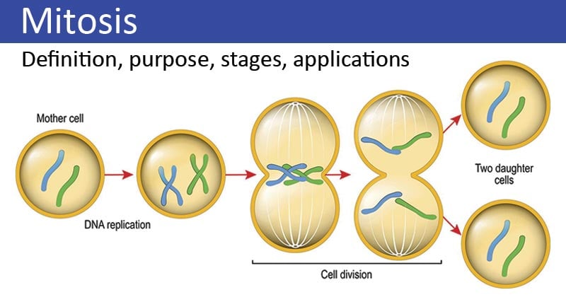
Image Source: Biology Wise.
Interesting Science Videos
Purpose of Mitosis
The process of mitosis is significant in both cell division as well as cell reproduction. Some of the major significances/purposes are given below:
- Continuous mitosis results in the increase in the number of cells enabling the organism to grow from a single cell to a complex living organism.
- Different cells in the body like the cells on the skin and red blood cells are continuously replaced by mitosis. About 5×10⁹ cells are formed per day in humans via mitosis.
- Mitosis is also involved in the repairmen and regeneration of body structures like in the starfish.
- In multiple organisms, mitosis is the method of asexual reproduction.
Read Also: Meiosis- definition, purpose, stages, applications with diagram
Stages of mitosis
Mitosis is a part of the cell cycle and is preceded by the S phase of interphase and usually followed or accompanied by cytokinesis. Replication of chromosomes and synthesis of proteins required for spindle fiber formation are formed prior to the onset of mitosis.

Figure: Stages of mitosis. Image Source: Wikipedia (Ali Zifan).
Mitosis is divided into the following phases based on the completion of one set of activities and the onset of the other.
1. Interphase
- Interphase is a part of the cell cycle where the cell copies its DNA as preparation for the M phase (mitotic phase).
- In interphase, metabolism of the cell increases, and it is often termed the most active phase of the cell cycle.
- A series of metabolic changes occur during this phase, all of which are divided into three subgroups.
G1-phase or Pre-DNA synthesis phase
- It is the longest phase of the cell cycle and is followed by the M phase of the previous cell cycle.
- It is also termed as the “resting phase” as no DNA synthesis takes place during this phase.
- However, during G1 phase, several cell organelles increase in size and cell rapidly synthesizes different types of RNA and proteins.
- Important events like the transcription of three types of RNAs, synthesis of regulatory proteins, enzymes required for DNA synthesis, and tubulin proteins along with other mitotic apparatus take place during this phase.
S-phase or DNA Synthesis phase
- S-phase involves the replication of nuclear DNA and the synthesis of histone proteins. The replication of cytoplasmic DNA can take place at any phase in the cell cycle.
- Thus, at the end of the S phase, each chromosome has two DNA molecules and a duplicate set of genes.
- This phase lasts for about 6-10 hours.
G2-phase or Post DNA synthesis phase

Figure: Late G2. Image Source: Khan Academy.
- G2 phase is termed the second gap phase or resting phase of the interphase.
- During this phase, the synthesis of RNA and proteins required for the cell continues.
- Cell division involves the enormous expenditure of energy, thus cell stores ATP in the G2.
- By the end of this phase, the cell enters the division or M-phase of the cell cycle.
2. Prophase
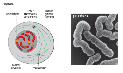
Figure: Prophase. Image Source: Wikipedia
- Prophase is the first stage of mitosis which is characterized by the appearance of thin-thread like condensing chromosomes.
- During prophase, the cell becomes spheroid while the cytoplasm becomes more refractile and viscous and pale.
- The chromosome in the prophase is composed of two coiled filaments, the chromatids, which are the result of the replication of DNA during the S phase.
- As prophase progresses, the chromatids become shorter and thicker, and two sister chromatids of each chromosome are held together by a special DNA-containing region, called the centromere.
- Similarly, the chromosomes approach the nuclear envelope, causing the central space of the nucleus to become empty.
- In the meantime, two pairs of centrioles surrounded by microtubules radiating in all directions migrate to opposite poles of the cell.
- Lastly, during prophase, the nucleolus gradually disintegrates, and this marks the end of prophase.
- However, in some primitive classes of plants and animals, the nuclear envelope does not dissolve during mitosis.
3. Prometaphase
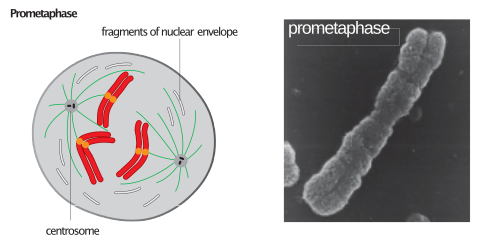
Figure: Prometaphase. Image Source: Wikipedia
- Prometaphase is initiated with the breakdown of the nuclear envelope, which enables the interaction of spindle fibers with the chromosomes.
- At this stage, the chromosomes are violently rotated and oscillated back and forth between the spindle poles because their centromeres are capturing the ends of microtubules and are being pulled by the captured microtubules.
- By the end of prometaphase, the sister chromatids are attached to the spindle fibers on the opposite ends and are held on the metaphase plate.
4. Metaphase
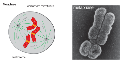
Figure: Metaphase. Image Source: Wikipedia
- During metaphase, the chromosomes are shortest and thickest.
- Their centromeres of the sister chromatids occupy the plane of the equator forming a metaphase plate, and the arms remain directed towards the poles.
- Two chromatids of a chromosome repulse each other with the microtubules remaining stationary and under tension.
5. Anaphase
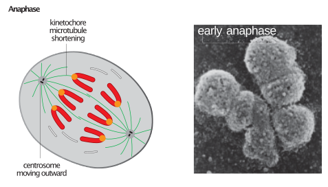
Figure: Anaphase. Image Source: Wikipedia
- The anaphase begins abruptly with the synchronous splitting of each chromosome into its sister chromatids, called daughter chromosomes, separating for the centromere.
- The splitting of each centromere during prophase is caused by an increase in cytosolic Ca2+.
- In anaphase, there is a movement of chromatids towards the pole due to the shortening of the microtubules.
- During their poleward migration, the centromeres remain forward so that the chromosomes characteristically appear U, V or J- shaped.
- Interzonal fibers expand and support the movement of chromosomes towards the pole.
- A total of 30 ATPs are required to carry chromosomes to the poles.
6. Telophase
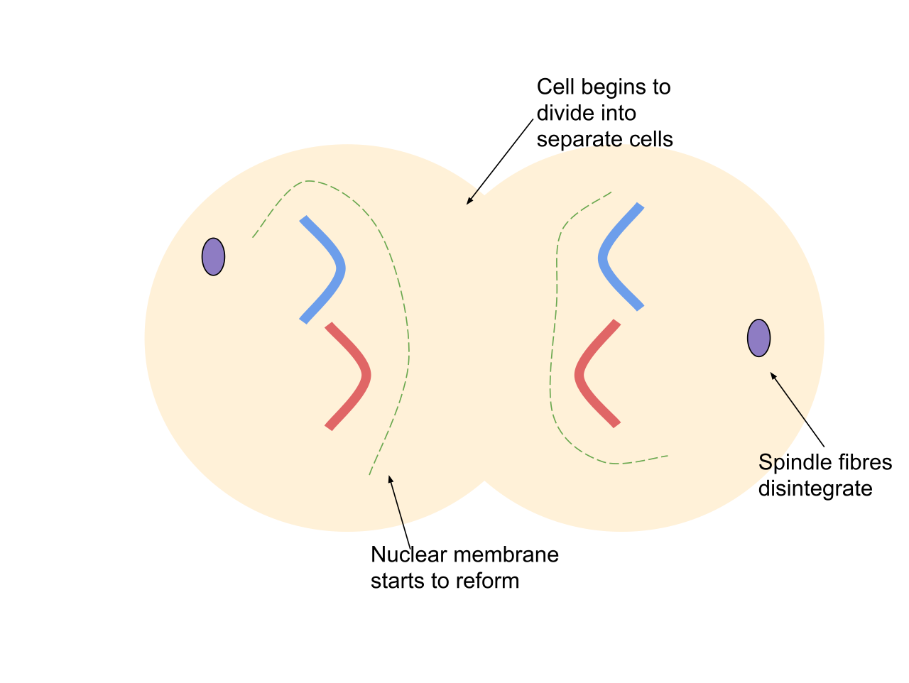
Figure: Telophase. Image Source: Wikipedia
- The end of the migration of the daughter chromosomes to the poles marks the beginning of the telophase
- During telophase, the events of prophase occur in reverse sequence.
- A nuclear envelope reassembles around each group of chromosomes to form two daughter nuclei.
- Events like the disappearance of mitotic apparatus, reduction in the viscosity of cytoplasm followed by synthesis of RNA take place during telophase.
- The chromosomes resume their long, slender, extended form and the nucleolus reappears at the end of telophase.
7. Cytokinesis
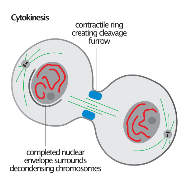
Figure: Cytokinesis. Image Source: Wikipedia
- Cytokinesis is the division of cytoplasm which is followed by mitosis, resulting in the formation of two separate daughter cells.
- Cytokinesis usually begins in anaphase and continues through telophase and into interphase.
- In animals, cytokinesis occurs through constriction and furrow formation.
- The first sign of cleavage in animal cells is constriction of the plasma membrane during anaphase.
- The constriction invariably occurs in the plane of the metaphase plate, at right angles to the long axis of the mitotic spindle apparatus.
- The constriction grows more in-depth from the outside to the inside, and ultimately a cell divides into two daughter cells.
- In plants, however, cytokinesis occurs by cell plate formation as constriction is not possible due to the presence of a rigid cell wall.
- Golgi apparatus arrange themselves on the equator to form phragmoplast, which later forms the cell plate in plants.
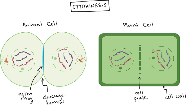
Figure: Cytokinesis in Animal Cell and Plant Cell. Image Source: Khan Academy.
Applications of Mitosis
Mitosis has been utilized for many lab-based techniques in molecular biology and biotechnology. Some of the common use of mitosis are:
- Cloning
- Cloning is a technique employed in biotechnology to produce identical copies of cells or DNA fragments.
- In cloning, the number of organisms is increased by the process of mitosis, which is then used in a wide array of biological experiments like fingerprinting.
- Tissue culture
- The growth of tissues or cells outside of the body of the organism in a liquid, semi-solid, or solid growth medium is called tissue culture.
- Tissue culture is based on the process of mitosis, where a cell undergoes division to form multiple tissues.
- Besides, tissue culture may lead to organ culture in various organisms.
- Stem cell regeneration
- Stem cells are a group of cells that can be directed to form specialized cells in the body.
- Stem cells can undergo mitosis to regenerate and repair diseased or damaged tissues in people.
Mitosis Video and Animation
References
- Verma PS and Agarwal VK (3005). Cell Biology, Genetics, Molecular Biology, Evolution, and Ecology. Multicolored Edition.
- Rastogi SC (2006). Cell and Molecular Biology. Second Edition. New Age International.
- http://www.authorstream.com/Presentation/aSGuest108184-1131832-application-of-mitosis/
- https://www.siyavula.com/read/science/grade-10-lifesciences/cell-division/03-cell-division-03


Thankyou very much, this was very helpful because I was constantly lookin gfor a site that has the explanation with diagrams. I give it a thumbs up.
Very helpful 🤝 really helped me complete my assignment briefly 🧘