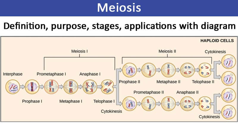
Meiosis is a type of cell division in sexually reproducing eukaryotes, resulting in four daughter cells (gametes), each of which has half the number of chromosomes as compared to the original diploid parent cell.
- The haploid cells become gametes, which by union with another haploid cell during fertilization defines sexual reproduction and formation of a new generation of diploid organisms.
- Meiosis occurs in the germ cells of sexually reproducing organisms.
- In both plants and animals, germ cells are localized in the gonads, but the time at which meiosis takes place varies among different organisms.

Figure: Overview of Meiosis. Image Source: Rice University (OpenStax).
Interesting Science Videos
Purpose of Meiosis
The process of meiosis is essential for all sexually reproducing organisms for the following reasons:
- The meiosis maintains a constant number of chromosomes in sexually reproducing organisms through the formation of gametes.
- By crossing over, the meiosis results in the exchange of the genes and, thus, causes the genetic variations among the species. These variations are the raw materials of the evolutionary process.
Read Also: Mitosis- definition, purpose, stages, applications with diagram.
Stages/Phases of Meiosis
- Meiosis is composed of two rounds of cell division, namely Meiosis I and Meiosis II.
- Each round of division contains a period of karyokinesis (nuclear division) and cytokinesis (cytoplasmic division).
Meiosis I

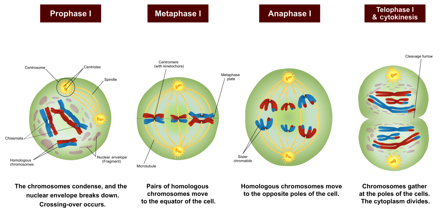
Figure: Phases of Meiosis I. Image Source: Wikipedia (Ali Zifan).
- The first meiotic division consists of prolonged prophase in which the homologous chromosomes come in close contact with each other and exchange hereditary material between them.
- Similarly, in the first meiotic division, the reduction of chromosome number takes place and, thus, two haploid cells are resulted by this division.
- The first meiotic division is also known as the heterotypic division.
- Meiosis I consists of the following steps:
Interphase
- Just like mitosis, meiosis also consists of a preparatory phase called interphase.
- The interphase is characterized by the following features :
- The nuclear envelope remains intact, and the chromosomes occur in the form of diffused, long, coiled, and indistinctly visible chromatin fibers.
- The DNA amount becomes double. Due to the accumulation of ribosomal RNA (rRNA) and ribosomal proteins in the nucleolus, the size of the nucleolus is significantly increased.
- In animal cells, a daughter pair of centrioles originates near the already existing centriole and, thus, an interphase cell has two pairs of centrioles.
- In the G2 phase of interphase, there is a decisive change that directs the cell toward meiosis, instead of mitosis.
- At the beginning of the first meiotic division, the nucleus of the dividing cell starts to increase in size by absorbing the water from the cytoplasm, and the nuclear volume increases about three folds.
Prophase I
Prophase I is the longest stage of the meiotic division. It includes the following substages:
Leptotene
- In the leptotene stage, the chromosomes become even more uncoiled and resemble a long thread-like shape, and they develop bead-like structures called chromomeres.
- The chromosomes at this stage remain directed towards centrioles, so the chromosomes in the nucleus appear like a bouquet in the animal cell. Therefore, this stage is also called the Bouquet Stage.
Zygotene or Synaptotene
- The zygotene stage begins with the pairing of homologous chromosomes, which is called synapsis.
- The paired homologous chromosomes are connected by a protein-containing framework called a synaptonemal complex.
- The synaptonemal complex helps to stabilize the pairing of homologous chromosomes and to facilitate recombination or crossing over.
- The synapsis might begin at one or more points along the length of the homologous chromosomes.
- Synapsis might start from the ends of the chromosomes and continues towards their centromeres (proterminal synapsis), or it might start at the centromere and proceed towards the ends (procentric pairing).
- In some cases, the synapsis occurs at various points of the homologous chromosomes (random pairing).
Pachytene
- In this stage, the pair of chromosomes become twisted spirally around each other and cannot be distinguished separately.
- In the middle of the pachytene stage, each homologous chromosome splits lengthwise to form two chromatids, but they continue to be linked together by their common centromere.
- The chromosomes at this point are termed bivalent because it consists of two visible chromosomes, or as a tetrad because of the four visible chromatids.
- This stage is particularly crucial as a critical genetic phenomenon called “ crossing over” takes place in this stage.
- The crossing over involves redistribution and mutual exchange of hereditary material between two homologous chromosomes.
- The enzyme endonuclease breaks the non-sister chromatids at the place of crossing over.
- After the breaking of chromatids, the interchange of chromatid segments takes place between the non-sister chromatids of the homologous chromosomes.
- Another enzyme, ligase, binds the broken chromatid segments with the non-sister chromatid.
- The process of mutual exchange of chromatin material between one non-sister chromatid of each homologous chromosome is known as the crossing over.
Diplotene
- The synaptonemal complex appears to be dissolved, leaving chromatids of the paired homologous chromosome physically joined at one or more localized points called
- In diplotene, chiasmata move towards the end of chromosomes in a zip like a manner.
Diakinesis
- In this stage, the bivalent chromosomes become more condensed and uniformly distributed in the nucleus.
- At this point, the nuclear envelope breaks down, and the nucleolus disappears.
- Further, the chiasmata reach the end of the chromosomes, and the chromatids remain attached until metaphase.
Metaphase I
- Metaphase I consists of spindle fiber attachment to chromosomes and chromosomal alignment at the equator.
- During metaphase I, the spindle fibers are attached with the centromeres of the homologous chromosomes, which are directed towards the opposite poles.
Anaphase I
- At anaphase I homologous chromosomes are separated from each other, and due to the shortening of chromosomal fibers or microtubules, each homologous chromosome with its two chromatids and undivided centromere move towards the opposite poles of the cell.
- Because during the chiasma formation, one of the chromatids has changed its counterpart, therefore, the two chromatids of a chromosome are not genetically identical.
Telophase I
- The onset of telophase I is defined by the movement of a haploid set of chromosomes at each pole.
- The nuclear envelope is formed around the chromosomes, and the chromosomes become uncoiled. The nucleolus reappears and, thus, two daughter nuclei are formed.
Cytokinesis I
- In animals, cytokinesis occurs by the constriction of the cell membrane while in plants, it occurs through the formation of the cell plate, resulting in the creation of two daughter cells.
Meiosis II

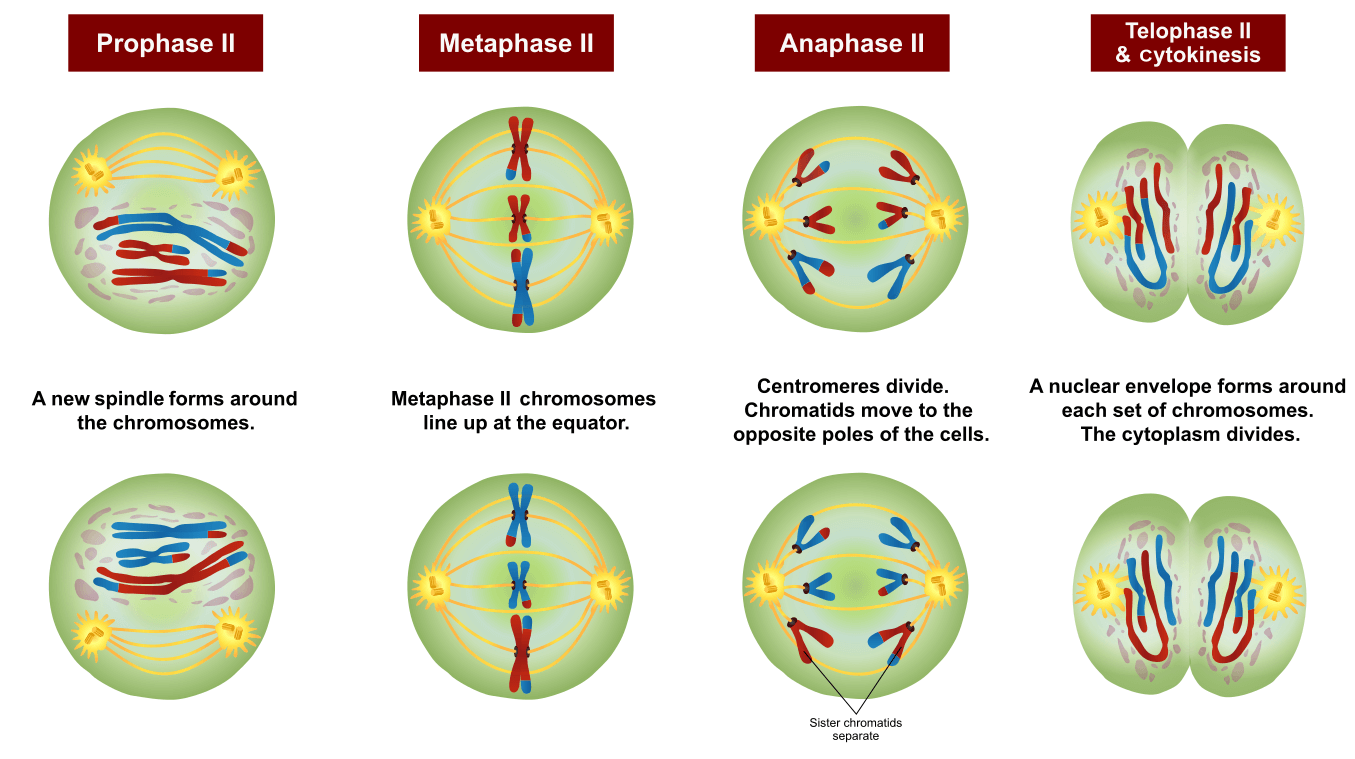
Figure: Phases of Meiosis II. Image Source: Wikipedia (Ali Zifan).
- In the second phase of the meiotic division, the haploid cell divides mitotically and results in four haploid cells. This division is also known as the homotypic division.
- This division does not include the exchange of the genetic material and the reduction of the chromosome number as in the first meiotic division.
- Meiosis II consists of the following steps:
Prophase II
- In prophase II, each centriole divides, resulting in two pairs of centrioles.
- The centrioles move towards the opposite poles and the nuclear membrane, and the nucleolus disappears.
Metaphase II
- During metaphase II, the chromosomes get arranged on the equator of the cell through the spindle fibers.
- The centromere divides and, thus, each chromosome produces two daughter chromosomes.
- The spindle apparatus is attached to the centromere of each chromosome.
Anaphase II
- The daughter chromosomes move towards the opposite poles due to the shortening of chromosomal microtubules and the stretching of interzonal microtubules of the spindle.
Telophase II
- The chromatids migrate to the opposite poles and now known as chromosomes.
- The endoplasmic reticulum forms the nuclear envelope around the chromosomes, and the nucleolus reappears due to the synthesis of ribosomal RNA.
Cytokinesis II
- The process of cytokinesis is identical to cytokinesis I resulting in the division of cytoplasm for each of the four daughter cells formed.
Applications of Meiosis
Meiotic like mitosis is used for several lab-based technologies, some of which are given below:
Tissue culture
- Like mitosis, meiosis is also used in biotechnology to acquire a gametic condition in cells.
- Meiosis often accompanies mitosis to generate variation which aids in studies regarding evolutionary processes.
In-vitro gamete formation
- In various gamete failure-derived infertility issues, the embryonic stem cells are differentiated into germ-like cells through the meiotic division.
- These gametes are formed in-vitro via meiosis and are inserted into the individuals with such disorders.
Meiosis Video and Animation
References
- Verma PS and Agarwal VK (3005). Cell Biology, Genetics, Molecular Biology, Evolution and Ecology. Multicolored Edition.
- Rastogi SC (2006). Cell and Molecular Biology. Second Edition. New Age International.
- Eguizabal C, Montserrat N, Vassena R, et al. Complete meiosis from human induced pluripotent stem cells. Stem Cells. 2011;29(8):1186‐1195. DOI:10.1002/stem.672
- Ronchi VN (1995). Eguizabal C, Montserrat N, Vassena R, et al. Complete meiosis from human induced pluripotent stem cells. Stem Cells. 2011;29(8):1186‐1195. DOI:10.1002/stem.672.


Please, what are the certain things I need do to facilitate the method of my learning.i am a 100 level student in one of universities in my country,Nigeria. I am particularly interested in studying biology since I was in secondary school level,I had some very inspiring people who in particular tried so much for me just to make that I be into tertiary level. I wanted to study botany but as time went on,I changed up my mind on the course due to some personal circumstances
Studying botany as a university student, I surely have to enrol in regular study,not par-time.that is,the system of school that offered me admission is like as follows; it is only students Studying on the regular basis will be granted such opportunity to study courses like” Botany” otherwise no admission for such. Moreover, if a student is not a regular student would not be given admission to study botany.
Finally,I am a part time student studying biology, what can you offer me as a means of guidelines so as to find my study as easier as possible?