Prophase is the phase that follows the interphase and typically the first and longest phase in the cell cycle, for both mitosis and meiosis.
It is the phase of DNA unwinding and chromatin condensation to make the chromosomes visible.
Interesting Science Videos
What Happens in Prophase?
- During prophase, the separation of the DNA that was synthesized in the interphase phase takes place, forming two identical daughter cells.
- The chromatins formed by the combination of DNA and proteins during interphase, condense.
- During condensation, the chromatins coil and become compact forming visible chromosomes.
- The chromosomes formed are made up of an organized single piece of DNA.
- The chromosomes take up an X-shape which is known as sister chromatids.
- The sister chromatids are pairs of identical DNA copies of DNA that are joined together at a point known as a centromere.
- Mitotic spindles start to form at the opposite ends of the cell.
- They are made up of long proteins known as microtubules.
- The mitotic spindles function to separate the sister chromatids into two cells.
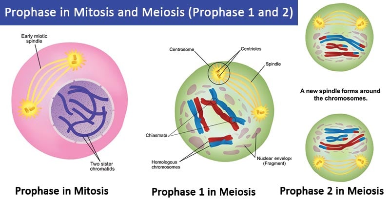
Image Source: Ali Zifan (Wikipedia) and Ali Zifan (Wikipedia).
Prophase in Mitosis
In animal cells, prophase is the first phase of mitosis and the second phase of mitosis in plant cells.
Condensation of chromosomes
- This is a mechanism that involves the condensing of the DNA that is synthesized during interphase.
- The interphase stage allows DNA replication which uniformly entangles (intertwines) the DNA molecules, for easy separation during mitosis.
- Chromosome condensation is the compacting of the chromatin into well-defined rod-shaped structures known as chromatins.
- Condensation allows structural reorganization to separate the identical sister chromatids, a mechanism known as chromatid resolution.
- Condensation forms sturdy, elastic chromosomes to prevent damage and breakage caused by the force of pulling microtubules and cytoplasmic drags experienced during mitosis.
- Chromosome condensation is done by the help of condensins complexes which include condensins and topoisomerase.
- Condensins are complex proteins that play a key role in the cell cycle, and majorly the separation of the chromosomes, chromosome condensation, maintaining the structural integrity of chromosomes, and the resolution of DNA topography during intertwining.
- Topoisomerase specifically topoisomerase 2 helps in the relaxation and unlinking of supercoils formed in interphase, cutting, and releasing of the DNA duplex.
- Chromosome condensation forms two sister chromatids which are X-shaped and joined at a point known as a centromere.
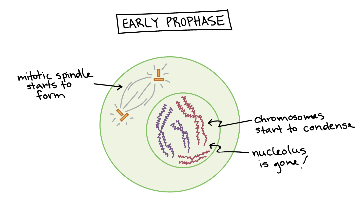
Image Source: Khan Academy.
Movement of centrosomes
- At the centromere are microtubules that function by recruiting tubulins.
- Tubulins act by incorporating microtubules to the nucleus.
- The centromeres that are replicated from the interphase move away towards the opposite poles of the cell.
- The moving is driven by motor proteins that are associated with the centromere.
- The centromere associated motor proteins track the centromere movement and the formation of spindles during chromatin separation.
- The motor proteins convert chemical energy in the form of ATP to mechanical energy to move along the microtubules.
- This helps the centromeres to move to opposite poles.
Formation of mitotic spindles – mitotic spindles are fibrous
- The microtubules of the interphase hold the replicated centrosomes during separation.
- On separation, the centrosomes move to opposite poles of the cell, mediated by the radial microtubules known as Kinetochore, found on each centrosome.
- Additionally, both centrosomes have interpolar microtubules that interact with each other and join the sets of microtubules to form the basic structure of the mitotic spindles.
- Cells that lack centrioles, the chromosomes nucleate the microtubule assembly into the mitotic apparatus.
- The formation of mitotic spindles in plants is a bit different.
- The microtubules gather at the opposite poles of the cell and start to form the spindle apparatus at the foci.
- Eventually, the mitotic spindles separate the sister chromatids as the cell cycle continues.
- Generally, the mitotic spindles are made up of hundreds to thousands of fibrous microtubules and microtubule-associated proteins (MTP) which function in nucleation, transportation, dynamics, and cross-linking of microtubule networks.
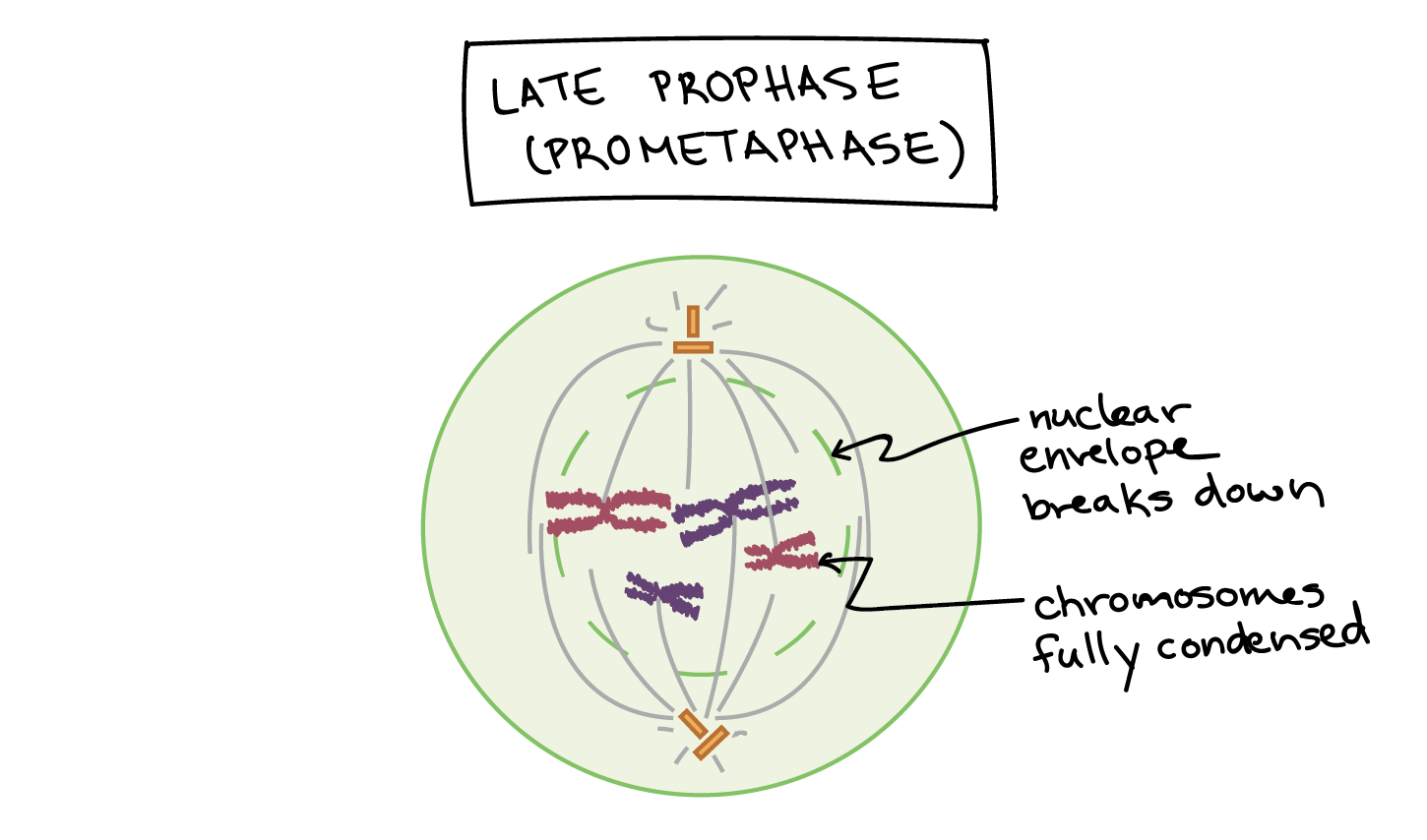
Image Source: Khan Academy.
Beginning of nucleoli breakdown
- During the late prophase (prometaphase), part of the nucleus where the synthesis of ribosomes takes place to disappear.
- This is an indication that the nucleus is starting to breakdown.
- The cell then redirects cellular energy from the cell’s’ metabolism to cell division mechanisms.
- This is the stage where the final chromosome condensation takes place making them very compact
- The nuclear envelope also breaks down releasing the chromosomes.
- The mitotic spindles also continue growing further and some of the interpolar microtubules start to capture the chromosomes.
Prophase in Meiosis
- Meiosis is a rather long process than that of mitosis because it takes place in two cycles involving the separation of chromosomes.
- The process is longer due to the phases of prophase which takes place in two phases i.e prophase I and prophase II.
- Prophase I is quite complex which involves the pairing up of the homologous chromosomes and the exchange of genetic information. It defines the difference between mitosis and meiosis.
- Prophase II is very similar to the mitotic prophase.
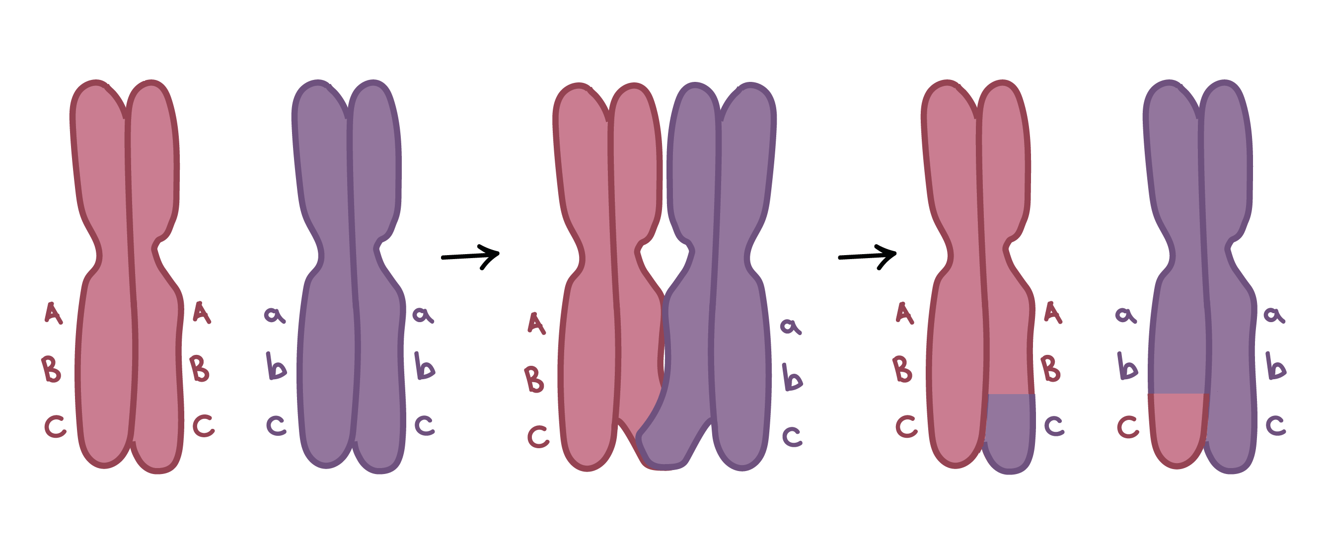
Image Source: Khan Academy.
Prophase I
This is the phase of homologous chromosome pairing and exchange of DNA to form recombinant chromosomes. There are five phases that define prophase I.
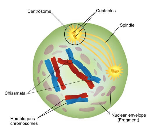
Figure: Prophase 1 of Meiosis. Image Source: Wikipedia.
These include:
Leptotene- This is the first stage of prophase I and the shortest phase of prophase I.
- It the phase of a replicated chromosome condensation
- The chromosomes condense and become compact and visible hence making it possible to distinguish between sister chromatids.
- The chromosomes appear like strings with beads and the beads are known as chromomeres.
- Each of the sister chromatids gets attached to the nuclear envelopes.
Zygotene- also known as zygonema.
- This is the phase where homologous chromosomes associate closely to forms pairs of chromosomes, a mechanism known as synapsis. The pairs of chromosomes have four chromatids (tetrads).
- The synaptic association forms up and down the chromosome creating several points of contact known as synaptonemal complex which has a zipper-like structure created by coils of the chromatids.
- Synapsis is facilitated by the synaptonemal complex by holding the aligned chromosomes together.
- After synapsis of the homologous pairs, they form four chromatids which are known as tetrads or bivalents (two pairs).
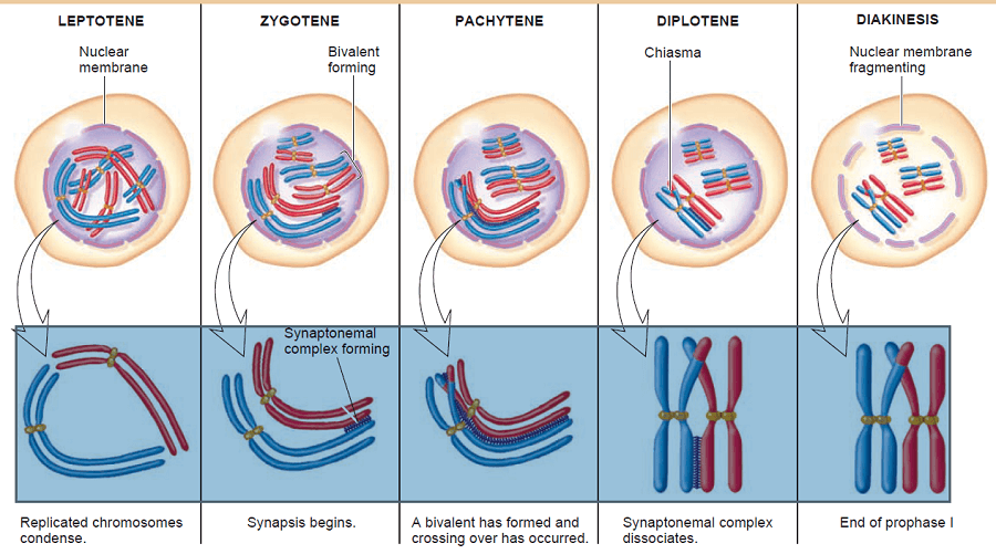
Figure: Prophase 1 stages of Meiosis. Image Source: Toppr.
Pachytene- This is the phase where the cross over of genetic materials takes place between non-sister chromatids i.e pairs of homologous chromosomes. This forms chiasmata.
- Crossing over is achieved by synapsis by the attachment created by the chromatid in a homologous chromosome.
- The synapses complete the crossing over of genetic information, creating a variation in the genetic materials due to the exchange between the mother and father genetic elements.
- Separation of sister chromatids occurs and the homologous chromosomes remain attached, hence a thick complex is formed known as the synaptonemal complex.
- The synaptonemal complex allows the formation of chiasma which function by allowing the crossing over of alleles within small regions of the chromosomes.
Diplotene- This is the stage of synaptonemal complex disappearance while the homologous pairs remain attached at the chiasmata.
- The disintegration of the synaptonemal complex occurs between the two chromosomal arms causing repulsion of the arms.
- This causes the chromosomes to move apart from each other while still being held by the chiasmata.
- The chromosomes then start to uncoil slowly and the chiasmata can be visualized microscopically and they can be seen moving close to the ends of the chromatids. This process is known as terminalization.
Diakinesis- The fifth and final phase of prophase I. It sets up the cell for metaphase.
- It takes place after the chromatids have condensed and the sister chromatids are bivalent of tetrad as visualized under a microscope.
- The chiasmata finally arrive at the end of the chromatid arms of the chromosomes finalizing terminalization.
- the Chromosomes become more condensed but remain connected by the chiasmata and can not move further toward the poles.
- At this stage the nucleolus and the nuclear envelope dissolves allowing the centrioles (the centrosome forming microtubules) that form the mitotic spindle, to migrate freely along with the remaining spindles formed during mitosis.
Prophase II
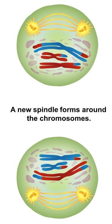
Figure: Prophase 2 of Meiosis. Image Source: Wikipedia.
- This phase involves chromosomal condensation and breaking down of the nuclear envelope.
- It also involves the moving apart of the centrosomes with spindle forming in between them
- These spindles which are basically microtubules start to capture the chromosomes.
- The distinguishing factor about this prophase and that of mitosis are that prophase II takes place with a haploid number of chromosomes while the mitotic prophase takes place with a diploid number of chromosomes.
- De-condensing of the chromosomes takes place in telophase I.
Prophase of Mitosis Video Animation
Prophase I of Meiosis Video Animation
Prophase II Video Animation
References and Sources
- https://www.ncbi.nlm.nih.gov/pmc/articles/PMC4962293/
- https://pubmed.ncbi.nlm.nih.gov/13351623/
- http://www.phschool.com/science/biology_place/biocoach/mitosisisg/prophase.html
- https://www.nature.com/scitable/definition/prophase-189/
- https://study.com/academy/lesson/mitosis-i-the-mitotic-spindle.html#:~:text=The%20mitotic%20spindle%20is%20a,organizing%20center%20during%20cell%20division.
- https://www.khanacademy.org/science/biology/cellular-molecular-biology/mitosis/a/phases-of-mitosis
- https://bio.libretexts.org/Bookshelves/Human_Biology/Book%3A_Human_Biology_(Wakim_and_Grewal)/07%3A_Cell_Reproduction/7.3%3A_Mitotic_Phase_-_Mitosis_and_Cytokinesis
- https://www.sciencedirect.com/topics/medicine-and-dentistry/mitosis-spindle
- https://teaching.ncl.ac.uk/bms/wiki/index.php/Meiosis_prophase_1#:~:text=Diakinesis%20is%20the%20final%20step,most%20condensed%20form%20during%20diakinesis.
- thebiologydictionary/prophase
- https://science.sciencemag.org/content/301/5634/785/tab-figures-data
- http://www.phschool.com/science/biology_place/biocoach/meiosis/proi.html#:~:text=During%20prophase%20I%2C%20they%20coil,give%20rise%20to%20genetic%20recombination.
- https://ib.bioninja.com.au/higher-level/topic-10-genetics-and-evolu/101-meiosis/stages-of-prophase.html
- https://www.nature.com/scitable/topicpage/meiosis-genetic-recombination-and-sexual-reproduction-210/
- https://www2.le.ac.uk/projects/vgec/highereducation/topics/cellcycle-mitosis-meiosis
- https://teaching.ncl.ac.uk/bms/wiki/index.php/Meiosis_prophase_1
- https://en.wikipedia.org/wiki/Meiosis
- https://en.wikipedia.org/wiki/Prophase#Mitotic_prophase


I need you to help me understanding this subject