Cytokinesis is a physical process of cell division, that normally takes place after mitosis.
Cytokinesis is the physical division of the cell cytoplasm, the cell membrane, and cell organelles in eukaryotic cells to produce two distinct cells at the end of the cell cycle in both mitosis and meiosis.
- In most cells, cytokinesis is initiated during the anaphase stage and ends in telophase, a phase where the chromosomes are completely segregated.
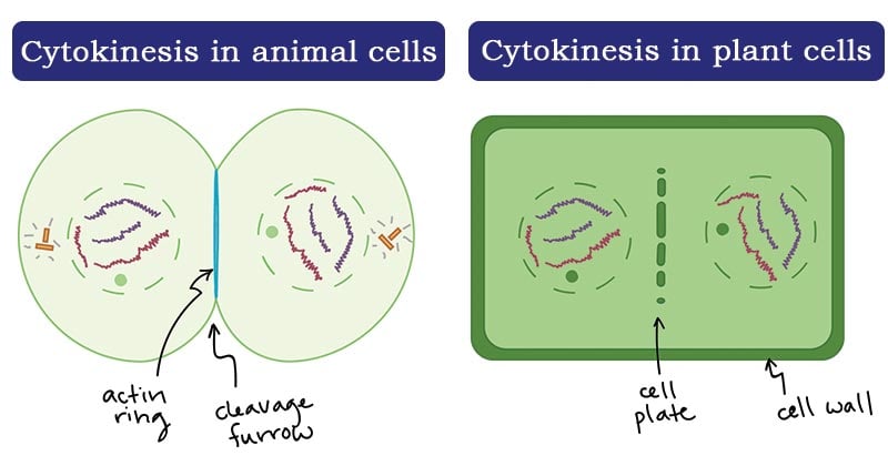
- In animal cells, cytokinesis is achieved when a contractile ring of the cell microtubules form a cleavage furrow that divides the cell membrane into half. The microtubules used during cytokinesis are those generated during the initial stages of division and they contribute to the restructuring of the new cell.
- In the plant cell, a cell plate is formed that divides the cell into two.
- Additionally, cytokinesis only takes place ones the separation of chromosomes is complete. This ensures that each daughter cell receives a full set of chromosomes along with the complete elements of the cytoplasm and cell organelles.
Interesting Science Videos
What happens during cytokinesis?
- Cytokinesis is similar in both plant and animal cells, however, it varies by the completion of the mechanism of the formation of two daughter cells from a parent cell, each with a set of separated chromosomes and halved cytoplasm and cell organelles.
- The division process of the cell generally entails the formation of a cleavage furrow, which divided the cells almost equally.
- Cell division may be symmetrical or asymmetrical, where one cell takes a majority of the cytoplasm.
- For example, spermatogenesis, a meiosis cell division process is symmetrical cytokinesis where the newly formed sperm cells are equal in size and content, while biogenesis is a typical example of asymmetrical cytokinesis, producing a large cell and 3 polar bodies.
- The cleavage furrow in animal cells is determined by the positioning of the mitotic spindles while in plant cells, the cleavage furrow is independent of the mitotic spindles.

Figure: Cytokinesis in plant and animal cells. Image Source: Khan Academy.
Generally, cytokinesis takes place in four stages:
Initiation and formation of the cleavage furrow
- During cytokinesis, the initial physical change observed is the appearance of the cleavage furrow on the cell surface.
- The furrow starts to deepen, spreading around the cell until it completely divides into two.
Contraction and constriction
- This is also known as abscission.
- The accomplishment of cytokinesis in animal cells in by the contractile ring, which is a ring that is made up of actin and myosin and regulatory proteins formed under the surface of an animal cell during cell division.
- These rings have the ability to contract and constrict the cell pinching it into two.
Membrane Insertion
- Concurrently, a new membrane is formed and inserted into the cell membrane, next to the contractile ring through the fusion of the intracellular vesicles.
- The new membrane enables the cell to increase as the cytoplasmic division takes place.
Completion of cytokinesis by forming two fully developed daughter cells.
Cytokinesis in animal cells

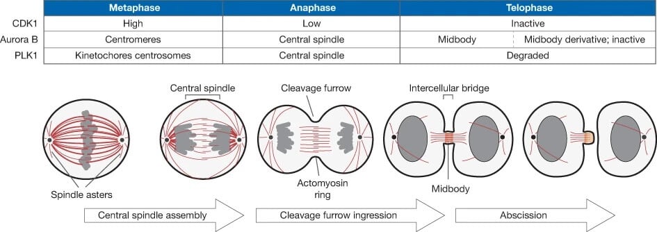
Figure: A schematic representation showing the reorganization of an animal cell progressing through the different stages of cytokinesis. Red, microtubules; grey, chromosomes. Image Source: Nature Cell Biology.
- The cytokinesis process in the animal cell is attributed to the role of the contractile ring. The contractile ring is held together by the microtubules of the mitotic spindles.
- These microtubules and cell signals determine the location of the contractile ring and therefore they direct the plane of cell division, known as the division plane.
- The cleavage furrow forms around the division plane which eventually pinches off separating the cell into two cells.
- This is followed by a process of contraction and constriction by the contractile ring, made up of actin, myosin, and regulatory proteins.
- Actin and myosin are formed during interphase assembled in a cortical network o the filaments filled with actin and myosin. As cell division continues, actin filaments are reorganized while the myosin filaments accumulate during anaphase to form the contractile rings.
- The contractile ring is positioned by the actin-myosin and regulatory proteins and they also act as the motor proteins, allowing the contraction of the muscle cells.
- The muscle cells which are packed with actin filaments are pulled together by myosin proteins to form an actin-myosin ring, which plays a major role in the exclusion of the cytoplasm and the cell organelles.
- After the exclusion of the cytoplasm and the organelles, the ring and the microtubules are left behind forming the midbody structure, which eventually divides.
- The cellular proteins cut and fusion of the plasma membrane are shut, while the extracellular elements that hold the cell together get dissolved, separating the cells.
- The separated cells may remain associated linked by the cytoplasm at bridges known as the gap junctions, which are formed by remains of the endoplasmic reticulum trapped by the midbody structures.
Cytokinesis in plant cells

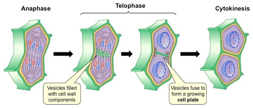
Figure: Cytokinesis in plant cells. Image Source: BioNinja.
- The major difference between an animal cell and a plant cell is that plants are made up of an extra-rigid cell wall, and therefore, a special kind of microtubules are involved in the completion of cytokinesis. These are known as the phragmoplasts.
- Phragmoplasts are vesicular spindle microtubules formed by Golgi vesicles during telophase on the metaphase plate, carrying vesicles and cellular elements such as cellulose to the new cell wall.
- When these Golgi vesicles fuse at the center next to the cell wall, they form the cell plate, the site of plant cytokinesis.
- The more the vesicles fuse, the cell plate continues to enlarge, emerging at the periphery of the cell wall of the cell.
- The phragmoplasts carry vesicles of the cell wall to the newly formed cell plate.
- Accumulated enzymes, structural proteins, and glucose molecules between the membranes by the Golgi apparatus during interphase contribute to the formation of the new cell wall, while the Golgi membranes are incorporated and form part of the plasma membrane.
- The cellulose carried by the phragmoplast interact and combine forming the complex and strong rigid matrix of the plant cell wall.
- After the cell plate divides the cell, the plasma membrane seals off separating the two newly formed daughter cells.
- In between the two cells, trapped endoplasmic reticulum forms the plasmodesmata, space, or gap which allows the passage of molecules from one cell to another and signaling of cells for cell communication.
Applications of Cytokinesis
- Cytokinesis studies have enabled the construction of block-cytokinesis micronuclei cytosome assay to study human lymphocytes.
- Failed cytokinesis can lead to tumorigenesis which has enhanced cancer studies in the understanding of oncogenesis and drug targets to unfulfilled cytokinesis processes.
References and Sources
- 1% – https://www.thoughtco.com/cytokinesis-in-a-cell-cycle-373541
- 1% – https://www.researchgate.net/publication/226631723_Molecular_Analysis_of_the_Cell_Plate_Forming_Machinery
- 1% – https://www.ncbi.nlm.nih.gov/pmc/articles/PMC2789570/
- 1% – https://quizlet.com/200759728/bio-121-chapter-12-flash-cards/
- 1% – https://courses.lumenlearning.com/boundless-biology/chapter/the-cell-cycle/
- <1% – https://www.thoughtco.com/daughter-cells-defined-4024745
- <1% – https://www.sciencedirect.com/topics/neuroscience/cytokinesis
- <1% – https://www.majordifferences.com/2013/10/difference-plant-cell-vs-and-animal-cell.html
- <1% – https://www.genome.gov/genetics-glossary/Plasma-Membrane
- <1% – https://www.cell.com/current-biology/pdf/S0960-9822(11)01205-X.pdf
- <1% – https://www.annualreviews.org/doi/abs/10.1146/annurev.cellbio.17.1.351
- <1% – https://study.com/academy/lesson/actin-filaments-function-structure-quiz.html
- <1% – https://quizlet.com/11000697/molecular-biology-of-the-cell-chapter-17-part-3-flash-cards/
- <1% – http://absuriani.my/BOOK%20CHAPTER/2018%20chapter%201.pdf
