Centrosome was first observed and discovered by Eduard van Beneden and Theoder Boveri in the years 1883 and 1888 respectively. The centriole duplication process or pattern was studied at first by Etienne de Harven in 1950 and Joseph G. Gall also in 1950 but independently.
Interesting Science Videos
What is Centrosome?
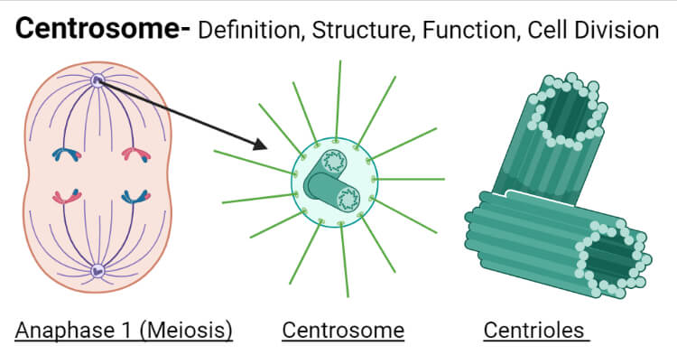
Centrosomes are cellular components that appear like a disc in shape and are internally built by microtubule components and play important role in cell division.
- Centrosomes are double or a pair of centrioles along with microtubule-organizing center (MTOC) with the pericentriolar matrix.
- The centrioles in the centrosome are arranged in pairs and perpendicularly i.e. forming the right angle between them.
- Centrioles are present at the base of cilia and flagella so they are also called basal bodies.
- The centrosome is present in an animal cell, not the plant cell or fungi. The plant cell contains a microtubule-organizing center.
- But they are also present in male gametes of bryophytes, seedless vascular plants, charophytes, ginkgo, and cycads.
- Development of cancer can occur in the case of non-functional centrosomes in animals.
- It is present in the cytoplasm at the central region i.e. near of nucleus hence called centrosome or centrioles.

Centrosome Structure
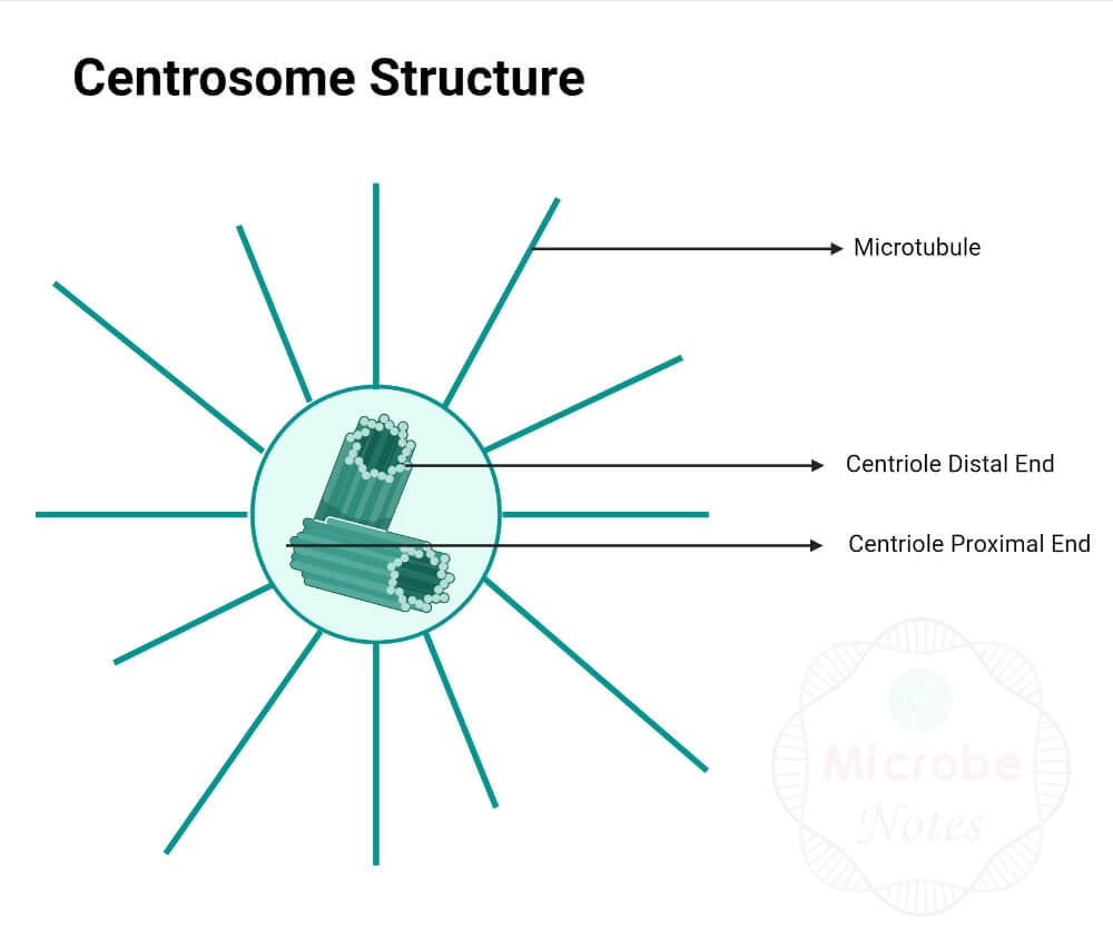
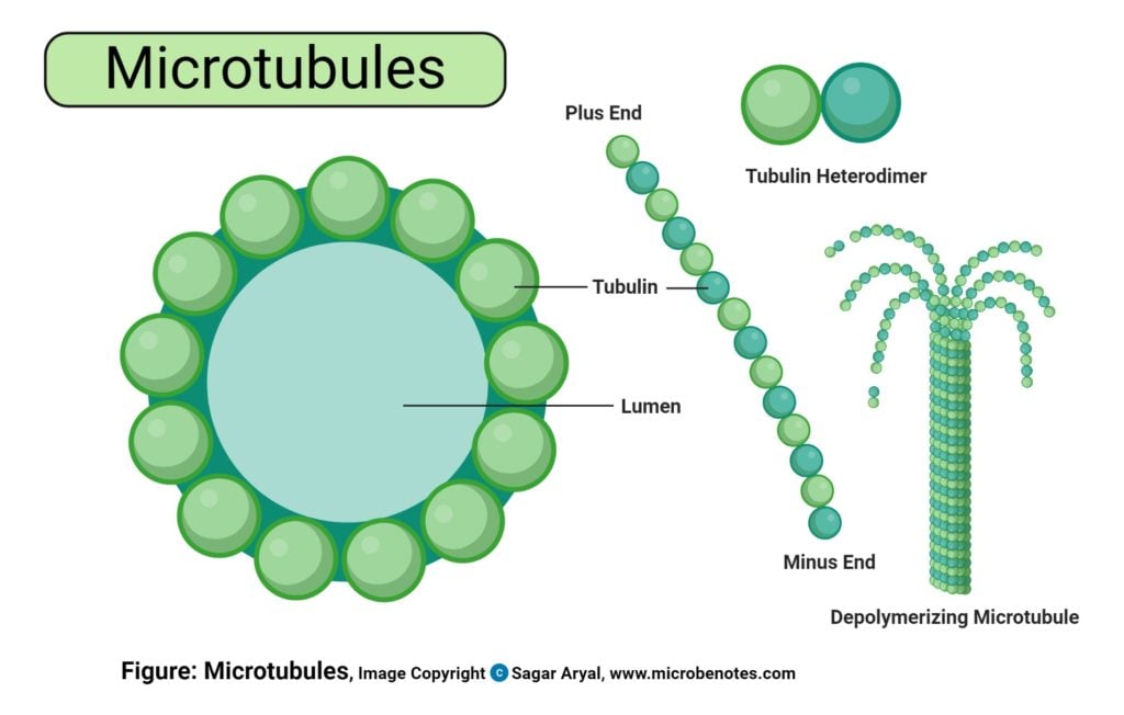
- Centrosome is composed of microtubules that are short i.e. length of about 500 nm and their width or diameter of about 200 nm.
- They are arranged in nine sets (in each centriole) where each set comprises three microtubules or tubulins which is also termed as 9+3 arrangement.
- The two centrioles are oriented in 90 degrees to one another.
- The centrioles are surrounded by the pericentriolar matrix.
- The pericentriolar matrix comprises dense cytosolic structures and proteins such as gamma-tubulins, pericentrin, ninein, etc.
- The centrioles and pericentriolar matrix as a whole are called the centrosome.
- The arrangement of microtubules in the centrosome is called cart-wheel structure.
- In the cart-wheel model, the nine microtubules(triplets) linked with each other are arranged in a circle and there is one microtubule(duplet) at the center connected to the nine microtubules by proteins called spokes and this arrangement appears like a cartwheel.

Centrosome Functions
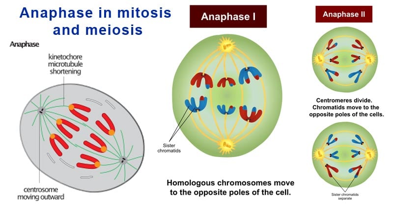
- They take part in cell division for the formation of spindle fibers necessary for the movement of chromosomes towards opposite poles during the separation of sister chromatids.
- They are also involved in the cellular organization i.e. by the formation or use of microtubules to prepare cytoskeleton.
- Its position in the cell determines the position of other organelles and the nucleus as it is involved in the formation of the cytoskeleton.
Centrosomes and Cell Division
- Each cell contains a centrosome near the nucleus. As the separation of sister chromatids requires two centrosomes arranged at two opposite poles the centrosome goes through a division process during cell division.
- In the G1 phase of cell division, initiation of duplication begins in which the centrioles which were arranged in pairs get separated.
- In the S phase, another centriole formation starts from each mother centriole.
- The complete formation of two centrosomes from a single mother centriole gets completed in the G2 phase of cell division.
- The centrosomes then get separated and move towards opposite poles of the nucleus during metaphase after the disintegration of the cell membrane.

Four types of microtubules arise from the centrosome based on the direction of their movement and arrangement.
- Astral Microtubules: They have directed away from the metaphase plate and help in the orientation of the centrosome during cell division.
- Chromosomal Microtubules: They are attached to the arms of the chromosome and protect chromatids from disruption during separation.
- Kinetochore Microtubules: They are attached to the kinetochore and separate the sister chromatids by the opposite pull force from both sides during anaphase.
- Polar Microtubules: They elongate to assist the separation but are not attached to chromosomes.

Controversy Over Necessity
- The centrosome is not present in plant cells but still, they can go through cell division by the presence of a microtubule-organizing center (MTOC).
- So the basic requirement of cell division is microtubule.
- The centrosome is not that vital for the survival or division of the cell.
- But microtubules are most required and if by any means microtubules are organized in the cell they can go through the division process.
- In past studies, centrosome was considered most required or vital for cell division.
- Recent researches show that even if centrosomes are disrupted or not present as in plants, fungi, and some animal cells, the cell continues to divide properly without any kind of delay or interruption. After research in 1961 by a scientist named Mazia, it was found that mitotic centers were actively functional in the presence or absence of centrosome.
- So it can be said that centrosomes just aid in cell division but are not most required for it.
Centrosomes FAQs
Where is centrosome located?
Centrosome is found in the cytoplasm at the central region, near of nucleus.
Does animal cell have centrosome?
Yes, the centrosome is present in an animal cell.
Does plant cell have centrosome?
No, the centrosome is absent in plant cell.
Does fungi have centrosome?
No, the centrosome is absent fungi.
References
- Sluder G. (2014). One to only two: a short history of the centrosome and its duplication. Philosophical transactions of the Royal Society of London. Series B, Biological Sciences, 369(1650), 20130455. https://doi.org/10.1098/rstb.2013.0455
- Joukov, V., & De Nicolo, A. (2019). The Centrosome and the Primary Cilium: The Yin and Yang of a Hybrid Organelle. Cells, 8(7), 701. https://doi.org/10.3390/cells8070701
- Bettencourt-Dias, M., Glover, D. Centrosome biogenesis and function: centrosomics brings new understanding. Nat Rev Mol Cell Biol 8, 451–463 (2007). https://doi.org/10.1038/nrm2180
- Bettencourt-Dias, M. Q&A: Who needs a centrosome?. BMC Biol11, 28 (2013). https://doi.org/10.1186/1741-7007-11-28.
