Dendritic cells are key antigen-presenting cells of the immune system that inform other effector immune cells to fight against invasive pathogens.
- These are also called professional antigen-presenting cells as these present antigens or parts of antigen to the receptors on different immune cells in order to elicit an immune response against them.
- Dendritic cells represent a distinct type of white blood cells capable of alerting the immune system about the presence of infections and activating the innate and adaptive immune response.
- The term ‘dendritic cell’ was given due to the tree-like or dendritic appearance of the cell. These are also considered the sentinels of the immune system as these play an essential role in the initiation of both innate and adaptive immune responses.
- As a part of antigen presentation, dendritic cells also process antigens to ensure the association between them and the major histocompatibility complex molecules.
- Dendritic cells can only perform their function after a complete maturation process initiated by direct exposure to Toll-like receptor ligands.
- Dendritic cells are easy to modify, and the modification of the cells is determined by signals which in turn depend on the local microenvironment.
- Besides antigen presentation, dendritic cells also work to maintain immune tolerance by ensuring that effector T cells are not produced against the normal or self-antigens under normal conditions.
- Dendritic cells exist in two distinct forms; immature and mature dendritic cells.
- Immature dendritic cells are present in peripheral tissues where they patrol for pathogens and antigens. Immature dendritic cells are defined by their inefficiency to present antigens to MHC receptors, but they do secrete few cytokines and express some ligands.
- The trigger for the activation of dendritic cells is brought by the encounter of pathogen-derived TLR ligands, intracellular sensors, or proinflammatory molecules.
- The maturation of dendritic cells involves the activation of lysosomes and antigen-processing machinery, which enhances the efficiency of peptide-MHC production.
Interesting Science Videos
Structure of Dendritic cells

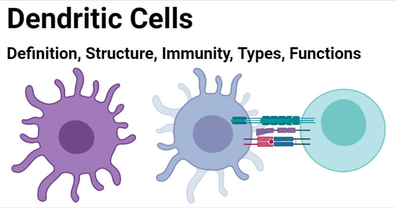
- Dendritic cells are larger antigen-presenting cells with large cytoplasmic projections that are similar in structure to dendrites of nerve cells.
- The cells are irregular in shape with phase-dense granules, an irregular nucleus, and a small nucleolus.
- The projections from the cell extend in many directions from the cell body, which are involved in patrolling for invading pathogens.
- The distinctive dendrite formation by dendritic cells is an important feature for the morphological identification of DC in a blood sample.
- The cytoplasm of dendritic cells does not have any filaments, but cell organelles like mitochondria and Golgi complex can be observed.
- Similarly, dendritic cells, as different stages of maturation, have different types of granules. The size and occurrence of granules in dendritic cells are diverse, but the most common granules occurring in dendritic cells are melanin granules.
How Dendritic cells work against pathogens? (Immunity)
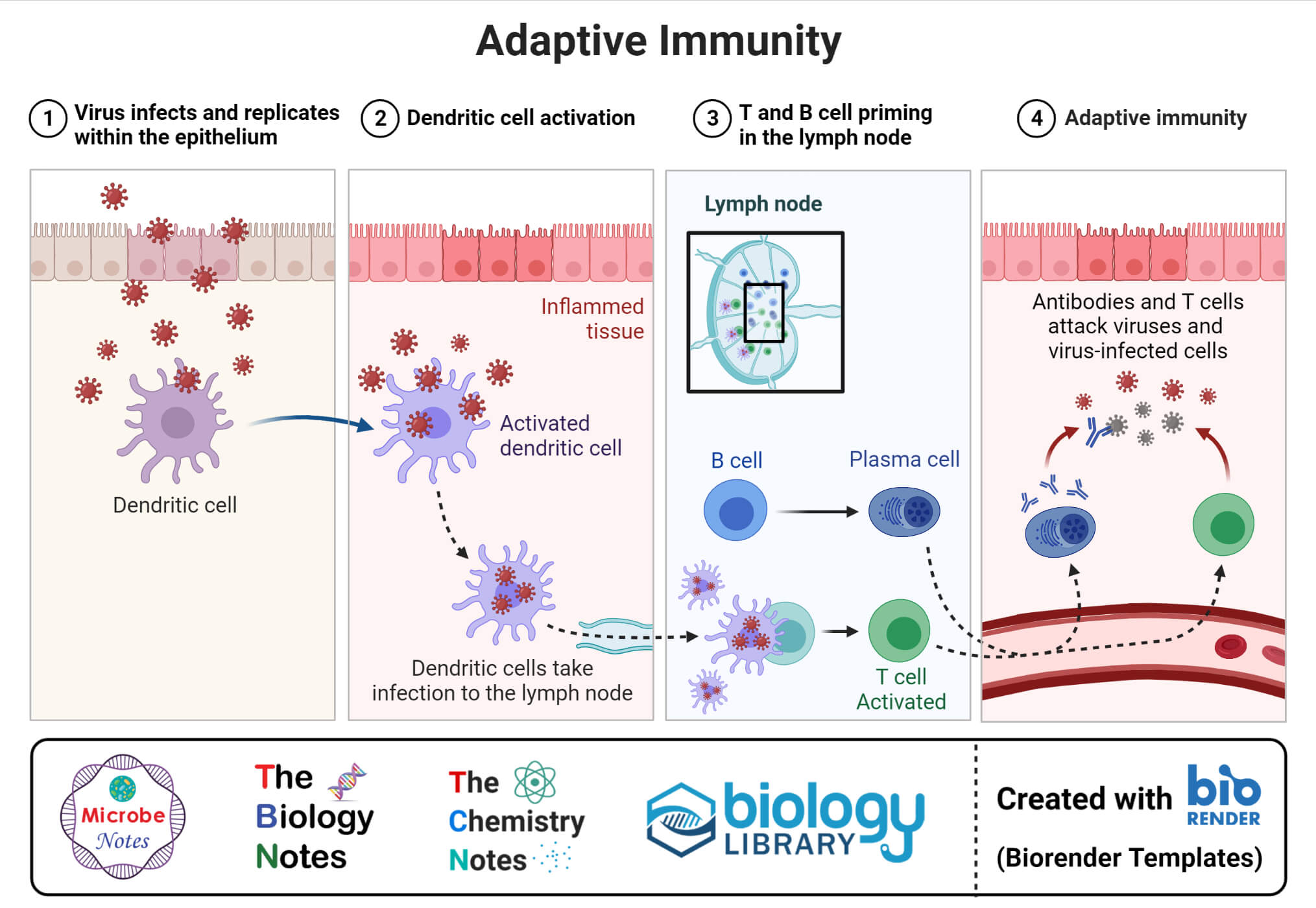
Dendritic cells occur in almost all types of tissue in the human body, where they act as a link between the innate and adaptive immune systems. The dendritic cells in these tissues can exist as either mature cells or immature cells. The immature dendritic cells undergo maturation in the presence of either antigens or cytokines or pathogen-associated molecular patterns (PAMPs). Maturation of such cells activates the metabolic, cellular, and gene transcription of the cells, causing the cells to migrate from peripheral tissues to T-dependent areas in lymphoid organs. The process of maturation causes the loss of adhesive structures, reorganization of the cytoskeleton, and increase in motility. It also leads to a decrease in their endocytic activity and an increased expression of MHC-II and costimulatory molecules.
1. Adaptive immune response

- The mature dendritic cells express a high level of the chemokine receptor CCR7 and cytokines, both of which are important for T-cell activation.
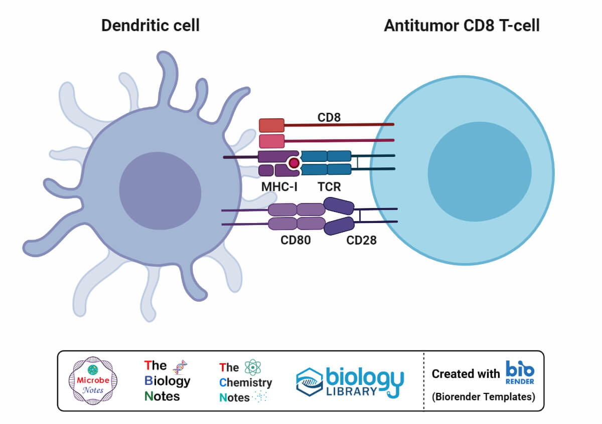
- The interaction between the dendritic cells and T cells forms the basis of antigen-specific immune responses. The interaction also induces the differentiation of T cells into different T helper subsets.
- Dendritic cells also have a unique characteristic of cross-presentation as these can trigger responses against intracellular antigens from different cell types.
- The activation of a T cell response by dendritic cells has been attributed to a combination of factors like a high level of expression of cell membrane costimulatory proteins.
- Dendritic cells also play an important role in directing an appropriate type of immune response against the invading microorganisms.
- These cells express different polarizing signals depending on the type of receptors linked with the cell surface.
- Dendritic cells also regulate B-cell responses as DCs can form T-independent short-lived clusters with B cells for their activation.
2. Innate immune response
- Even though the activation of adaptive immune response is the primary mechanism of dendritic cells, studies have revealed that these cells are important during the early phases of the immune response in innate immunity.
- Dendritic cells activate NK cells in lymph nodes by the secretion of cytokines like IL-2 and INF-γ that are essential for NK cell proliferation.
- The mechanism of DC-mediated NK cell activation is an important pathway for the activation of cells of innate immunity.
- Dendritic cells activated in the presence of microorganisms can also induce NK-cell cytotoxic function.

Types of Dendritic cells
Dendritic cells are a heterogeneous group of cells consisting of multiple groups and subsets. The following are subsets of dendritic cells found in primates;
1. Plasmacytoid DCs
- Plasmacytoid DCs are dendritic cells characterized by the expression of B220 and PDCA1. These resemble plasma cells, hence the name.
- These cells are important mediators of an immune response against viral particles as a result of the production of a large amount of type I interferons.
2. Conventional DCs
- Conventional DCs are dendritic cells that are further divided into subsets; CD8α+ and CD11b+.
- These cells can process and present antigens to T cells by the secretion of cytokines like IL-2. The CD8a+ DCs are involved in the priming of CD8+ T cells, whereas the CD11b+ cDCs prime the CD4 T cells.
3. Migratory DCs
- Migratory DCs are found in lymphoid organs like the liver, gut, skin, and aorta, consisting of two other sets of DCs; CD103+ and CD103-.
- These cells are called migratory DCs as these cells transfer from tissue to lymphoid organs.
4. Monocyte-DCs
- Dendritic cells derived from monocytes form a new subset of dendritic cells during inflammation.
- Monocytes that differentiate into dendritic cells are monocytes that express CCR2 which migrate from the bone marrow to the site of inflammation.
- These monocyte-DCs provide innate protection to the cells of the innate immune system during bacterial infections.

Functions of Dendritic cells
The following are some of the functions of dendritic cells;
- Dendritic cells are professional antigen-presenting cells that perform the most important function of presenting antigens to different receptors on different immune cells for their activation.
- Dendritic cells are responsible for eliciting an immune response in T-cells and are also involved in the differentiation of T-helper cells.
- In the innate immune system, dendritic cells are involved in the activation of natural killer cells by the secretion of different cytokines.
- Dendritic cells have also been observed to be involved in the functional control of regulatory T cells.
References
- Peter J. Delves, Seamus J. Martin, Dennis R. Burton, and Ivan M. Roitt(2017). Roitt’s Essential Immunology, Thirteenth Edition. John Wiley & Sons, Ltd.
- Judith A. Owen, Jenni Punt, Sharon A. Stranford (2013). Kuby Immunology. Seventh Edition. W. H. Freeman and Company.
- Satthaporn S, Eremin O. Dendritic cells (I): Biological functions. J R Coll Surg Edinb. 2001 Feb;46(1):9-19. PMID: 11242749.
- Snell, R.S. An electron microscopic study of the dendritic cells in the basal layer of guinea-pig epidermis. Zeitschrift für Zellforschung 66, 457–470 (1965). https://doi.org/10.1007/BF00334726
- Sahu R, Bethunaickan R, Singh S, Davidson A. Structure and function of renal macrophages and dendritic cells from lupus-prone mice. Arthritis Rheumatol. 2014 Jun;66(6):1596-607. doi: 10.1002/art.38410. PMID: 24866269; PMCID: PMC4547797.
- Wesa, A. K., & Storkus, W. J. (2007). Killer dendritic cells: mechanisms of action and therapeutic implications for cancer. Cell Death & Differentiation, 15(1), 51–57. doi:10.1038/sj.cdd.4402243
- Vyas, Jatin M. “The dendritic cell: the general of the army.” Virulence vol. 3,7 (2012): 601-2. doi:10.4161/viru.22975
- Liu, K.. “Dendritic Cells.” Encyclopedia of Cell Biology (2016): 741–749. doi:10.1016/B978-0-12-394447-4.30111-0
- Granucci F, Foti M, Ricciardi-Castagnoli P. Dendritic cell biology. Adv Immunol. 2005;88:193-233. doi: 10.1016/S0065-2776(05)88006-X. PMID: 16227091.
- Patente, Thiago A et al. “Human Dendritic Cells: Their Heterogeneity and Clinical Application Potential in Cancer Immunotherapy.” Frontiers in immunology vol. 9 3176. 21 Jan. 2019, doi:10.3389/fimmu.2018.03176
- Zhou, Haibo, and Li Wu. “The development and function of dendritic cell populations and their regulation by miRNAs.” Protein & cell vol. 8,7 (2017): 501-513. doi:10.1007/s13238-017-0398-2
- Mbongue, Jacques et al. “The role of dendritic cells in tissue-specific autoimmunity.” Journal of immunology research vol. 2014 (2014): 857143. doi:10.1155/2014/857143.
