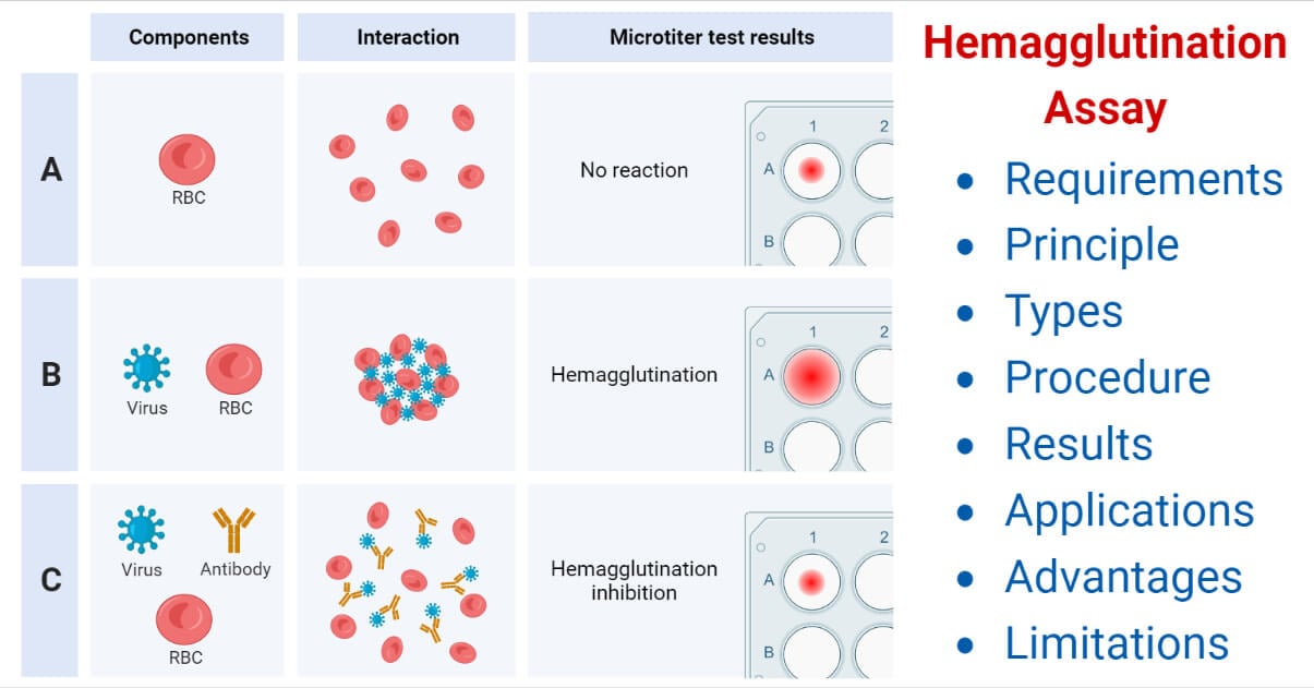
The antigen and antibody reaction in which the antigen-antibody complex formed is visible in the form of clumps is called agglutination.
It occurs on the surface of the cells or components involved as antigens are expressed on their surface. It occurs between insoluble antigens and soluble antibodies.
Agglutination reaction is of two types:
- Direct agglutination: It includes slide agglutination, tube agglutination, coombs’ test, and heterophile agglutination test.
- Passive agglutination: It includes latex agglutination, hemagglutination test and, cooaglutination test.
Hemagglutination is a type of passive agglutination reaction.
Hemagglutination assay (HA) is a type of immunoassay in which erythrocytes are used as carrier particles and are commonly preferred for serological diagnosis of various infections.
George Hirst was the person to discover hemagglutination tests. He was an American virologist.
Interesting Science Videos
Requirements
- RBC suspension: It is coated with antigens specific to the antibody to be detected or antibodies specific to antigens to be detected; hence called a carrier particle. RBCs can be of humans, sheep, chicks, etc.
- Serum or blood sample
- Microtitre plates such as 96 well-microtitre (V-bottomed) plate
- Diluent: Phosphate Buffered Saline (PBS)
- Negative and Positive Control Samples
- RDT kits (generally with slides, reagents, and control) are available in case of Rapid Hemagglutination assay
Principle of Hemagglutination Assay
The primary theme of the hemagglutination test is that when any antigens present on the surface of Red Blood Cells come in contact with any complementary antibody and vice-versa, they combine to agglutinate and form noticeable clumps, which can be observed clearly distinguishing the positive test from the negative one.
Types of Hemagglutination Assay
It is of mainly two types based on the methodology:
A. Rapid Hemagglutination Assay
As the name suggests, in around one minute, this test can determine the presence of a haemagglutinating agent. Hence, it can also be called Rapid Diagnosis Test (RDT). The negative and positive control samples must be tested only once when testing multiple samples.
Whenever a haemagglutination test is performed, the settling pattern of the red blood cell suspension must be tested. This is done by combining diluent with red blood cells and allowing them to settle.
i. The diluent should be dispensed.
ii. Add red blood cells and gently shake them to combine.
iii. Allow the red blood cells to settle before examining the pattern
iv. Examine the cells to see if they are setting in a normal pattern and have no auto-agglutination. In the micro-agglutination assay, there will be a distinct button of cells and an even suspension with no signs of clumping in the rapid assay.
Procedure of Rapid Hemagglutination Assay
- Place four separate drops of 10% chicken red blood cells on a glass slide or the provided one in the kit.
- Add one drop of each control and test sample to each drop of blood along with PBS. To dispense each sample, use separate tips, pipettes, or a flamed loop.
- At first, PBS is dropped, followed by control and unidentified samples.
- It should be mixed for one minute by rotating the slide or tile.
- Observe the result and compare it with the positive and negative control provided in the kit to analyze the result.
B. Micro-hemagglutination Assay
This method is useful for testing the presence or absence of haemagglutinin in allantoic fluid from many embryonated eggs. It is a more time-consuming method than RDT. Red blood cells are dissolved in a 1% solution. Cells settle faster in V-bottom plates, and the slight difference between positive and negative results is greater than in U-bottom plates.
Procedure of Micro-hemagglutination Assay
- Fill out a recording sheet with information about the samples being tested. The samples and controls will be placed in the wells indicated on this sheet.
- Take the sample of about 50 ml with a micropipette and dispense it into a well of the microwell plate. Use a different tip for each sample to prevent contamination of samples.
- Place negative and positive controls on one of the plates.
- Pour 50 mL of PBS into each well. These wells will serve as auto-agglutination controls for red blood cells.
- Fill each well with 25 mL of 1% red blood cells.
- Gently tap the plate’s sides to mix. Cover the plate with a plate cover.
- Let the plate stand for about 40 minutes, and observe/record the data.
Result Interpretation of Hemagglutination Assay
The appearance of clumps in the case of agglutinated suspension can indicate a positive test in all tests. It can be compared with the positive control set to analyze properly. A positive test suggests that the respective sample is contaminated with antibodies or antigens related to a pathogen.
Note: The clumps can be observed at the bottom of the well in the case of Micro-well and on the surface in the case of RDT.

Applications of Hemagglutination Assay
- It can be used to detect the humoral immune response of the body against any disease or infective agents.
- It can be used for determining different blood cell types or groups.
- It can detect and quantify viral infections such as paramyxovirus, influenza, etc.
- Different rapid diagnosis test kits based on hemagglutination are designed. For e.g. an RDT kit for detecting HbsAg in case of Hepatitis B infection.
- It can also detect various bacterial infections, such as syphilis.
Advantages of Hemagglutination Assay
- Simple to perform
- Not as expensive as many other tests
- Types of equipment used are generally easily available.
- Fast interpretation of results (as in the case of RDT)
Limitations of Hemagglutination Assay
- Faults in incubation time and concentration of RBC may lead to wrong results.
- Different factors considered in the reaction must be specific. Their non-specificity may result in an incorrect result.
- Determination of quantitative values and result interpretation require qualified individuals.
- Result interpretation is made manually without any digital data, so the different observers might have errors or fluctuations in analysis.
References
- S. Miyaishi, F. Moriya, (2005) Encyclopedia of Analytical Science (Second Edition)
Parija S.C., (2009), Textbook of Microbiology and Immunology, 2nd edition, Elsevier, a division of Reed Elsevier India Private Limited, pg. 108 - Sukhadeo B. Barbuddhe and Deepak B. Rawool, Methods in Microbiology, 2020
- Townsend, A., Rijal, P., Xiao, J. et al. A haemagglutination test for rapid detection of antibodies to SARS-CoV-2. Nat Commun 12, 1951 (2021). https://doi.org/10.1038/s41467-021-22045-y
- Thangavelu, C. P., & Koshi, G. (1980). Micro-indirect hemagglutination test for detection of antibodies to the Ibc protein of group B Streptococcus. Journal of clinical microbiology, 12(1), 1–6. https://doi.org/10.1128/jcm.12.1.1-6.1980
