Agglutination is an antigen-antibody reaction in which a particulate antigen combines with its antibody in the presence of electrolytes at a specified temperature and pH resulting in the formation of visible clumping of particles.
It occurs optimally when antigens and antibodies react in equivalent proportions. This reaction is analogous to the precipitation reaction in that antibodies act as a bridge to form a lattice network of antibodies and the cells that carry the antigen on their surface. Because cells are so much larger than a soluble antigen, the result is more visible when the cells aggregate into clumps.

When particulate antigens react with specific antibody, antigen-antibody complex forms visible clumping under optimum PH and temperature. Such a reaction is called agglutination. Antibodies that produce such reactions are called agglutinins.
Interesting Science Videos
What is Agglutination?
Agglutination is the visible expression of the aggregation of antigens and antibodies. Agglutination reactions apply to particulate test antigens that have been conjugated to a carrier. The carrier could be artificial (such as latex or charcoal particles) or biological (such as red blood cells). These conjugated particles are reacted with patient serum presumably containing antibodies. The endpoint of the test is the observation of clumps resulting from that antigen-antibody complex formation. The quality of the result is determined by the time of incubation with the antibody source, amount and avidity of the antigen conjugated to the carrier, and conditions of the test environment (e.g., pH and protein concentration). Various methods of agglutination are used in diagnostic immunology and these include latex agglutination, flocculation tests, direct bacterial agglutination, and hemagglutination.
Agglutination differs from precipitation reaction in that since agglutination reaction takes place at the surface of the particle involved, the antigen must be exposed and be able to bind with the antibody to produce visible clumps. In agglutination reactions, serial dilutions of the antibody solution are made and a constant amount of particulate antigen is added to serially diluted antibody solutions. After several hours of incubation at 37°C, clumping is recorded by visual inspection. The titer of the antiserum is recorded as the reciprocal of the highest dilution that causes clumping. Since the cells have many antigenic determinants on their surface, the phenomenon of antibody excess is rarely encountered.
Prozone Phenomenon
The condition of excess antibody, however, is called a prozone phenomenon. At a high concentration of antibody, the number of epitopes are outnumbered by antigen-binding sites. This results in the univalent binding of antigen by antibody rather than multivalently and thus, interferes in the crosslinking of antigen (Lattice formation).
Occasionally, antibodies are formed that react with the antigenic determinants of a cell but does not cause any agglutination. They inhibit the agglutination by the complete antibodies added subsequently. Such antibodies are called blocking antibodies. Anti-Rh antibodies and anti-brucella antibodies are few examples of such blocking antibodies.
Agglutination tests are easy to perform and in some cases are the most sensitive tests currently available. These tests have a wide range of applications in the clinical diagnosis of non- infectious immune disorders and infectious diseases. Agglutination reactions have a wide variety of applications in the detection of both antigens and antibodies in serum and other body fluids. They are very sensitive and the result of the test can be read visually with ease.
Types of Agglutination Reactions
Agglutination reactions can be broadly divided into three groups:
- Active/Direct agglutination
- Passive agglutination
- Hemagglutination
1. Active Agglutination
Agglutination reactions where the antigens are found naturally on a particle are known as direct agglutination. In active agglutination, direct agglutination of particulate antigen with specific antibody occurs. Direct bacterial agglutination uses whole pathogens as a source of antigen. It measures the antibody level produced by a host infected with that pathogen. The binding of antibodies to surface antigens on the bacteria results in visible clumps Active agglutination can be of following types:
- Slide/Tile agglutination: Basic type of agglutination reaction that is performed on a slide. Identification of bacterial types represents a classic example of a slide agglutination. In this method suspension of unknown antigen is kept on slide and a drop of standardized antiserum is added or vice versa. A positive reaction is indicated by formation of visible clumps. E.g. Widal test, RPR test.
- Tube agglutination: It is agglutination test performed in tube and standard quantitative technique for determination of antibody titre. In this method serum is diluted in a series of tubes and standard antigen suspensions (specific for the suspected disease) are added to it. After incubation, antigen-antibody reaction is indicated visible clumps of agglutination.
- Heterophile agglutination test: This test depends on demonstration of heterophilic antibodies in serum present in certain bacterial infections.
- Antiglobulin (Coombs) test: This’ test was devised by Coombs, Mourant, and Race for detection of incomplete anti-Rh antibodies that do not agglutinate Rh+ erythrocytes in saline. When serum containing incomplete anti-Rh antibodies is mixed with Rh+ erythrocytes in saline, incomplete antibody antiglobulin coats the surface of erythrocytes but does not cause any agglutination. When such erythrocytes are treated with antiglobulin or Coombs serum (rabbit antiserum against human gamma globulin), then the cells are agglutinated. Coombs test can be direct as well as indirect.
In direct method, the sensitization of red blood cells (RBCs) with incomplete antibodies takes place in vivo. Cell-bound antibodies can be detected by this test in which antiserum against human immunoglobulin is used to agglutinate patient’s RBC. In indirect method, the sensitization of RBCs with incomplete antibodies takes place in vitro. Patient’s serum is mixed with normal red cells and antiserum to human immunoglobulin. Agglutination occurs if antibodies are present in serum. Coombs test is used for detection of anti-Rh antibodies and incomplete antibodies in brucellosis and other diseases.
2. Passive Agglutination
Passive agglutination employs carrier particles that are coated with soluble antigens. In this either antibody or antigen is attached to certain inert carrier thereby, particles or cells gets agglutinated when corresponding antigen or antibody reacts. Latex particles, Carbon particles, Bantonite etc. are used as inert carriers. E.g. Antigens coated in latex particles used in ASO test. When the antibody instead of antigens is adsorbed on the carrier particle for detection of antigens, it is called reverse passive agglutination.
- Latex Agglutination: It employs latex particles as carrier of antigen or antibodies. In latex agglutination, many antibody or antigen molecules are bound to latex beads (particles), which increases the number of antigen-binding sites. If corresponding antigen or antibody is present in a test specimen, antigen antibody bind and form visible, cross-linked aggregates. Latex agglutination can also be performed with the antigen conjugated to the beads for testing the presence of antibodies in a serum specimen.
3. Hemagglutination Test
RBCs are used as carrier particles in hemagglutination tests. RBCs of sheep, human, chick, etc. are commonly used in the test. When RBCs are coated with antigen to detect antibodies in the serum, the test is called indirect hemagglutination (IHA) test. Hemagglutination uses erythrocytes as the biological carriers of bacterial antigens, and purified polysaccharides or proteins for determining the presence of corresponding antibodies in a specimen. When antibodies are attached to the RBCs to detect microbial antigen, it is known as reverse passive hemagglutination (RPHA).

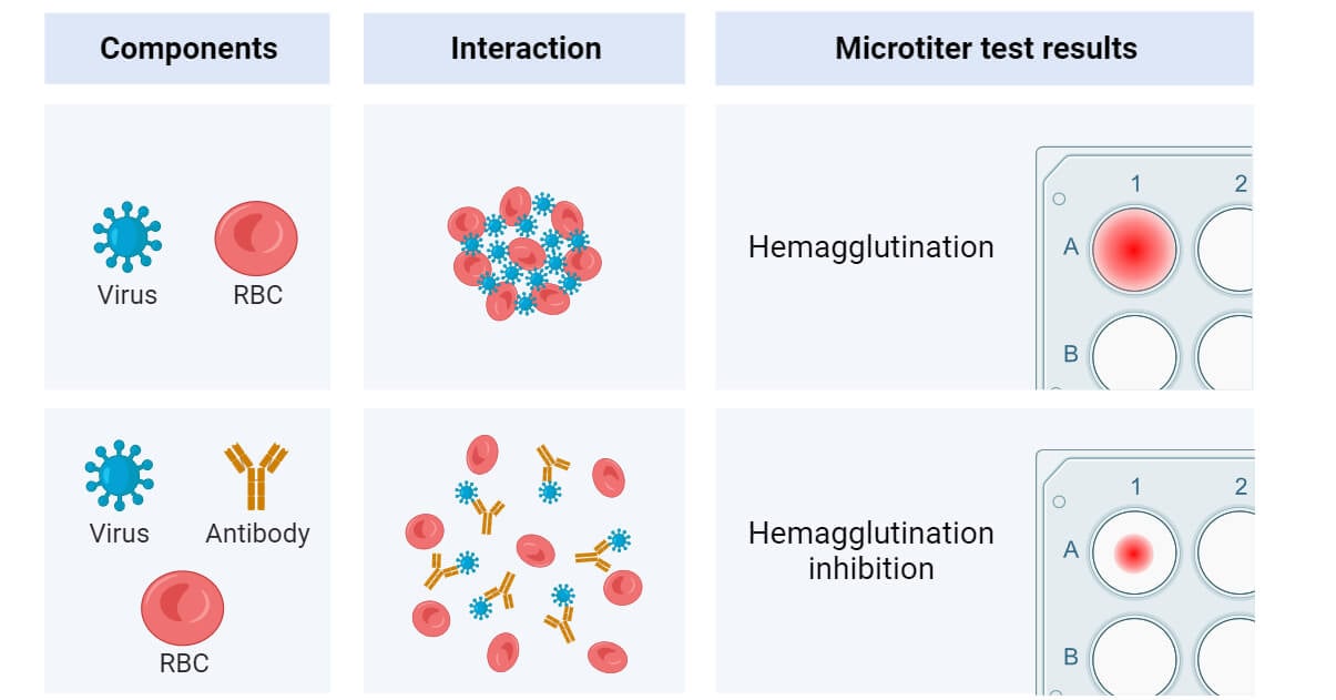
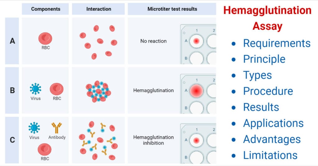
Viral hemagglutination: Many viruses including influenza, mumps, and measles have the ability to agglutinate RBCs without antigen–antibody reactions. This process is called viral hemagglutination. This hemagglutination can be inhibited by antibody specifically directed against the virus, and this phenomenon is called hemagglutination inhibition.
Coagglutination test: Coagglutination is a type of agglutination reaction in which Cowan I strain of S. aureus is used as carrier particle to coat antibodies. Cowan I strain of S. aureus contains protein A, an anti-antibody, that combines with the Fc portion of immunoglobulin, IgG, leaving the Fab region free to react with the antigen present in the specimens. In a positive test, protein A bearing S. aureus coated with antibodies will be agglutinated if mixed with specific antigen. The advantage of the test is that these particles show greater stability than latex particles and are more refractory to changes in ionic strength.
Uses of Coagglutination test
- Detection of cryptococcal antigen in the CSF for diagnosis of cryptococcal meningitis;
- Detection of amoebic and hydatid antigens in the serum for diagnosis of amoebiasis and cystic echinococcosis,
- Grouping of streptococci and mycobacteria and for typing of Neisseria gonorrhoeae.
Applications of Agglutination Reactions
- Cross-matching and grouping of blood.
- Identification of Bacteria. E.g. Serotyping of Vibrio cholera, Serotyping of Salmonella Typhi and Paratyphi.
- Serological diagnosis of various diseases. E.g Rapid plasma regains (RPR) test for Syphilis, Antistreptolysin O (ASO) test for rheumatic fever.
- Detection of unknown antigen in various clinical specimens. E.g. detection of Vi antigen of Salmonella Typhi in the urine.
Further Readings
- https://bio.libretexts.org/Bookshelves/Microbiology/Book%3A_Microbiology_(Boundless)/12%3A_Immunology_Applications/12.2%3A_Immunoassays_for_Disease/12.2E%3A__Agglutination_Reactions
- https://www.brainkart.com/article/Types-of-Agglutination-Reactions—Antigen-Antibody-Reactions_20188/
- https://iscnagpur.ac.in/study_material/dept_zoology/2.2_ANM_agglutination_reaction.pdf
- http://www.biosciencenotes.com/agglutination/
- http://microbiologyonlinenotes.com/agglutination/
- http://www.brainkart.com/article/Agglutination—Antigen-Antibody-Reactions_17920/

thanks so much keep it up
Thanks