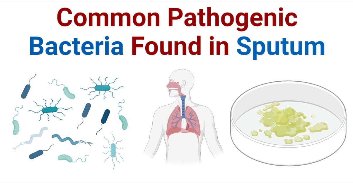
Sputum is thick mucus secreted by cells in the lower respiratory tract including bronchi, bronchioles, and lungs. It is also called phlegm. It is a commonly collected sample for laboratory diagnosis of respiratory tract infections (RTIs), especially lower respiratory tract infections (LRTIs). Sputum is usually sterile, but it can be contaminated with normal flora of the upper respiratory tract and oral cavity. LRTI is an infection of the trachea, bronchi, and lungs. It is the 5th leading cause of death worldwide. Several viruses, bacteria, fungi, and a few parasites are found responsible for RTIs.

Interesting Science Videos
List of Bacteria Isolated From Sputum
| Gram-Positive Bacteria | Gram-Negative Bacteria | Others Bacteria |
| Staphylococcus spp.* Streptococcus spp.* Enterococcus spp. Mycobacterium spp. Micrococcus spp. Corynebacterium spp. | Klebsiella spp.* Haemophilus spp.* Pseudomonas spp.* Neisseria spp.* Legionella spp.* Chlamydia spp.* Yersinia spp. Proteus spp. Moraxella spp. Acinetobacter spp. Stenotrophomonas spp. E. coli Enterobacter spp. Bordetella pertussis Serratia spp. | Mycoplasma spp.* |
Gram +ve and Gram -ve Bacteria Found in Sputum
Streptococcus spp. in Sputum
- Gram-positive
- anaerobic, and facultative anaerobic,
- catalase-negative
- cocci bacteria
- family Streptococcaceae
- arrangement in the form of a chain of spheres
- normal flora of the upper respiratory tract, oral cavity, GI tract, and skin
S. pneumoniae is the most common pathogenic bacteria isolated in the sputum. It is the major cause of community-acquired pneumonia.
S. agalactiae (Group B Streptococcus) is also associated with upper respiratory tract infections like sore throat and tonsillitis, and rarely in LRTIs. Although it is isolated in sputum as a pathogen, most of the time it can be contaminants.
S. pyogenes is also detected in case of about 2 – 5% of pneumonia in sputum culture. It is a fatal type of pneumonia.
S. anginosus, although being normal flora of the oropharynx, is rarely associated with LTRIs and hence rarely isolated as a pathogen in sputum culture.
Viridans Streptococci are also identified as pathogens in sputum culture. Often, the presence of the Viridans group is neglected as contamination of normal flora of the oropharynx, but in some cases, they are found responsible for pneumonia.
Haemophilus spp. in Sputum
Haemophilus is a genus of Gram-negative, coccobacilli (pleomorphic), aerobic or facultative anaerobic Gammaproteobacteria in the family Pasteurellaceae. They are normal flora of the upper respiratory tract, oral cavity, lower GI tract, and vagina. However, some species are pathogenic.
H. influenzae is one of the most common pathogens causing pneumonia. They are frequently isolated in sputum culture. H. parainfluenzae is another species that is reported in a few sputum cultures.
Klebsiella spp. in Sputum
Klebsiella is a genus of Gram-negative, rod-shaped, and facultative anaerobic and capsule-forming coliform bacteria in the family Enterobacteriaceae.
K. pneumoniae is the most common species in this genus responsible for hospital-acquired pneumonia. It is responsible for about 11.8% of hospital-acquired pneumonia cases.
K. oxytoca is also rarely isolated in sputum culture.
Pseudomonas spp. in Sputum
Pseudomonas is a genus of Gram-negative, rod-shaped, aerobic, gammaproteobacteria in the family Pseudomonadaceae. They are mainly associated with hospital-acquired infections.
P. aeruginosa is the main species involved in Pseudomonas pneumonia, pharyngitis, and other RTIs. They are commonly detected in patients with a hospital stay or medical respiratory catheters.
Chlamydia spp. in Sputum
Chlamydia is a genus of Gram-negative, non-motile, coccoid obligate intracellular parasitic bacteria in the family Chlamydiaceae. Chlamydia pneumoniae and Chlamydia psittaci are commonly responsible species for respiratory infection, hence are isolated in sputum culture. C. pneumoniae is responsible for most atypical pneumonia.
Staphylococcus spp. in Sputum
Staphylococcus is a genus of Gram-positive, catalase-positive, cocci bacteria belonging to the family Staphylococcaceae, typically known for producing grape-like clusters under a microscope. They are normal flora of the skin.
S. aureus is responsible for several cases of pulmonary infection in adults. It may be community-acquired or hospital-acquired pneumonia and other respiratory tract infections. MRSA is the nastiest strain of S. aureus responsible for severe cases of hospital-acquired pneumonia.
Enterococcus spp. in Sputum
Enterococcus is a genus of Gram-positive, facultatively anaerobic, lactose fermenting cocci (diplococci) bacteria in the family Enterococcaceae having the capacity to tolerate bile salt concentrations up to 40%. E. faecalis is associated with both community-acquired and hospital-acquired LRTIs.
Mycobacterium spp. in Sputum
Mycobacterium is a genus of Gram-positive (Acid-fast bacilli), rod-shaped, aerobic Actinobacteria in the family Mycobacteriaceae with characteristic mycolic acid content in cell-wall making them acid-fast.
M. tuberculosis complex responsible for pulmonary tuberculosis is commonly reported in sputum. TB is a highly contagious RTI caused by the M. tuberculosis complex responsible for about 2 million death per year globally.
M. tuberculosis is the most common species isolated in pulmonary TB. M. africanum, M. bovis, M. canettii, M. microti, M. pinnipedii, M. pinnipedii, M. orygis, and M. mungi are other rarely detected species in sputum of TB patients.
Micrococcus spp. in Sputum
Micrococcus is a genus of Gram-positive, mostly non-motile, strictly aerobic cocci in clusters in the family Micrococcaceae. They are mostly normal flora and their presence is usually reported as contamination. But, rarely M. luteus is responsible for Micrococcus pneumonia and is isolated in sputum.
Corynebacterium spp. in Sputum
Corynebacterium is a genus of Gram-positive, rod-shaped (club-shaped), aerobic bacteria in the family Corynebacteriaceae. C. propinquum and C. striatum are reported in the sputum of some patients with pneumonia.
Neisseria spp. in Sputum
Neisseria is a genus of Gram-negative, aerobic, or facultative anaerobic cocci (and diplococci) Betaproteobacteria in the family Neisseriaceae. It is commensal to the nasopharynx and sometimes they are found in the mucosal lining of the vaginal tract.
N. meningitidis, N. sica, N. flavescens, and N. catarrhalis (now called Branhamella catarrhalis) are Neisseria species reported in sputum culture. They are associated with pneumonia and bronchiectasis.
Legionella spp. in Sputum
Legionella is a genus of Gram-negative, rod-shaped or coccobacilli, motile, aerobic Gammaproteobacteria in the family Legionellaceae.
L. pneumophila is responsible for an illness called Legionellosis which is one of the top 3 commonly identified pathogens in community-acquired pneumonia. It is responsible for about 90% of human cases of legionella pneumonia.
L. micdadei, L. bozemanae, L. longbeachae, and L. dumoffii are responsible for rare cases of legionella pneumonia.
Yersinia spp. in Sputum
Yersinia is a genus of Gram-negative, coccobacilli, facultative anaerobic Gammaproteobacteria in the family Yersiniaceae.
Y. enterocolitica and Y. pestis are responsible for pneumonic plague and pneumonia. Hence, they are isolated in sputum culture.
Proteus spp. in Sputum
Proteus is a genus of Gram-negative, rod-shaped, aerobic, and facultative anaerobic motile bacteria in the family Enterobacteriaceae best known for their swarming colonies. P. mirabilis is associated with a few cases of LRTIs including pneumonia.
Moraxella spp. in Sputum
Moraxella is a genus of Gram-negative, coccobacilli (bacilli or diplococci in some), strictly aerobic Gammaproteobacteria in the family Moraxellaceae. M. catarrhalis is a normal inhabitant of the upper respiratory tract. In case of immunosuppressed condition and chronic chest infection, they may migrate to the lower respiratory tract and develop tracheobronchitis and pneumonia.
Acinetobacter spp. in Sputum
Acinetobacter is a genus of Gram-negative, rod-shaped, strict aerobic Gammaproteobacteria in the family Moraxellaceae. Acinetobacter hospital-acquired pneumonia and Acinetobacter ventilator-associated pneumonia are commonly reported. They are also responsible for community-acquired pneumonia.
A. baumnaii and A. calcoaceticus are the main species in this genus that are isolated in sputum culture as a pathogen for LTRIs.
Stenotrophomonas spp. in Sputum
Stenotrophomonas is a genus of Gram-negative, rod-shaped, aerobic, or facultative anaerobic Gammaproteobacteria in the family Xanthomonadaceae.
S. maltophilia is an opportunistic pathogen in this genus responsible for rare cases of LTRIs in immunosuppressed patients. Mostly it is associated with hospital-acquired pneumonia.
Bordetella pertussis in Sputum
Bordetella is a genus of Gram-negative, rod-shaped (coccobacilli), aerobic, encapsulated Betaproteobacteria in the family Alcaligenaceae. B. pertussis is pathogenic species responsible for whooping cough.
Serratia spp. in Sputum
Serratia is a genus of Gram-negative, rod-shaped, facultative anaerobic Gammaproteobacteria in the family Yersiniaceae producing a characteristic red pigment.
S. marcescens and S. ficaria are pathogenic species isolated in sputum. They are involved in pneumonia and LRTIs.
Other Bacteria Found in Sputum
Mycoplasma spp. in Sputum
These are bacteria lacking cell-wall, hence can’t be classified based on Gram staining (but stains pink). Mycoplasma is a normal inhabitant of the upper respiratory tract but is often involved in some RTIS. M. pneumoniae is one of the common causes of pneumonia and LRTIs, mostly in young adults.
References
- Peng, Z., Zhou, J. & Tian, L. Pathogenic characteristics of sputum and bronchoalveolar lavage fluid samples from patients with lower respiratory tract infection in a large teaching hospital in China: a retrospective study. BMC Pulm Med 20, 233 (2020). https://doi.org/10.1186/s12890-020-01275-8
- Yang, K., Kruse, R. L., Lin, W. V., & Musher, D. M. (2018). Corynebacteria as a cause of pulmonary infection: a case series and literature review. Pneumonia (Nathan Qld.), 10, 10. https://doi.org/10.1186/s41479-018-0054-5
- Cukic V. (2013). The Most Common Detected Bacteria in Sputum of Patients with the Acute Exacerbation of COPD. Materia socio-medica, 25(4), 226–229. https://doi.org/10.5455/msm.2013.25.226-229
- Akuzawa, N., & Kurabayashi, M. (2016). Bacterial Pneumonia Caused by Streptococcus pyogenes Infection: A Case Report and Review of the Literature. Journal of clinical medicine research, 8(11), 831–835. https://doi.org/10.14740/jocmr2737w
- Cameron, S., Lewis, K. E., Huws, S. A., Hegarty, M. J., Lewis, P. D., Pachebat, J. A., & Mur, L. (2017). A pilot study using metagenomic sequencing of the sputum microbiome suggests potential bacterial biomarkers for lung cancer. PloS one, 12(5), e0177062. https://doi.org/10.1371/journal.pone.0177062
- Sarkar, T. K., Murarka, R. S., & Gilardi, G. L. (1989). Primary Streptococcus viridans pneumonia. Chest, 96(4), 831–834. https://doi.org/10.1378/chest.96.4.831
- Kaye, M. G., Fox, M. J., Bartlett, J. G., Braman, S. S., & Glassroth, J. (1990). The clinical spectrum of Staphylococcus aureus pulmonary infection. Chest, 97(4), 788–792. https://doi.org/10.1378/chest.97.4.788
- Ashurst JV, Dawson A. Klebsiella Pneumonia. [Updated 2022 Feb 2]. In: StatPearls [Internet]. Treasure Island (FL): StatPearls Publishing; 2022 Jan-. Available from: https://www.ncbi.nlm.nih.gov/books/NBK519004/
- Malvisi, L., Taddei, L., Yarraguntla, A., Wilkinson, T., Arora, A. K., & AERIS Study Group (2021). Sputum sample positivity for Haemophilus influenzae or Moraxella catarrhalis in acute exacerbations of chronic obstructive pulmonary disease: evaluation of association with positivity at earlier stable disease timepoints. Respiratory research, 22(1), 67. https://doi.org/10.1186/s12931-021-01653-8
- Pillai, A., Mitchell, J. L., Hill, S. L., & Stockley, R. A. (2000). A case of Haemophilus parainfluenzae pneumonia. Thorax, 55(7), 623–624. https://doi.org/10.1136/thorax.55.7.623
- Becker Y. Chlamydia. In: Baron S, editor. Medical Microbiology. 4th edition. Galveston (TX): University of Texas Medical Branch at Galveston; 1996. Chapter 39. Available from: https://www.ncbi.nlm.nih.gov/books/NBK8091/
- Kanabalan, R. D., Lee, L. J., Lee, T. Y., Chong, P. P., Hassan, L., Ismail, R., & Chin, V. K. (2021). Human tuberculosis and Mycobacterium tuberculosis complex: A review on genetic diversity, pathogenesis and omics approaches in host biomarkers discovery. Microbiological research, 246, 126674. https://doi.org/10.1016/j.micres.2020.126674
- Enterococcal-associated Lower Respiratory Tract Infections: A Case Report and Literature Review. Infection 2009; 37: 60–64 DOI 10.1007/s15010-007-7123-7. s15010-007-7123-7.pdf (springer.com)
- Souhami, L., Feld, R., Tuffnell, P. G., & Feller, T. (1979). Micrococcus luteus pneumonia: a case report and review of the literature. Medical and pediatric oncology, 7(4), 309–314. https://doi.org/10.1002/mpo.2950070404
- Yang, K., Kruse, R.L., Lin, W.V. et al. Corynebacteria as a cause of pulmonary infection: a case series and literature review. Pneumonia 10, 10 (2018). https://doi.org/10.1186/s41479-018-0054-5
- Gris, P., Vincke, G., Delmez, J. P., & Dierckx, J. P. (1989). Neisseria sicca pneumonia and bronchiectasis. The European respiratory journal, 2(7), 685–687.
- Huang, L., Ma, L., Fan, K., Li, Y., Xie, L., Xia, W., Gu, B., & Liu, G. (2014). Necrotizing pneumonia and empyema caused by Neisseria flavescens infection. Journal of thoracic disease, 6(5), 553–557. https://doi.org/10.3978/j.issn.2072-1439.2014.02.16
- Chahin, A., & Opal, S. M. (2017). Severe Pneumonia Caused by Legionella pneumophila: Differential Diagnosis and Therapeutic Considerations. Infectious disease clinics of North America, 31(1), 111–121. https://doi.org/10.1016/j.idc.2016.10.009
- Portnoy, D., & Martinez, L. A. (1979). Yersinia enterocolitica septicemia with pneumonia. Canadian Medical Association journal, 120(1), 61–62.
- Cleri, D. J., Vernaleo, J. R., Lombardi, L. J., Rabbat, M. S., Mathew, A., Marton, R., & Reyelt, M. C. (1997). Plague pneumonia disease caused by Yersinia pestis. Seminars in respiratory infections, 12(1), 12–23.
- Okimoto, N., Hayashi, T., Ishiga, M., Nanba, F., Kishimoto, M., Yagi, S., Kurihara, T., Asaoka, N., & Tamada, S. (2010). Clinical features of Proteus mirabilis pneumonia. Journal of infection and chemotherapy : official journal of the Japan Society of Chemotherapy, 16(5), 364–366. https://doi.org/10.1007/s10156-010-0059-3
- Ahmad, N., Cheong, Y. M., & Tahir, H. M. (1994). Isolation of Moraxella catarrhalis from sputum specimens of Malaysian patients. The Malaysian journal of pathology, 16(1), 63–67.
- Hunt, J. P., Buechter, K. J., & Fakhry, S. M. (2000). Acinetobacter calcoaceticus pneumonia and the formation of pneumatoceles. The Journal of trauma, 48(5), 964–970. https://doi.org/10.1097/00005373-200005000-00027
- Hartzell, J. D., Kim, A. S., Kortepeter, M. G., & Moran, K. A. (2007). Acinetobacter pneumonia: a review. MedGenMed : Medscape general medicine, 9(3), 4.
- Gibb, J., & Wong, D. W. (2021). Antimicrobial Treatment Strategies for Stenotrophomonas maltophilia: A Focus on Novel Therapies. Antibiotics (Basel, Switzerland), 10(10), 1226. https://doi.org/10.3390/antibiotics10101226
- Soane, M. C., Jackson, A., Maskell, D., Allen, A., Keig, P., Dewar, A., Dougan, G., & Wilson, R. (2000). Interaction of Bordetella pertussis with human respiratory mucosa in vitro. Respiratory medicine, 94(8), 791–799. https://doi.org/10.1053/rmed.2000.0823
- Kerr, J. R., & Matthews, R. C. (2000). Bordetella pertussis infection: pathogenesis, diagnosis, management, and the role of protective immunity. European journal of clinical microbiology & infectious diseases : official publication of the European Society of Clinical Microbiology, 19(2), 77–88. https://doi.org/10.1007/s100960050435
- Gul, M., Dogan, E., Kirecci, E., Ucmak, H., Dirican, E., & Karadag, A. (2011). Serratia ficaria isolated from sputum specimen. Acta microbiologica et immunologica Hungarica, 58(3), 235–238. https://doi.org/10.1556/AMicr.58.2011.3.7
- Khanna, A., Khanna, M., & Aggarwal, A. (2013). Serratia marcescens- a rare opportunistic nosocomial pathogen and measures to limit its spread in hospitalized patients. Journal of clinical and diagnostic research : JCDR, 7(2), 243–246. https://doi.org/10.7860/JCDR/2013/5010.2737
