Interesting Science Videos
Transitional epithelium definition
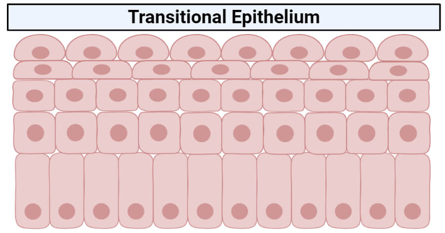
Transitional epithelium is a type of stratified epithelium consisting of multiple layers of cells where the shape of the cell changes according to the function of the organ. The epithelium has a varying appearance as they appear cubical or round when in a relaxed state, except the apical layer which seems to be flattened when stretched. This epithelium is almost limited to the urinary system, which is why it is also termed as “urothelium”.
Structure of the transitional epithelium
- Transitional epithelium is an epithelial tissue which in a relaxed state appears as a stratified cuboidal epithelium.
- The cells in the transitional epithelium are pear-shaped or round, but as tissue is stretched, cells become flattened, giving the appearance of stratified squamous epithelium.
- The cells in the basal layer appear cuboidal or columnar, but the cells in the apical layer become flattened depending on the degree of extension.
- The layers of cells in the epithelium are divided into three groups.
- The lowermost layer is the basal layer, which is directly attached to the basement membrane. This membrane is responsible for providing the constant renewal of cells to the upper layers.
- The cells in the basal layer are rich is cytoplasmic proteins that bind to form tonofibrils, which then converge with hemidesmosomes to build an effective connection with the basement membrane.
- The cells in the basal layer are rich in mitochondria as they require more energy for the renewal of the epithelium.
- The middle layer, called the intermediate layer, consists of highly proliferative, rapidly dividing cells that provide rapid regeneration in times of injury or damage to the existing cells.
- The cells in this layer are abundant in the Golgi apparatus that aids in the transport of proteins, like keratin to the superficial layers.
- The cells of the superficial layer are highly keratinized, which provides a barrier against salts and water.
- The apical layer, called the superficial layer, lines the lumen, and protects the underlying layer of cells against harmful waste material pathogens from the lumen.
- Some of the cells in the superficial layer are covered with microvilli and are provided with a mucus coating.
- The cells in the epithelium are connected to each other via gap junctions and desmosomes. These structural elements allow the epithelium to extend; however, it also causes the cells to become fragile.
- Like all other epithelial tissues, the transitional epithelium is also avascular with no supply of blood vessels. The cells in this epithelium rely on the blood vessels of the adjacent connective tissues for oxygen, nutrients, and excretion.
- However, the cells do have a distinct nerve supply.
Functions of the transitional epithelium
Based on the structure of the cell and its composition, the transitional epithelium performs two main functions, which are:
1. Permeability barrier
- Due to the presence of large deposits of keratin in the cells, the tissue provides an excellent impermeability towards the water as well as other molecules.
- The cells in the tissue are highly resistant against osmotic pressure which prevents desiccation even in times when the cells are fully stretched.
- Toxins and chemicals are also prevented from re-entering the bloodstream.
- A classic example of this function is observed in the urinary system, where even when the hypertonic urine is present in the lumen, the cells in the urothelium are not desiccated.
2. Volume control
- The second important function of this epithelium is the ability to allow the organs to change their shape and increase in volume by stretching as the fluid pressure increases.
- In the excretory system, when the amount of fluid in the urinary bladder and ureters increases, the cells in the superficial layer stretch changing the shape from round to flat.
- The stretching increases the volume of the organs while protecting the underlying tissue against the exposure to the toxins in the urine.
Location and examples
- The most prominent example of transitional epithelium is the urothelium.
- As the urothelium, the transitional epithelium lines the urinary bladder, ureters, and parts of the urethra.
- Similarly, the lining of the prostatic urethra in the male reproductive system is also lined with the transitional epithelium, which is continuous with the urothelium of the urinary bladder.
References and Sources
- Mescher AL (2016). Basic Histology. Fourteenth Edition. McGraw-Hill Education.
- Tortora GJ and Derrickson B (2017). Principles of Physiology and Anatomy. Fifteenth Edition. John Wiley & Sons, Inc.
- Waugh A and Grant A. (2004) Anatomy and Physiology. Ninth Edition. Churchill Livingstone.
- 2% – https://en.wikipedia.org/wiki/Transitional_epithelium
- 2% – https://biologydictionary.net/transitional-epithelium/
- 1% – https://www.sciencedirect.com/science/article/pii/B978012802926800001X
- 1% – https://www.sciencedirect.com/science/article/pii/0002939461919511
- 1% – https://www.researchgate.net/publication/235443030_BLOOD-TISSUE_BARRIERS_Morphofunctional_and_Immunological_Aspects_of_the_Blood-Testis_and_Blood-Epididymal_Barriers
- 1% – https://www.quia.com/jg/838874list.html
- 1% – https://training.seer.cancer.gov/anatomy/urinary/
- 1% – https://quizlet.com/14186293/anatomy-chapter5-flash-cards/
- 1% – https://en.wikipedia.org/wiki/Keratinocyte
- <1% – https://www.ncbi.nlm.nih.gov/pmc/articles/PMC3579394/
