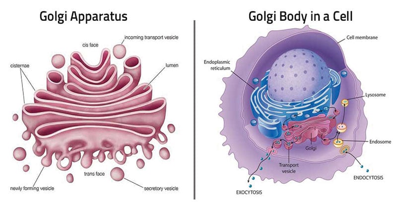
- The Golgi apparatus or the Golgi body or Golgi complex or simply Golgi is a cellular organelle present in most of the cells of the eukaryotic organisms.
- It is referred to as the manufacturing and the shipping center of the cell.
- Golgi is involved in the packaging of the protein molecules before they are sent to their destination. These organelles help in processing and packaging the macromolecules like proteins and lipids that are synthesized by the cell and hence act as the ‘post office’ of the cell.
- Golgi apparatus was discovered in the year 1898 by an Italian biologist Camillo Golgi.

Figure: Diagram of the Golgi Apparatus
Interesting Science Videos
Structure of Golgi Apparatus
- Under the electron microscope, the Golgi apparatus is seen to be composed of stacks of flattened structures that contain numerous vesicles containing secretory granules.
- The Golgi apparatus is morphologically very similar in both plant and animal cells. However, it is extremely pleomorphic: in some cell types it appears compact and limited, in others spread out and reticular (net-like).
- Typically, however, Golgi apparatus appears as a complex array of interconnecting tubules, vesicles, and cisternae.
A. Cisternae
- It is the simplest unit of the Golgi apparatus is the cisterna.
- Cisternae (about 1 μm in diameter) are central, flattened, plate-like or saucer-like closed compartments that are held in parallel bundles or stacks one above the other.
- In each stack, cisternae are separated by a space of 20 to 30 nm which may contain rod-like elements or fibers.
- Each stack of cisternae forms a dictyosome which may contain 5 to 6 Golgi cisternae in animal cells or 20 or more cisternae in plant cells.
- Each cisterna is bounded by a smooth unit membrane (7.5 nm thick), having a lumen varying in width from about 500 to 1000 nm.
- The margins of each cisterna are gently curved so that the entire dictyosome of the Golgi apparatus takes on a bow-like appearance.
- The cisternae at the convex end of the dictyosome comprise proximal, forming or cis-face and cisternae at the concave end of the dictyosome comprise the distal, maturing or trans-face.
B. Tubules
- A complex array of associated vesicles and anastomosing tubules (30 to 50 nm diameter) surround the dictyosome and radiate from it. In fact, the peripheral area of the dictyosome is fenestrated (lace-like) in structure.
C. Vesicles
The vesicles (60 nm in diameter) are of three types:
(i) Transitional vesicles are small membrane limited vesicles which are thought to form as blebs from the transitional ER to migrate and converge to cis face of Golgi, where they coalesce to form new cisternae.
(ii) Secretory vesicles are varied-sized membrane-limited vesicles that discharge from margins of cisternae of Golgi. They, often, occur between the maturing face of Golgi and the plasma membrane.
(iii) Clathrin-coated vesicles are spherical protuberances, about 50 μm in diameter and with a rough surface. They are found at the periphery of the organelle, usually at the ends of single tubules, and are morphologically quite distinct from the secretory vesicles. The clathrin-coated vesicles are known to play a role in intracellular traffic of membranes and of secretory products, i.e., between ER and Golgi, as well as, between the GELR region and the endosomal and lysosomal compartments.
Functions of Golgi Apparatus
1. Golgi vesicles are often, referred to as the “traffic police” of the cell. They play a key role in sorting many of the cell’s proteins and membrane constituents, and in directing them to their proper destinations.
- To perform this function, the Golgi vesicles contain different sets of enzymes in different types of vesicles— cis, middle and trans cisternae—that react with and modify secretory proteins passing through the Golgi lumen or membrane proteins and glycoproteins that are transiently in the Golgi membranes as they are en route to their final destinations.
- The Golgi apparatus hence acts as the assembly factory of the cell where the raw materials are directed to the Golgi apparatus before being passed out from the cell.
2. In animals, the Golgi apparatus is involved in the packaging and exocytosis of the following materials :
- Zymogen of exocrine pancreatic cells;
- Mucus (=a glycoprotein) secretion by goblet cells of the intestine ;
- Lactoprotein (casein) secretion by mammary gland cells (Merocrine secretion) ;
- Secretion of compounds (thyroglobulins) of thyroxine hormone by thyroid cells;
- Secretion of tropocollagen and collagen ;
- Formation of melanin granules and other pigments; and
- Formation of yolk and vitelline membrane of growing primary oocytes.
3. It is also involved in the formation of certain cellular organelles such as plasma membrane, lysosomes, acrosome of spermatozoa and cortical granules of a variety of oocytes.
4. They are also involved in the transport of lipid molecules around the cell.
5. The Golgi complex also plays an important role in the production of proteoglycans. The proteoglycans are molecules that are present in the extracellular matrix of the animal cells.
6. It is also a major site of synthesis of carbohydrates. These carbohydratres include the synthesis of glycosaminoglycans, Golgi attaches to these polysaccharides which then attaches to a protein produced in the endoplasmic reticulum to form proteoglycans.
7. The Golgi involves in the sulfation process of certain molecules.
8. The process of phosphorylation of molecules by the Golgi requires the import of ATP into the lumen of the Golgi.
9. In plants, Golgi apparatus is mainly involved in the secretion of materials of primary and secondary cell walls (e.g., formation and export of glycoproteins, lipids, pectins and monomers for hemicellulose, cellulose, lignin, etc.)
References
- Verma, P. S., & Agrawal, V. K. (2006). Cell Biology, Genetics, Molecular Biology, Evolution & Ecology (1 ed.). S .Chand and company Ltd.
- Stephen R. Bolsover, Elizabeth A. Shephard, Hugh A. White, Jeremy S. Hyams (2011). Cell Biology: A short Course (3 ed.).Hoboken,NJ: John Wiley and Sons.
- Alberts, B. (2004). Essential cell biology. New York, NY: Garland Science Pub.
- https://biology.tutorvista.com/animal-and-plant-cells/golgi-apparatus.html
- http://www.biologydiscussion.com/cell/golgi-complex/golgi-complex-structure-and-functions-with-diagram/36799

REALLY INFORMATIVE AND HUGE ARTICLE. LIKED IT AND PLEASE ALSO GIVE SOME EXTRA QUESTION BASED ON INFORMATION GIVEN IN THE ARTICLE AND ALSO DEEPLY EXPLAINED SO, THANK YOU FOR PROVIDING THIS AND HELPING ALL THOSE WHO WERE AND ARE SEARCHING FOR DEEPLY EXPLAINED GOLGI APPARATUS ARTICLE. THANK YOU, THANK YOU, THANK YOU!!!
Helpful
Well explained article
I didn’t get what i really want 😞😞
osm 😊
A well researched article.