Interesting Science Videos
What is Periodic Acid-Schiff (PAS) Staining?
Periodic-Acid Schiff (PAS) staining technique is used in histochemistry and histological studies to demonstrate the presence of carbohydrates and carbohydrate compounds such as polysaccharides, mucin, glycogen, and fungal cell wall components. It has been used to detect glycogen in tissues such as the skeletal muscles, the liver, the cardiac muscles. It uses formalin-fixed, paraffin-embedded tissue sections, or/and frozen tissue sections.
Principle of Periodic Acid-Schiff (PAS) Staining
The periodic acid of Schiff’s reaction with carbohydrates is an oxidative process where some polysaccharides react with the periodic acid producing an oxidized compound, an aldehyde. Aldehydes are revealed by the red or pink or magenta coloration due to the fixation of the colorless Schiffs fuchsin. PAS is linked to its association with diastase enzymes which are responsible for the convention of starch to maltose and sequentially to glucose. During glucose conversion, the stain appears pink which defines the intra or extracellular mucins persistence. Hematoxylin or methyl green is used to stain the nuclei.
Preparation of Solutions and Reagents
Periodic Acid solution (0.5%)
- Periodic acid- 0.5g
- Distilled water- 100ml
Testing Schiff’s Reagent
- 10 ml of 37% formalin
- Add Schiff’s reagent to be tested.
- A good Schiffs reagent turns red-purple in color while a poor Schiffs reagent will have a delayed reaction producing deep blue-purple coloration
Mayer’s Hematoxylin Solution
Procedure for Periodic Acid-Schiff (PAS) Staining
- Deparaffinize and hydrate water.
- Oxidation: Add 0.5% of the Periodic acid solution for oxidation for 5 minutes.
- Rinsing: In distilled water.
- Aldehydration: Place the stain in Schiff reagent for 15 minutes, which turns light pink.
- Washing: Using lukewarm tap water, wash the stain for 5 minutes, turning it dark pink.
- Counterstaining: Add Mayer’s Hematoxylin for 1 minute.
- Wash with running tap water for 5 minutes.
- Dehydrate and mount with a synthetic mount medium.
Results and Interpretation

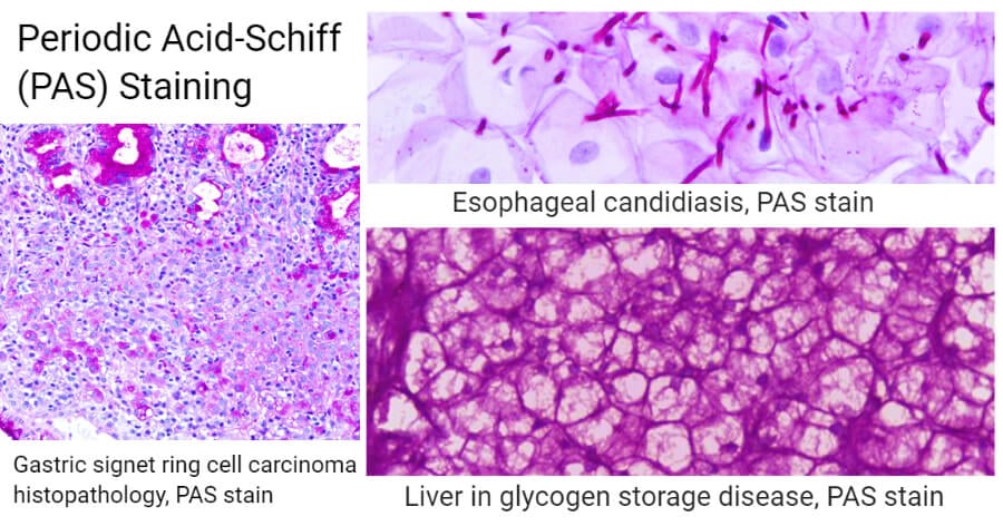
Image Source: Wikipedia.
- Glycogen, mucin, elastic fibers, basophilic granules of the pituitary gland, reticular fibers, thyroid colloids, basement membranes, bone cartilage, and other carbohydrate components stain pink or purple.
- The nuclei stains green or blue depending on the dye used.
- The background stains blue.
Applications of Periodic Acid-Schiff (PAS) Staining
- Cytology: This stain has also been used in undifferential identification of tumors hence used in the diagnosis of glandular carcinomas (adenocarcinoma).
- Pathology: It is also used in the diagnosis of liver and kidney pathologies.
- Fungal studies: It is used to demonstrate the fungal hyphae and yeast-forms of fungi in tissue samples to identify Candida albicans, Aspergillus fumigatus, and Cryptococcus neoformans infections.
- Gastrointestinal pathology: To detect the presence of mucins in the gastrointestinal tract.
- Lung studies: the stain studies the amorphous or granular globules of the pulmonary alveolar proteinosis.
- Skin: to study the eosinophilic globoid bodies or Kamino bodies.
- Muscle biopsies: to demonstrate glycogen components.
- To detect prostate cancer, pancreatic pathologies, effects of the small intestines, testis.
- Application in enzymatic cytochemistry for the detection of granules.
References and Sources
- 4% – https://www.ncbi.nlm.nih.gov/pubmed/12846888
- 3% – http://www.histology.leeds.ac.uk/what-is-histology/histological_stains.php
- 1% – https://www.sigmaaldrich.com/content/dam/sigma-aldrich/docs/Sigma/General_Information/2/395.pdf
- 1% – https://link.springer.com/article/10.1007/BF01003468
- 1% – http://www.pathologyoutlines.com/topic/stainspas.html
- 1% – http://www.ihcworld.com/_protocols/special_stains/masson_trichrome.htm
- 1% – http://aura.abdn.ac.uk/bitstream/handle/2164/8941/FUNK_0035_2016.pdf;sequence=1

Excellent