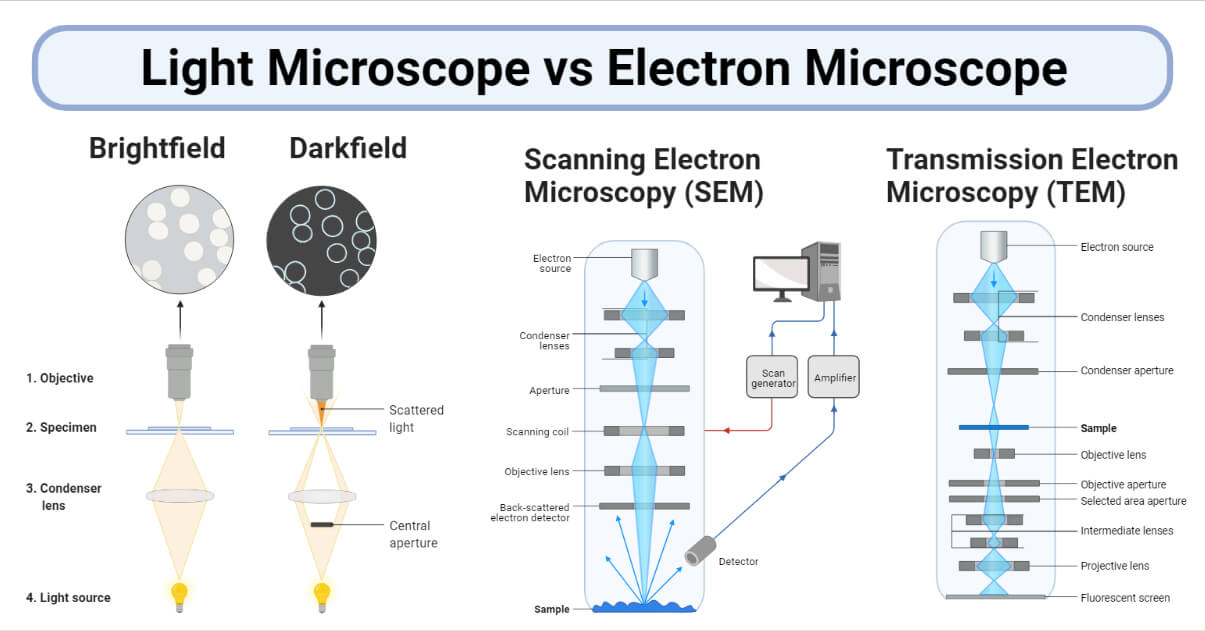
Both light microscopes and electron microscopes use radiation (light or electron beams) to form larger and more detailed images of objects which cannot be seen clearly through an unaided eye.

Interesting Science Videos
Light Microscope vs Electron Microscope (Table Form)
However, each of these microscopes has distinct features and is suitable for different purposes.
| S.N. | Character | Light Microscope | Electron Microscope |
| 1. | Alternatively known as | Optical microscope | Beam microscope |
| 2. | Invented by | It is believed that Dutch spectacles makers Zacharius Jansen and his father Hans were the first to invent the compound microscope in the 16th century. | In 1931 physicist Ernst Ruska and German engineer Max Knoll. |
| 3. | Illuminating source | Uses light (approx wavelength 400-700 nm) to illuminate the objects under view. | Uses a beam of electrons (approx equivalent wavelength 1 nm) to make objects larger for a detailed view. |
| 4. | Principle | The image is formed by the absorption of light waves. | The image is formed by scattering or transmission of electrons. |
| 5. | Structure | Light microscopes are smaller and lighter. | Heavier and larger in size. |
| 6. | Lenses used | Lenses are made of glass. | Lenses are made of electromagnets. |
| 7. | Vacuum | Not used under a vacuum | Operates under a high vacuum |
| 8. | Specimen type | Fixed or unfixed, stained or unstained, living or non-living. | Fixed, stained, and non-living. |
| 9. | Specimen observed | Both live and dead specimens can be observed. | Only dead specimens are possible to be observed. |
| 10. | Specimen preparation | Less tedious and simple. | It generally involves harsher processes, e.g. using corrosive chemicals. More skill required – both to prepare specimens and to interpret EM images (due to artifacts). |
| 11. | Preparation time | Specimen preparation takes usually a few minutes to hours. | Specimen preparation takes usually takes a few days. |
| 12. | Thickness of specimen | 5 micrometer or thicker | Ultra-thin, 0.1 micrometers or below |
| 13. | Dehydration of Specimen | Specimens need not be dehydrated before viewing. | Only dehydrated specimens are used. |
| 14. | Coating of specimen | Stained by colored dyes for proper visualization. | Coated with heavy metals to reflect electrons. |
| 15. | Mounting of specimen | Mounted on the glass slide. | Mounted on the metallic grid (mostly copper). |
| 16. | Focusing | Done by adjusting the lens position mechanically. | Done by adjusting the power of the electric current to the electromagnetic lenses. |
| 17. | Magnification power | Low magnification of up to 1,500x. | High magnification of up to 1,000,000x. |
| 18. | Resolving power | Low resolving power, usually below 0.30µm. | The high resolving power of up to 0.001µm, about 250 times higher than the light microscope. |
| 19. | Viewing of the image formed | Light microscope images can be viewed directly. Images are viewed by the eyes through the eyepiece. | Images are viewed on a photographic plate or zinc sulfate fluorescent screen. |
| 20. | Nature of Image formed | Poor surface view | Good surface view and internal details |
| 21. | Image Color | Colored images. | Electron microscopes produce grayscale (sometimes called “black and white”) images (except “false-color” electron micrographs). |
| 22. | Image dimension | Image plane “flat” (2D). | 2D only in a Transmission electron microscope (TEM); Scanning electron microscope (SEM) images give depth information that seems like 3D. |
| 23. | Living processes | Visualization of living processes such as microscopic pond life in action and even cell division is possible. | Living processes cannot be viewed. |
| 24. | Room settings | No special settings are required. | It must be used in a room where humidity, pressure, and temperature are controlled. |
| 25. | Simplicity in use | Simple to use | Users require technical skills |
| 26. | Electric Current | No need for high voltage electricity. | A high voltage electric current is required (50,000 V or above). |
| 27. | Filaments | No filaments are used. | Tungsten filaments are used to generate electrons. |
| 28. | Cooling System | Absent | Cooling system present to pacify the heat generated due to high voltage electric current. |
| 29. | Radiation leakage | No radiation risk. | Risk of radiation leakage. |
| 30. | Complexity | Less complex | Complex |
| 31. | Expense | Cheap to buy and has low maintenance costs. | Very expensive to buy as well as to maintain. |
| 32. | Suitability / Practicality | Suitable for most basic functions, and is very common in schools and other learning institutions. | Limited to specialized use such as research. |
| 33. | Advantages | Easy to use Cheap True color but sometimes require staining Live specimens | High resolution Provide detailed images of surface structures and interior structures High magnification 3D images |
| 34. | Disadvantages | Low resolution due to shorter wavelength of light (0.2nm) Low magnification The specimen used is thin. | Expensive Requires extensive training Sample must be dead Black and white/false-color image |
| 35. | Types/ Variants | Dark-field microscope Phase-contrast microscope Fluorescent microscope Confocal microscope Polarized microscope Differential interference contrast microscope | Transmission electron microscope (TEM) Scanning electron microscope (SEM) |
| 36. | Application | It is used for the study of detailed gross internal structure. | It is used in the study of the external surface, the ultrastructure of cells, and very small organisms. |
References
- Manandhar S. (2013). A practical approach to microbiology. Revised 2nd Edition. National Book Centre: Kathmandu.
- https://theydiffer.com/difference-between-a-light-microscope-and-an-electron-microscope/
- https://www.majordifferences.com/2013/10/difference-between-electron-microscope.html
- http://www.ivyroses.com/Biology/Techniques/light-microscope-vs-electron-microscope.php
- https://biodifferences.com/difference-between-light-microscope-and-electron-microscope.html
- https://www.easybiologyclass.com/light-microscope-vs-electron-microscope-similarities-differences-comparison-table/

Thank you, was very helpful
Thanks the contents were useful to me