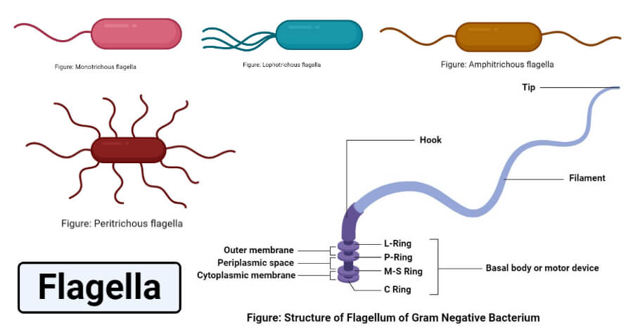
A flagellum or flagella is a lash or hair-like structure present on the cell body that is important for different physiological functions of the cell.
- The term ‘flagellum’ is the Latin term for whip indicating the long slender structure of the flagellum that resembles a whip.
- Flagella are characteristic of the members of the protozoan group Mastigophora, but also occur in different microscopic and macroscopic animals like bacteria, fungi, algae, and animals.
- Flagella are primarily essential as an organelle of locomotion in different organisms besides helping in gathering food and circulation.
- Flagella occurring in different living beings are different in structure, mechanism, movement, and even functions. A flagellum occurring in archaea is termed archaellum as it is quite different from the flagellum occurring in bacteria.
- Flagella are similar in structure with other hair-like protrusions called cilia but differ in number, occurrence, movement, and sometimes, functions.
- These appendages have been studied in different groups of animals for their functions of both movement and sensation as they can detect changes in the environmental composition and pH.
- The most commonly studied flagellum is the flagellum occurring in Chlamydomonas reinhardtii as a result of its size.
- Even though most flagella occur at the polar ends of the cells, their number and position differ as their composition and functions remain the same within a species.
Interesting Science Videos
Structure of Flagellum
The size structure and number of flagella are different in prokaryotes and eukaryotes. Even within prokaryotes, the bacterial flagellum is different from archaeal flagellum. Similarly, the composition and mechanism of flagella formation are also different and diverse. However, the basic structure of a flagellum consists of some structures that are most common in all domains of life.
The basic structure of a flagellum include the following structure;
1. Filament
- The filament is the most prominent part of a flagellum that represents about 98% of all the structures of a flagellum.
- The filaments extend from the hook-like structure present within the cytoplasm of the cell with an average length of 18 nm. The length of the filament differs in different groups of living beings like the filaments of bacterial flagella is 20 nm while that of archaea is 10-14 nm.
- Filaments can be observed outside the cell membrane specific flagellar staining methods. The movement of the filaments is controlled by the motor present in the cytoplasm.
- Filaments are self-assembling macromolecular structures composed of hook proteins and flagellins, and the number of flagellin and hook protein subunits might differ in different cells.
2. Hook or anchoring structures
- The flagellar hook is a short and curved tubular structure that connects the basal body or the flagellar motor to the long filament.
- The most important function of the hook is to transmit the motor torque to the helical filament so that it can move in a different orientation for different functions. Besides, it also plays an essential role in the assembly of the flagellum.
- The hook is composed of numerous hook protein subunits forming polymorphic supercoil structures.
- This structure is present near the cell membrane in all types of cells, but the shape and exact composition of the structure might differ between cells.
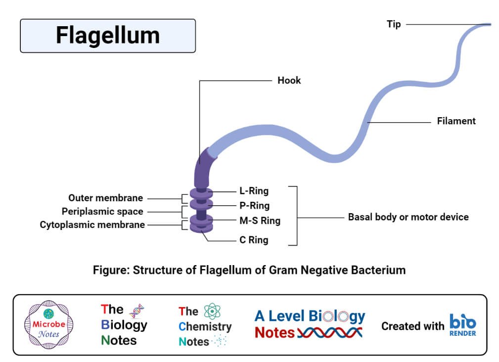
3. Basal body or motor device
- The basal body of a flagellum is the only structure of the flagellum that is present within the cell membrane. It is connected to the hook of the flagellum which then connects to the long filament.
- The basal body is a rod-shaped structure with a system of rings of microtubules. The component of the basal body differs in different types of cells.
- The rods present in the basal body act as a reversible motor that propels the filament in a different orientation for specific functions.
- The basal body is also essential for the transfer of flagellar proteins from the cytoplasm to the hook and filament part of the flagellum during assembly.
Flagella formation mechanism
The process of flagella formation and assembly begins with the formation of the FliF ring complex in the basal body. The process occurs in the cytoplasmic membrane and proceeds both inwards and outwards. Most of the studies related to the process of formation and assembly of the flagellum have been done on bacteria. As the flagellum comprises a complex membrane and structures composed of numerous proteins and their interactions.
The overall process of flagella formation and assembly can be described as below;
1. Formation of the basal body
- The process begins with the incorporation of FliF, which is an integral protein consisting of MS-ring into the cytoplasm.
- The MS-ring is essential as it determines the assembly of all the other structures of a flagellum.
- The FliF proteins assemble into a single ring that forms two adjacent loops spanning the cytoplasmic membrane.
- After the incorporation of FliF into the membrane, other proteins like FliG, FliM, and FliN are also incorporated into the cytoplasmic face of the MS-ring.
- These proteins are essential for various functions of the flagella like motility and the export of other flagellar component proteins.
- The inwards assembly of the basal body involves the formation of a C-ring, which is formed in the cytoplasmic space. Within the C-ring, a flagellar export apparatus is formed in order to export flagella axial proteins through the channel.
- A flagellum-specific type III protein export system binds and moves flagellar axial proteins into the central channel of the flagellum.
- The next step is the formation of the rod, which is a major component of the basal flagellar body. The rod comprises five proteins attached to the FliF ring at the proximal portion and to the hook at the distal end.
2. Formation of the hook
- Proteins are translocated through the rod into the hook so that the hook grows up to the length of 55 nm, which might differ in different types of cells.
- Hook formation is induced by a protein FlgD which is lacking in the completed flagella. As hook assembly begins, the rod cap protein is replaced by hook capping proteins.
- The FlgD is necessary for the polymerization of subunits into an α-helical structural arrangement.
- The hook capping protein, FlgE, is exported from the cytoplasm through the central channel from base to tip.
3. Filament assembly
- The assembly of filament occurs in the presence of the HAP2 pentamer complex that caps the distal end of the filaments as the filament monomers are assembled.
- The cap is essential to prevent the diffusion of subunits from the filament and induce a conformational change to enable polymerization.
Types of Flagella
1. Bacterial flagella
- Bacterial flagella are helically coiled structures that are slightly longer than the archaeal and eukaryotic flagella.
- They are thinner than eukaryotic flagella.
- The diameter is around 20 nanometers.
- The number of flagella in bacteria depends on different species that are primarily involved in locomotion. In some cases, the flagella can act as sensory structures and detect changes occurring in the environment.
- The length of the flagella might also be different as the filaments are longer than that of archaea and generally possess a left-handed helix with swimming motility as a result of counter-clockwise movement.
2. Archaeal flagella
- Flagellum in archaea is a unique motility apparatus that is different in composition but similar in assembly to bacterial flagellum.
- Flagella occur in almost all the main groupings of the domains like halophiles, methanogens, and thermophiles.
- Archaeal flagella are different from bacterial flagella in diameter as archaeal filaments are thinner.
- The proteins in the flagella are arranged in a right-handed helix, resulting in the clockwise rotation of the flagella. The speed of the archaeal flagella is more as the clockwise rotation pushes the cell.
- The hooks in the archaeal flagella are variable in length between the species than those occurring in bacteria.
- Different studies have also demonstrated that archaeal flagellar switching mechanism and sensory control is different from that of bacterial flagella.
3. Eukaryotic flagella
- Flagella in eukaryotes commonly occur in many algae and some animal cells like sperms.
- Eukaryotic flagella are mostly associated with cell motility, cell feeding, and reproduction in eukaryotic animals. In some algae, these also function as sensory antennae.
- Eukaryotic flagella are different from bacterial flagella in architecture, composition, mechanism, and assembly. Eukaryotic flagella are composed of several hundred different proteins, whereas bacterial flagella contain about 30 proteins.
- Similarly, eukaryotic flagella are dependent on constrained dynein-dependent microtubules sliding for their motility, whereas bacterial flagella are controlled by a rotary motor present at the basal body.
- Eukaryotic flagella are formed from microtubule-based centriole where the proteins are targeted from which the axoneme extends. The centriole in a eukaryotic cell is often considered the basal body of the flagella.
Bacterial flagella arrangement
1. Monotrichous
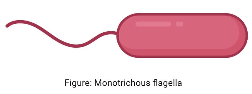
- The monotrichous arrangement of flagella is the presence of a single flagellum in each cell. If the flagellum is present at the polar end, it is called a monotrichous polar distribution.
- The mechanism of movement of monotrichous flagella is simple and coordinated by different chemoreceptors that induce motility of the cell.
- Different sensory receptors can sense changes in the environment resulting in a transmembrane electrochemical gradient of ions which powers the bacteria flagella motor.
- The thrust generated in the motor is transmitted to the hook and to the filament, causing counterclockwise rotation of the flagella.
- The counterclockwise movement of the flagella causes the cell to move forward or ‘run’.
- The change in the direction of the cell in a monotrichous arrangement is due to the counterclockwise movement of the flagella. This movement pulls the bacteria backward and allows reorientation.
- Examples of a monotrichous arrangement of flagella can be observed in bacteria like Vibrio cholerae, Campylobacter spp., Caulobacter crescentus, etc.
2. Lophotrichous
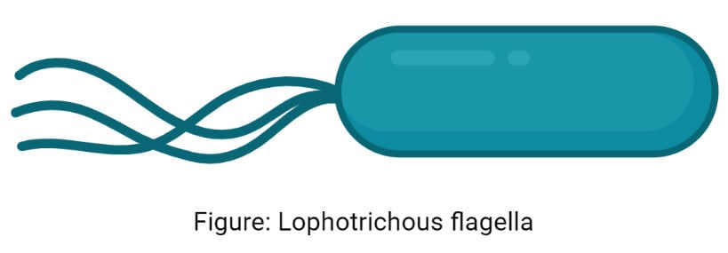
- Lophotrichous arrangement of flagella is the presence of multiple flagella at the same point in the cell. Most of the time, these flagella occur at the polar end of the cell.
- The bases of these flagella are often surrounded by a region of the cell membrane called the polar organelle.
- The mechanism of movement of lophotrichous flagella is similar to that of monotrichous flagella in that it is induced by various changes in the surrounding environment.
- As there are multiple flagella involved in the movement of lophotrichous flagella, all the flagella need to move in the same direction and with the same speed to ensure smooth movement.
- The flagella are controlled by different motors, but since all the motors are triggered by similar stimuli, the thrust generated by the flagella occurs together.
- The change in direction is obtained by the subsequent separation of the flagella as a result of the stimuli present due to changes in the environment.
- Examples of a lophotrichous arrangement of flagella include Spirillium, Pseudomonas fluorescens, etc.
3. Amphitrichous

- Amphitrichous distribution of flagella is the presence of either a single flagellum or multiple flagella at either polar ends of the cell.
- In the case of multiple flagella at the ends, the flagella are present at a region of the cell membrane called the polar organelle.
- As compared to the mechanism of movement of other arrangements of flagella, not much is known about the working of the amphitrichous arrangement.
- Different studies have indicated that the two flagella operate differently as each of the flagella can move in only one direction in a monotrichous or lophotrichous manner.
- Another study had revealed that the two flagella work simultaneously but rotate in opposite directions.
- The mechanism of movement is, however, similar to that of other arrangements.
- Some examples of bacteria with amphitrichous flagella are Campylobacter jejuni, Rhodospirillum rubrum, and Magnetospirillum.
4. Peritrichous
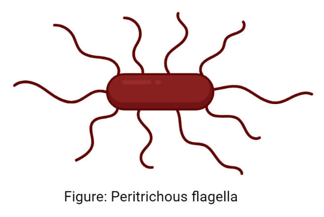
- Peritrichous arrangement of flagella is the arrangement where flagella are present throughout the body of the cell, all of which are directed in different ways.
- In the peritrichous arrangement, the flagella form a bundle that moves the cell towards the stimuli through the ‘run’ movement of the cell.
- In the case of repellent, a phosphorylation cascade is prepared that change the phosphorylation status of the regulator, CheY.
- The activated regulator then directly interacts with the motor switch proteins, casing the flagella to rotate in the clockwise direction.
- The interaction causes the destruction of bundles and separation of the flagella, which changes the speed and direction of the movement.
- The Brownian motion of the cell enables them to reorient until a next stimulus is detected randomly.
- The examples of peritrichous arrangement of flagella include Escherichia coli, Bacillus subtilis, Salmonella, and Klebsiella.
Functions of Flagella
- Flagella are the primary structures of locomotion in many bacteria so that bacteria can move towards the most favorable environment. The movement of bacteria occurs in response to various stimuli which enables them to adapt to different environmental conditions. In eukaryotic cells like sperm, flagella are essential for motility and eventually fertilization.
- Flagella play an important role in the colonization of tissue surfaces as a virulence factor to invade host tissue and develop within them.
- These are also important for non-pathogenic colonization of surfaces like a plant, soil, or animal surfaces.
- In some bacteria, flagella are involved in the nutrient and waste exchange by disturbing the nutrient-poor, waste-rich shell present within the bacteria.
- In alkaliphilic bacteria, flagella are sodium-driven which enables the reentry of sodium into the cytoplasm to maintain the neutral cytoplasmic pH.
Examples of Flagella
1. Flagella in Helicobacter pylori
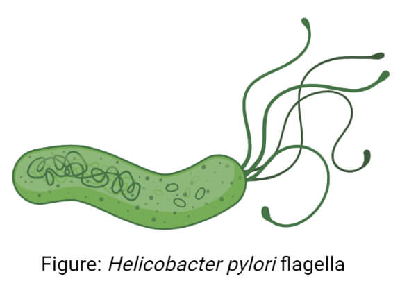
- Helicobacter pylori is a flagellated bacterium that uses the flagella for its propulsion through the tissue surface.
- The bacteria contain about 4-8 unipolar flagella important virulence factors for different diseases caused by the bacteria.
- H. pylori flagella result in swimming motility or swarming motility as they can move through the liquid and semisolid media.
- The flagella influence different physiological activities of the cell-like inflammation, immune evasion, and colonization in H. pylori.
2. Flagellum in human sperm cell
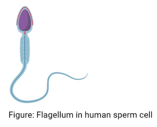
- The flagellum in a sperm cell is essential for motility and in vivo fertilization in humans.
- Failure to propel the cell and move the flagella might result in the loss of fertilization during sexual reproduction in humans.
- The core of the flagella is composed of microtubules arranged in a 9+2 arrangement with structures like dynein regulatory complex, radial spokes, and dynein arms.
- It is also important for the penetration of sperm into the egg during fertilization as it orients the sperm in a specific direction.
References
- Lodish H, Berk A, Zipursky SL, et al. Molecular Cell Biology. 4th edition. New York: W. H. Freeman; 2000. Section 19.4, Cilia and Flagella: Structure and Movement. Available from: https://www.ncbi.nlm.nih.gov/books/NBK21698/
- Nikhil A. Thomas, Sonia L. Bardy, Ken F. Jarrell, The archaeal flagellum: a different kind of prokaryotic motility structure, FEMS Microbiology Reviews, Volume 25, Issue 2, April 2001, Pages 147–174, https://doi.org/10.1111/j.1574-6976.2001.tb00575.x
- Vonderviszt F, Namba K. Structure, Function and Assembly of Flagellar Axial Proteins. In: Madame Curie Bioscience Database [Internet]. Austin (TX): Landes Bioscience; 2000-2013. Available from: https://www.ncbi.nlm.nih.gov/books/NBK6250/
- Samatey, F., Matsunami, H., Imada, K. et al. Structure of the bacterial flagellar hook and implication for the molecular universal joint mechanism. Nature 431, 1062–1068 (2004). https://doi.org/10.1038/nature02997
- Francesco Sala (1972) Structure and Function of Bacterial Flagella, Italian Journal of Zoology, 39:2, 111-118, DOI: 10.1080/11250007209430052
- Ng SY, Chaban B, Jarrell KF. Archaeal flagella, bacterial flagella and type IV pili: a comparison of genes and posttranslational modifications. J Mol Microbiol Biotechnol. 2006;11(3-5):167-91. doi: 10.1159/000094053. PMID: 16983194.
- Jonathan Moran, Paul G. McKean, Michael L. Ginger, Eukaryotic Flagella: Variations in Form, Function, and Composition during Evolution, BioScience, Volume 64, Issue 12, December 2014, Pages 1103–1114, https://doi.org/10.1093/biosci/biu175
- Nakamura, Shuichi, and Tohru Minamino. “Flagella-Driven Motility of Bacteria.” Biomolecules vol. 9,7 279. 14 Jul. 2019, doi:10.3390/biom9070279
- Moens S, Vanderleyden J. Functions of bacterial flagella. Crit Rev Microbiol. 1996;22(2):67-100. doi: 10.3109/10408419609106456. PMID: 8817078.
- Dmitry Apel, Michael G. Surette. Bringing order to a complex molecular machine: The assembly of the bacterial flagella. Biochimica et Biophysica Acta (BBA) – Biomembranes. Volume 1778, Issue 9. 2008. Pages 1851-1858. https://doi.org/10.1016/j.bbamem.2007.07.005.
- Tohru Minamino, Katsumi Imada. The bacterial flagellar motor and its structural diversity. Trends in Microbiology. Volume 23, Issue 5. 2015. Pages 267-274. https://doi.org/10.1016/j.tim.2014.12.011.

Hello Ma’am
You have given 9+2 microtubules arrangement in Bacterial flagella but it is written in many books and websites that prokaryotic flagella do not have 9+2 arrangement. Please clarify.
Hello,
Ya, you are correct. Most of the prokaryotic flagella do not have a 9+2 arrangement since they are thinner than eucaryotic flagella. This section has been updated in the note. Thanks for the correction.
Best,
Sagar