Interesting Science Videos
Centrioles Definition
- Eukaryotic cells contain two cylindrical, rod-shaped, microtubular structures, called centrioles, near the nucleus.
- They lack a limiting membrane and DNA or RNA and occur in most algal cells (a notable exception being red algae), moss cells, some fern cells, and most animal cells.
- They are absent in prokaryotes, red algae, yeast, cone-bearing and flowering plants (conifers and angiosperms) and some non-flagellated or non-ciliated protozoans (such as amoebae).
- Centrioles form a spindle of microtubules, the mitotic apparatus during mitosis or meiosis and sometimes get arranged just beneath the plasma membrane to form and bear flagella or cilia in flagellated or ciliated cells.
- When a centriole bears a flagellum or cilium, it is called the basal body.

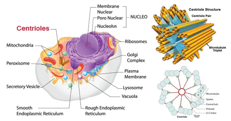
Figure: Diagram of Centrioles
Structure of Centrioles
- Centrioles and basal bodies are cylindrical structures which are 0.15–0.25 um in diameter usually 0.3–0.7 um in length, though, some are as short as 0.16 um and others are as long as 8um.
- They are visible under a light microscope, but the details of the centriole structure were revealed only under an electron microscope.
- Each cell has a pair of centrioles in the centrosome, a region near the nucleus. The members of each pair of centrioles are at right angles to one another.
- They are minute-sub-microscopic microtubular sub cylinders with a configuration of nine triplet fibrils and the ability to form their own duplicates, astral poles and basal bodies, without having DNA and a membranous covering.
- A centriole possesses a whorl of nine peripheral fibrils. Fibrils are absent in the center. The arrangement is, therefore, called 9 + 0. Fibrils run parallel to one another but at an angle of 40°. Each fibril is made up of three sub-fibers. Therefore, it is called a triplet fibril.
- The three sub-fibers are in reality microtubules joined together by their margins and, therefore, sharing the common walls made of 2-3 proto-filaments.
- Each sub-fiber has a diameter of 25 nm. From outside to inside the three sub-fibers of a triplet fibril are named as С, В and A. Sub-fibre A are complete with 13 proto-filaments while В and С sub-fibers are incomplete due to sharing of some microfilaments.
- The adjacent triplet fibrils are connected by С—A proteinaceous linkers. The center of centriole possesses a rod-shaped proteinaceous mass known as the hub. The hub has a diameter of 2.5 nm. From the hub, develops 9 proteinaceous strands towards the peripheral triplet fibrils. They are called spokes.
- Each spoke has a thickening called X before uniting with A sub-fiber. Another thickening known as Y is present nearby. It is attached both to X thickening as well as C – A linkers by connectives.
- Due to the presence of radial spokes and peripheral fibrils, the centriole gives a cartwheel appearance in T.S.
Functions of Centrioles
- Centrioles are involved in the formation of the spindle apparatus, which functions during cell division.
- The absence of centrioles causes divisional errors and delays in the mitotic process.
- A single centriole forms the anchor point, or basal body, for each individual cilium or flagellum.
- Basal bodies direct the formation of cilia and flagella as well.
References
- Verma, P. S., & Agrawal, V. K. (2006). Cell Biology, Genetics, Molecular Biology, Evolution & Ecology (1 ed.). S .Chand and company Ltd.
- Stephen R. Bolsover, Elizabeth A. Shephard, Hugh A. White, Jeremy S. Hyams (2011). Cell Biology: A short Course (3 ed.).Hoboken,NJ: John Wiley and Sons.
- Alberts, B. (2004). Essential cell biology. New York, NY: Garland Science Pub.
- Winey, M., & O’Toole, E. (2014). Centriole structure. Philosophical transactions of the Royal Society of London. Series B, Biological sciences, 369(1650), 20130457.
- http://www.biologydiscussion.com/cell/centrioles/centrioles-structure-and-functions-with-diagram/70541.

Its fabulous things very well displayed
nice concept. got all concepts clear