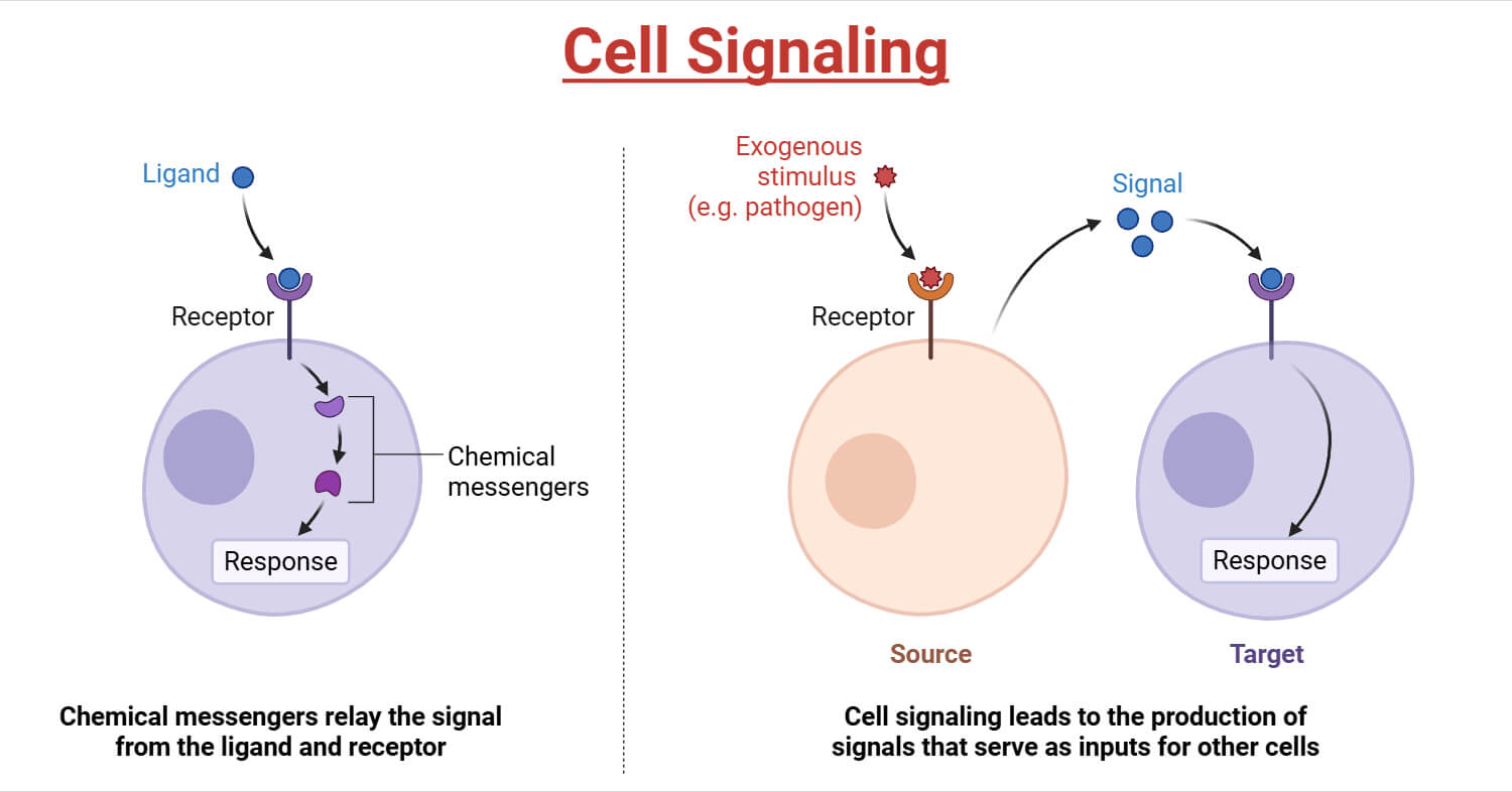
Cell signaling, or intracellular signal transduction, is a series of chemical reactions that work together to control cellular functions by activating a chain of biochemical events.
A simple intracellular signaling pathway is activated by an extracellular signal molecule as follows:
- The signal originates from the cellular environment when a signal molecule, like a growth factor or a hormone, binds to a specific protein receptor on or in the cell.
- The molecules get activated one after another, forming a cascade of such reactions, until the target protein is activated.
- One or more of these intracellular signaling proteins interact with a target protein, altering their conformation and performing cellular functions.
- Finally, the cellular function can be cell proliferation, altered gene expression, altered cell shape or movement, or even cell death.
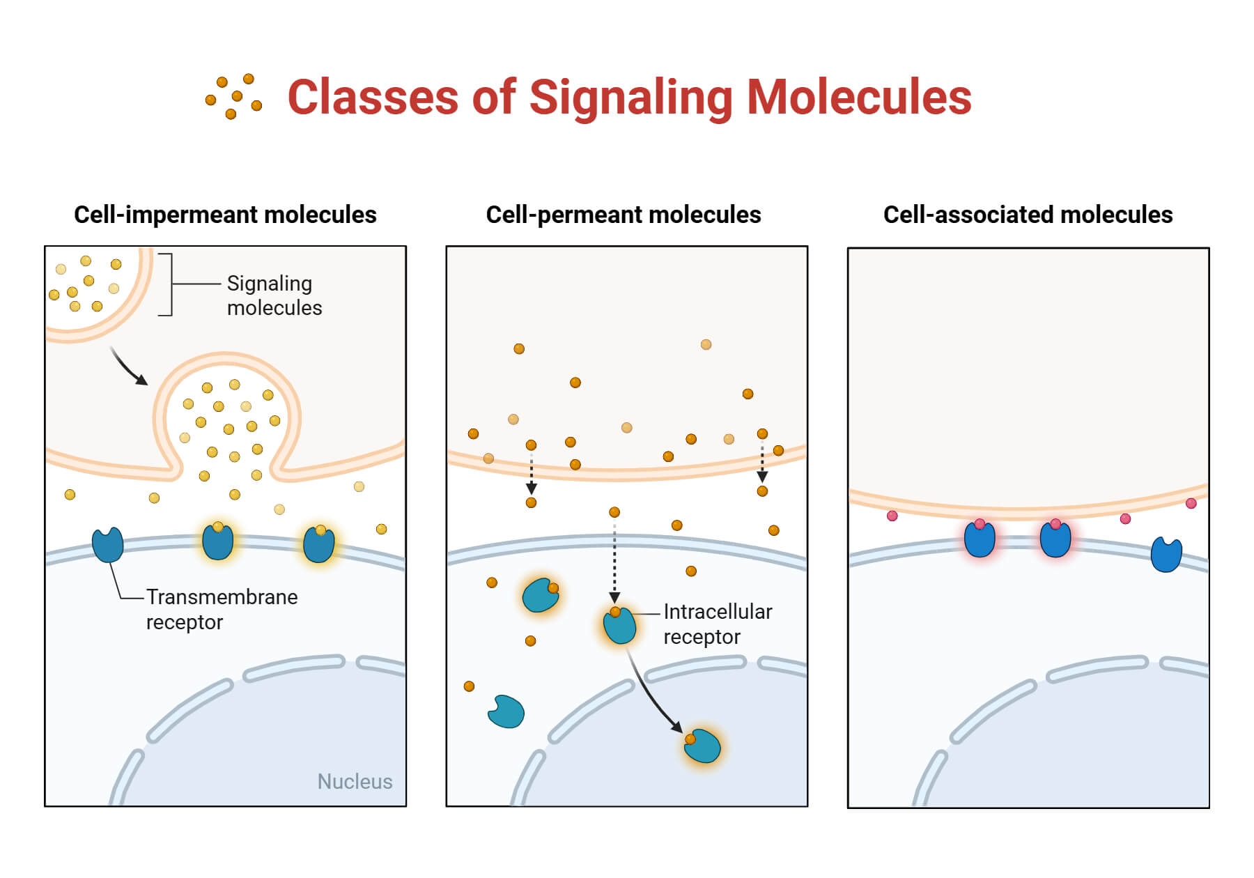
It is to be noted that the signaling molecules are mostly hydrophilic and are unable to directly cross the plasma membrane, therefore, they bind to cell-surface receptors. In contrast, smaller signal molecules diffuse across the plasma barrier and bind to intracellular receptors inside the target cell in the cytosol or the nucleus.
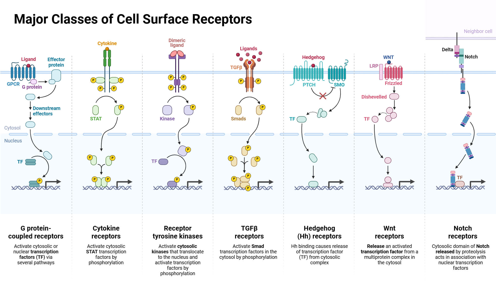
Each cell type displays a set of receptors that enable it to respond to a corresponding set of signal molecules produced by other cells. These signal molecules work in combination to regulate the cell’s behavior. An individual cell requires a multitude of signals to survive and more to divide and differentiate. When cells do not receive these signals, they are programmed to die, i.e., they undergo apoptosis.
Types of Cell Signaling
Contact-dependent signaling: The signaling cell directly interacts with the target cell via direct membrane-membrane contact.
Paracrine signaling: In paracrine signaling, the signaling cells release signal molecules in the extracellular space and locally activate the neighboring cells.
Synaptic signaling: In synaptic signaling, the neurons transmit cell signals electrically along their axons and release neurotransmitters at synapses. This leads to the excitation of other neurons, often far away from the cell body (soma).
Endocrine signaling: Endocrine signaling depends on the endocrine cells, which secrete hormones into the bloodstream, which are widely distributed throughout the body.
Autocrine signaling: In autocrine signaling, a group of identical cells produces a higher concentration of a secreted signal than a single cell. When this signal binds back to a receptor of the same cell type, it encourages the cells to respond coordinately as a group, generating a stronger signal.

Pathways of Cell Signaling
G Protein-Coupled Receptor
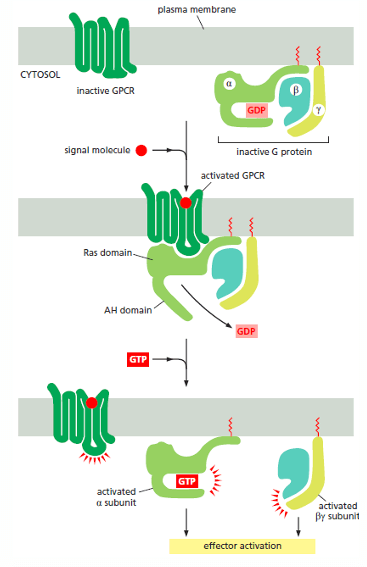
G protein-coupled receptors (GPCRs) are crucial membrane proteins containing an extracellular amino terminus, seven transmembrane a-helical domains, and an intracellular carboxy terminus. They are the fourth-largest superfamily in the human genome, responsible for a wide variety of signaling mechanisms relating to sight, smell, and taste. GPCR exists as a single polypeptide chain that constitutes back and forth across the lipid bilayer membrane, forming a cylindrical structure, with a ligand binding site at its center. Inactive G proteins are composed of a and y subunits, coupled by lipid molecules which bind to the plasma membrane and the GDP-bound α subunit.
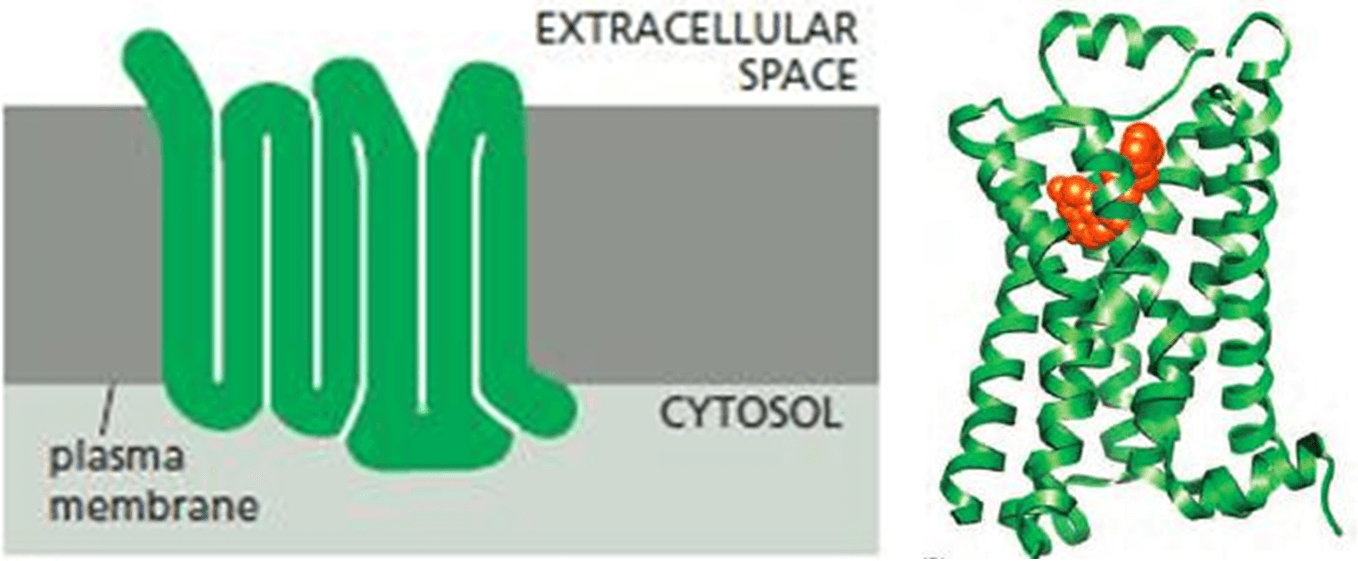
The activation of signal transduction via GPCR is as follows:
- When a signal molecule binds to the ligand binding region of GPCR, the conformation of the receptor changes.
- This allows for the receptor to bind and alter the conformation of the trimeric G protein.
- The Alpha Helical (AH) domain of the α subunit moves outward, opening up the nucleotide-binding site and promoting GDP dissociation.
- GTP then binds to the α subunit, inducing the dissociation of the α subunit from both the GPCR and the By complex. This is crucial for downstream signaling events.
- The By complex can activate or inhibit a wide variety of downstream signaling molecules and pathways, depending on the specific G protein and the cellular context.
This mechanism of GPCR signaling enables cells to respond to numerous external signals, including hormones, sensory stimuli, and neurotransmitters.
cAMP-dependent Pathway
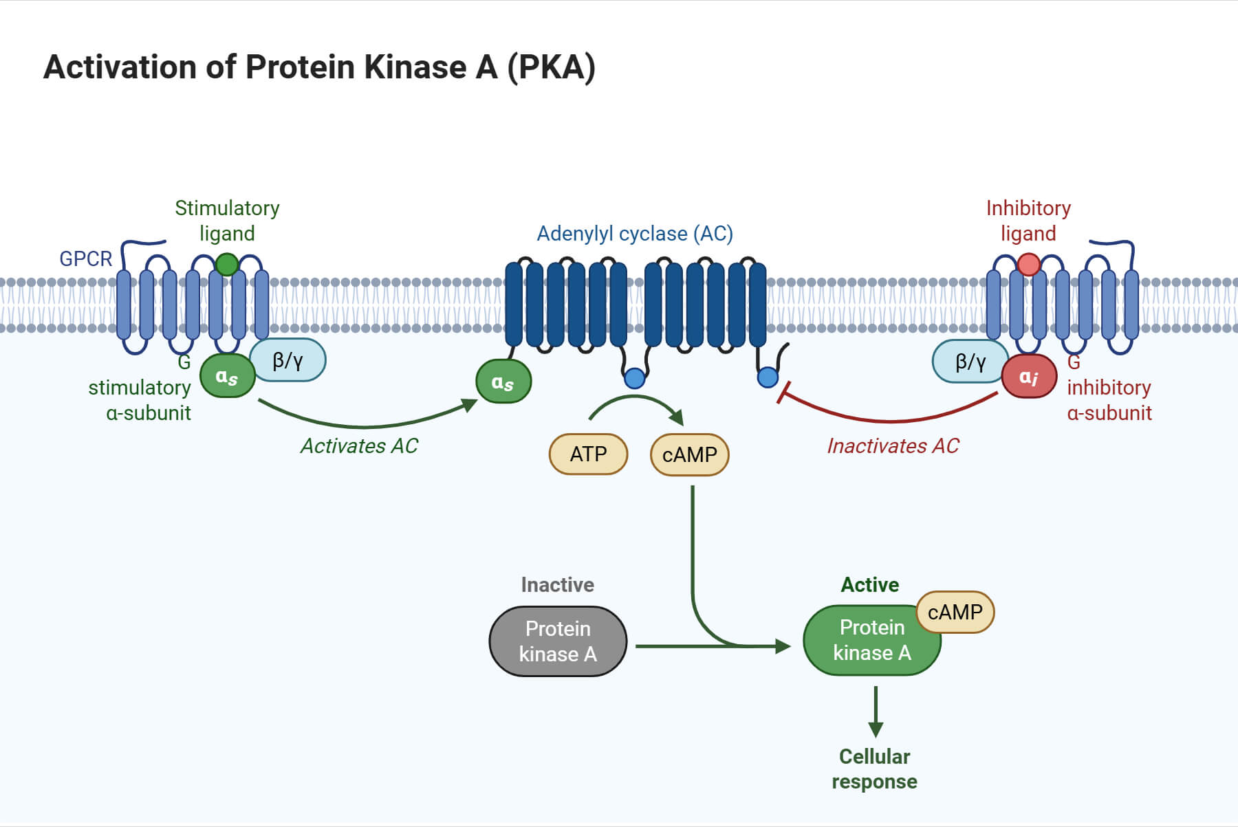
Cyclic AMP (cAMP) is the first intracellular cell signaling molecule elucidated in hormones. They are responsible for the breakdown of glycogen to glucose for muscular activity. Following this discovery and several others, cAMP has been regarded as a secondary messenger in hormonal signaling, the initial being the hormone itself. Cyclic AMP originates from ATP by the action of adenylyl cyclase and is degraded to AMP by cAMP phosphodiesterase. This extracellular signal enhances cAMP concentration by more than 20-fold in seconds. Most of the cAMP activity is governed by the action of cAMP-dependent protein kinase, i.e., protein kinase A.
Protein kinase A (PKA) is a family of serine-threonine kinases that has an inactive and an active form (heterodimer). The former is a tetramer consisting of two catalytic and two regulatory subunits. Cyclic AMP (cAMP) binds to the regulatory subunits, which separates them from the catalytic subunits. The catalytic subunits then become enzymatically active, phosphorylating serine residues on their target proteins.
Receptor Tyrosine Kinases (RTKs)
Receptor Tyrosine Kinase (RTKs) are enzyme-coupled receptors with their ligand-binding domain on the outer surface of the plasma membrane. They are responsible for the mediation of cell-to-cell communication. The cytosolic domain either has an intrinsic enzyme activity or associates directly with an enzyme.
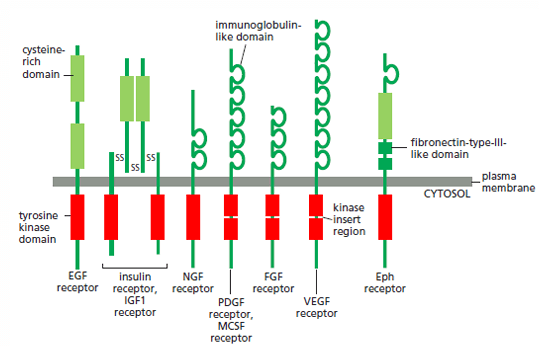
The RTK family is diverse, each with varied cellular function, comprising epidermal growth factor receptor (EGFR), platelet-derived growth factor receptors (PDGF), insulin receptors, etc. The protein and receptor family of some of the RTKs and their responses are mentioned below:
| Signal protein family | Receptor family | Responses |
| Epidermal growth factor (EGF) | EGF receptors | Stimulates cell growth, survival, proliferation, or differentiation |
| Insulin-like growth factor (IGF-1) | IGF-receptor-1 | Stimulates cell growth and survival in numerous cell types |
| Fibroblast growth factor (FGF) | FGF receptors | Stimulates the proliferation of numerous cell types; inhibits the differentiation of a few precursor cells |
| Vascular endothelial growth factor (VEGF) | VEGF receptors | Stimulates angiogenesis |
| Platelet-derived growth factor (PDGF) | PDGF receptors | Stimulates growth, survival, proliferation, and migration of various cell types |
Activation of RTKs by dimerization
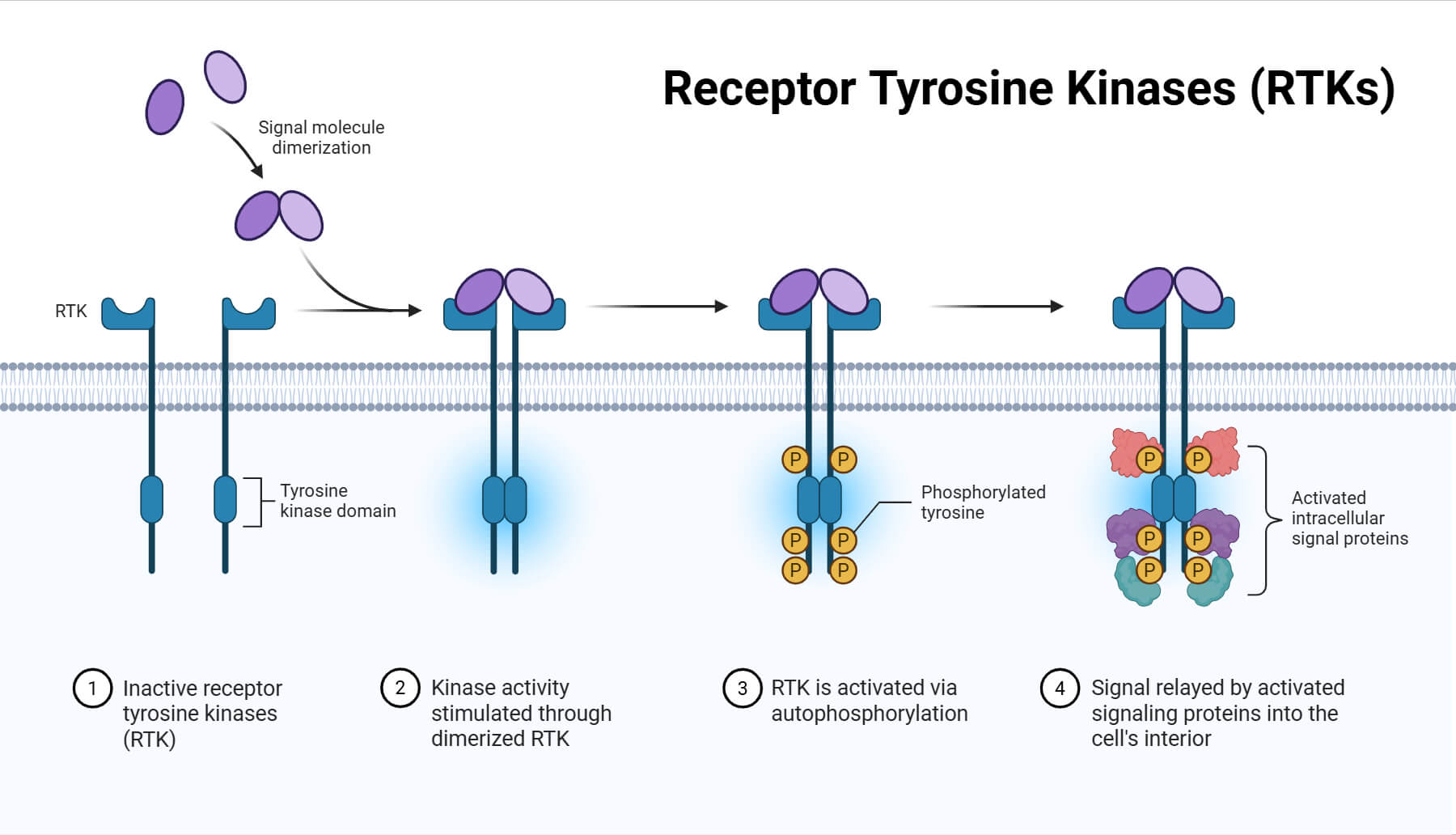
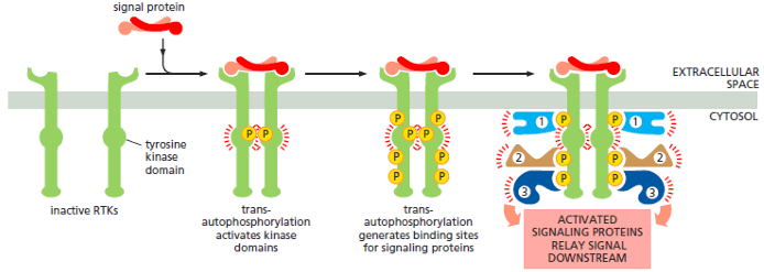
Most RTKs are monomers in which the internal kinase domain is inactive. The binding of a signal protein brings two monomers together to form a dimer. Then the proximity of the dimer leads the two kinase domains to phosphorylate each other, with two different effects, which are as follows:
- First, phosphorylation of some tyrosine residues in the kinase promotes the complete activation of the domains.
- Then phosphorylation at tyrosine in different parts of the receptors generates docking sites for intracellular signaling proteins. This results in the formation of large signaling complexes that can be used to send signals along multiple signaling pathways.
Ras protein Activation by RTK
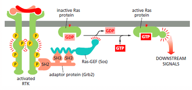
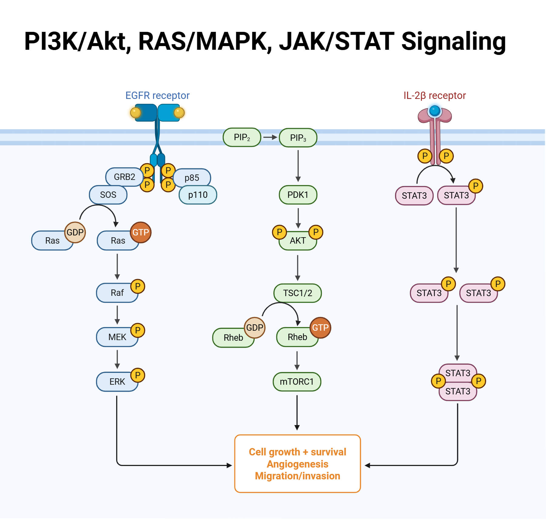
RTKs are responsible for the activation of numerous other inactive proteins, such as the Ras protein. When RTKs get activated, an adaptor protein known as Grb2 recognizes the activation via SH2 domain. This coordination recruits the Sos protein through two SH3 domains. Sos stimulates the inactive Ras protein to replace its bound GDP with GTP, activating Ras to relay the signal downstream.
JAK-STAT Signaling Pathway
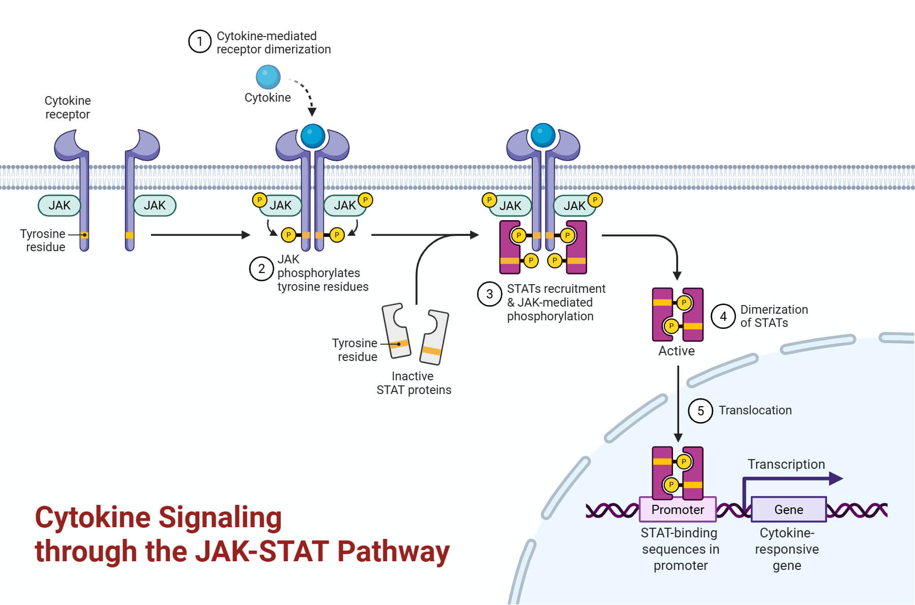
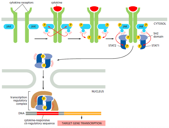
JAK-STAT Pathway is exclusive for immune system responses of inflammation, infection, or even cell death. The pathway initiates from cytokine receptors, which are activated by local mediators (cytokines) or hormones such as growth hormone and prolactin. Once the cytokines bind to the receptor, the two separate receptor polypeptide chains dimerize or orient themselves to a preformed dimer structure. The associated JAKs are brought together to phosphorylate each other on tyrosine. Upon this activation, they phosphorylate the receptors to generate binding sites for the SH2 domains of the STAT proteins.
Similarly, JAKs also phosphorylate the STAT proteins, which dissociate from the receptor to form dimers. The dimers transfer into the nucleus and form a transcription regulatory complex to induce gene expression.
The table below represents some of the extracellular signal proteins that act through the JAK-STAT signaling pathway:
| Signal Protein | Response |
| Interferon-Y (IFN-Y) | Activates macrophage |
| Growth Hormone | Stimulates growth by inducing IGF1 production |
| Prolactin | Stimulates milk production |
| Erythropoietin | Stimulates the production of erythrocytes |
Conclusion
Cell signaling is an integral part of how cells survive, proliferate, differentiate, form tissues and organs, maintain physiological harmony, respond to signals, and eventually die. Understanding these mechanisms is essential for studying large-scale coordination in multicellular organisms. The article is a brief overview of a few signaling pathways, such as GPCR, cAMP, RTKs, and the JAK-STAT pathway.
References
- Alberts, B., Johnson, A., Lewis, J., Raff, M., Roberts, K., & Walter, P. (2002). Molecular Biology of the Cell (4th ed.). Garland Science.
- Cell Communication and signaling, evidence of design. (n.d.). Reasonandscience.Catsboard.Com. Retrieved April 8, 2025, from https://reasonandscience.catsboard.com/t2181-cell-communication-and-signaling-evidence-of-design
- Cell Signalling- Types, Stages & Functions of Cell Signalling. (n.d.). BYJUS. Retrieved April 8, 2025, from https://byjus.com/biology/cell-signalling/
- Cooper, G. M. (2000a). Pathways of Intracellular Signal Transduction. In The Cell: A Molecular Approach. 2nd edition. Sinauer Associates. https://www.ncbi.nlm.nih.gov/books/NBK9870/
- Cooper, G. M. (2000b). Signaling Molecules and Their Receptors. In The Cell: A Molecular Approach. 2nd edition. Sinauer Associates. https://www.ncbi.nlm.nih.gov/books/NBK9924/
- Definition of signaling pathway- NCI Dictionary of Cancer Terms- NCI (nciglobal,ncienterprise). (2011, February 2). [nciAppModulePage]. https://www.cancer.gov/publications/dictionaries/cancer-terms/def/signaling-pathway
- Introduction to cell signaling (article). (n.d.). Khan Academy. Retrieved April 8, 2025, from https://www.khanacademy.org/science/biology/cell-signaling/mechanisms-of-cell-signaling/a/introduction-to-cell-signaling
