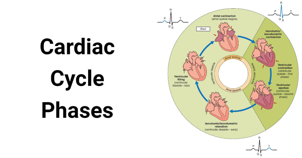
The cardiac cycle is a continuous closed sequence of events that results in the continuous and systematic contraction and relaxation of the chambers of the heart.
It includes all the events that occur in one heartbeat. It involves the complete contraction and relaxation of the atria and ventricles ensuring efficient blood circulation in arteries and veins in a synchronized manner.
The heart continuously completes the cardiac cycle under the control of the cardiac action potential. Approximately 0.8 seconds are needed for one cardiac cycle to be completed all together.

The human cardiac cycle can be broken down into four main phases; the atrial systole, the ventricular systole, the atrial diastole, and the ventricular diastole. There is an intermediary phase called the protodiastole phase marking the end of the systole and the beginning of the diastole phase.
Phases of Cardiac Cycle
Interesting Science Videos
1. Atrial Systole
This phase is also known as presystole or the last rapid filling phage or atrial kick and is the phase when the atria contract to pump the blood out of the atria into the ventricles. The impulse generated by the natural pacemaker, the sinoatrial (SA) node, is transmitted via the internodal tract all over the right atrial wall and through Bachmann’s bundle to the left atrial wall triggering the contraction of the atrial walls. Approximately 20 to 30% of the remaining atrial blood is driven into the ventricle during this stage. This amount of blood forced during the atrial systole is called the atrial kick or the atrial contribution. This step takes roughly 0.11 seconds to complete.
2. Ventricular Systole
It is the stage when the ventricle contracts expelling the blood outside of the ventricles. The Purkinje fibers relay the cardiac impulses all over the ventricular wall stimulating the ventricular wall to contract. This step is completed in about 0.3 seconds.
The ventricular systole can be further subdivided into three sub-phases:
a. Isovolumetric Ventricular Contraction
It is the first stage of ventricular systole when the muscle tension increases and the ventricles begin to contract without any change in the volume of the ventricles. In this stage, the pressure inside the ventricle increases and exceeds the atrial pressure. This pressure difference causes the two atrioventricular (AV) valves (the mitral valve and the tricuspid valve) to close. The two semilunar (SL) valves (the aortic and the pulmonary valves) are also closed in this stage, so the blood can’t leave the ventricle. This results in increasing the internal pressure of the ventricles without changing their volume. This stage lasts for about 0.05 seconds.
b. Rapid Ventricular Ejection
In this stage, the SL valves open due to pressure differences in the ventricles and the major arteries forcing about 70% of the ventricular blood in high pressure out of the ventricles through the aorta and the pulmonary arteries within about 0.13 seconds. The pressure inside the left ventricle will be higher, about 80 mm of Hg, than in the right ventricle (about 8 mm of Hg). Hence, the blood is rushed more forcefully in the aorta than in the main pulmonary artery.
c. Reduced Ventricular Ejection
In this stage, the remaining 30% of the blood is ejected from the ventricles. This stage usually lasts for about 0.09 seconds. The ventricular pressure will have decreased by this time, and the blood will have reached the smaller arteries.
3. Protodiastole
It is the intermediary stage which indicates the end of systole and the beginning of the diastole stage. The ventricular ejection will have completely reduced the ventricular pressure making it lower than the blood pressure inside the major arteries. These pressure differences will close the SL valves. This stage lasts for about 0.04 seconds.
4. Atrial Diastole
It is the stage when the atria are filled. During this stage, the AV valves are closed and the superior and the inferior vena cava pours in the deoxygenated blood collected from the body tissue to the right atrium; whereas the pulmonary veins bring the re-oxygenated (purified) blood to the left atrium. This stage lasts about 0.7 seconds and overlaps the ventricular systole phage. This stage occurs immediately before the ventricular diastole stage.
5. Ventricular Diastole
It is the stage when the blood is passed to the ventricles from the atria increasing the ventricular pressure and volume of the ventricles. This stage lasts for about 0.5 seconds and can be further subdivided into the following sub-phases:
a. Isovolumetric Ventricular Relaxation
It is the first stage of ventricular diastole when the muscle tension in the ventricular wall decreases without any change in the volume of the ventricles. This reduced pressure causes the semilunar valves to completely close. The AV valves are also closed in this stage so blood can’t flow in the ventricles and the volume of the ventricles remains the same. However, the rapid pressure drop will make the atrial pressure higher and the ventricular pressure lower which triggers the next stage. This stage usually lasts about 0.08 seconds.
b. Rapid Ventricular Filling
It is the stage when the AV valves open rushing about 70% of the atrial blood into the ventricles. This stage lasts for about 0.11 seconds.
c. Reduced Ventricular Filling
It is the stage when about 20% of the atrial blood enters the ventricles at a slower rate. This stage usually lasts for about 0.19 seconds.
d. Last Rapid Ventricular Filling
It is the last stage of ventricular diastole that coincides with the atrial systole phase. In this phase, the remaining 10% of the blood is passed to the ventricle. This stage lasts for about 0.1 seconds.
References
- Ross & Wilson Anatomy & Physiology in Health and Illness. 13th ed. Churchill Livingstone Elsevier. ISBN 978-0-7020-7276-5
- Pollock JD, Makaryus AN. Physiology, Cardiac Cycle. [Updated 2022 Oct 3]. In: StatPearls [Internet]. Treasure Island (FL): StatPearls Publishing; 2023 Jan-. Available from: https://www.ncbi.nlm.nih.gov/books/NBK459327/
- The Cardiac Cycle – SimpleMed – Learning Medicine, Simplified
- Cardiac Cycle,Phases of Cardiac Cycle,Cardiac Cycle & ECG (medicosite.com)
- Cardiac cycle explained: cardiac cycle phases, ECG, graph (physiosunit.com)
- Cardiac Cycle – Definition, Phases and Quiz | Biology Dictionary
- Phases of the Cardiac Cycle When the Heart Beats (thoughtco.com)
- 17.4D: Cardiac Cycle – Medicine LibreTexts
- The cardiac cycle – Structure and function of the heart – Higher Human Biology Revision – BBC Bitesize
- 19.3 Cardiac Cycle – Anatomy & Physiology (oregonstate.education)
- Cardiac Cycle- Physiology, Diagram, Phases of the Cardiac Cycle (byjus.com)
- Cardiac cycle phases: Definition, systole and diastole | Kenhub
- Cardiac cycle. (2023, April 4). In Wikipedia. https://en.wikipedia.org/wiki/Cardiac_cycle
CV Physiology | Cardiac Cycle – Rapid Filling (Phase 6)

Sir/ maddam I would like to suggest that stem cell therapy should introduce this all systems to help all needy persons that’s why nutritionist course came but not affordable by all as this is ment according to age diseases cost earnings to it became thanks sir/ maddam