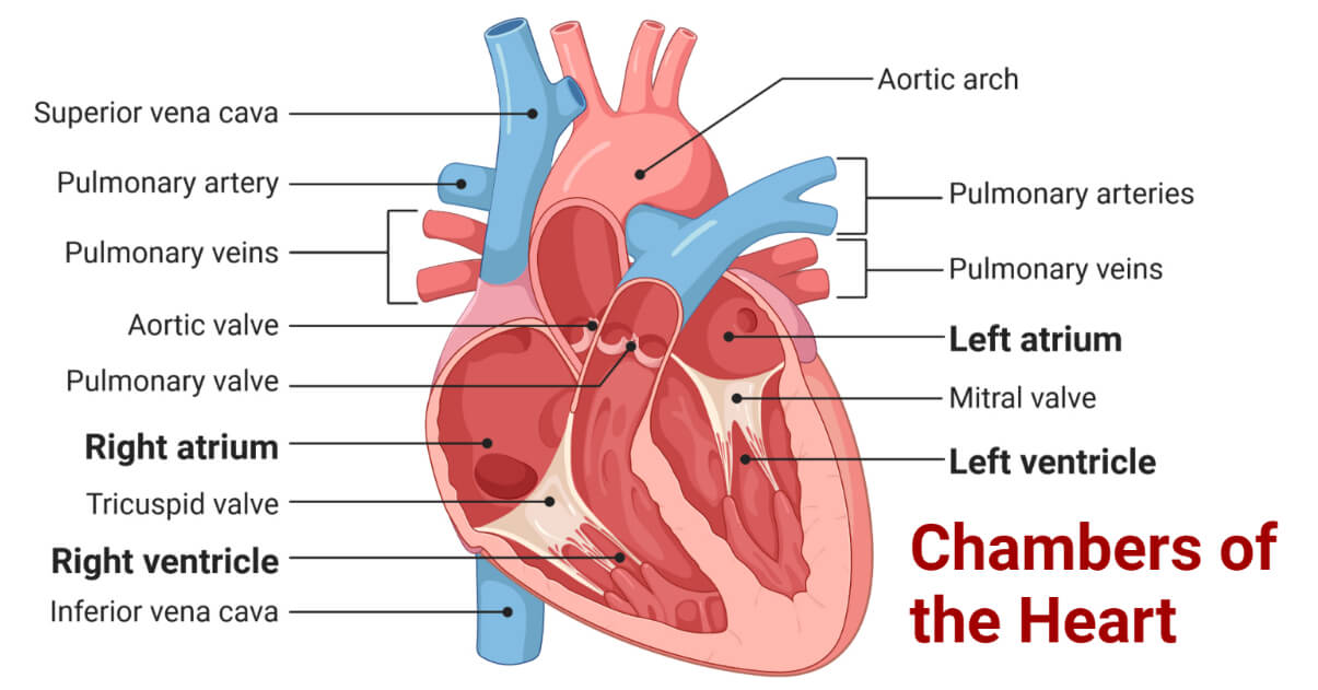
The heart is internally divided into several compartments, well separated by septum and valves, called the chambers of the heart. The human heart has four chambers, two auricles, and two ventricles.

- Based on anatomic location and the presence of either oxygenated or deoxygenated blood, the heart is divided into the right heart (with deoxygenated blood) and the left heart (with oxygenated blood) by septum (atrial and ventricular septum). Each side contains one auricle (atrium) at the top and one ventricle at the base separated by the atrioventricular septum and a specific valve.
- These chambers are lined with smooth endothelial cells of the endocardium, providing a smooth surface for free flotation of blood without adherence and formation of blood clots.
- These chambers collect the deoxygenated blood via the vena cava, pump the blood for re-oxygenation and collect the oxygenated blood via pulmonary vessels, and pump it all over the body via systemic vessels.
Interesting Science Videos
What are Auricles (Atria)?
Auricles, also known as atria, are the two uppermost blood-receiving chambers of the heart that receives the blood from the veins. These are smaller and thin-walled as compared to the ventricles as they don’t need to generate the force of contraction to pump the blood out of the heart. There are two atria, the right, and the left atrium, separated by the interatrial septum. They receive blood from the venous system and pass it to the ventricles during the atrial systole.
- Right Atrium
It is the uppermost chamber at the right side of the heart that receives the deoxygenated blood from the body via two large veins, the superior and the inferior vena cava. The internal surface of the right atrium is divided into two parts, the sinus venarum and the atrium proper, by a muscular ridge called the crista terminalis. It is separated from the right ventricle by the atrioventricular septum containing a tricuspid valve. It collects the deoxygenated blood and supplies it to the right ventricle.
- Left Atrium
It is the upper left chamber of the heart that receives the oxygenated blood from the lungs via the main pulmonary vein. Its interior is also divided into two parts, the inflow port, and the outflow part. It is separated from the left ventricle by the atrioventricular septum containing the mitral valve. It collects the reoxygenated blood and supplies it to the left ventricle.
What are Ventricles?
Ventricles are the two lowermost blood-pumping chambers of the heart that receives the blood from the atria and pumps it outside the heart through the arteries. Ventricles are comparatively thick-walled and muscular so that they can generate enough muscular force to pump blood out of the heart. The two ventricles, the right and the left ventricles are separated from each other by the interventricular septum and from the atria by the atrioventricular septum.
- Right Ventricle
It is located just beneath the right atrium and it receives the deoxygenated blood collected by the right atrium through the tricuspid valve. It is anatomically divided internally into portions, the inflow and the outflow portion by the supraventricular crest. It sends the blood into the pulmonary arteries that take the blood to the lungs for purification.
- Left Ventricle
It is the largest and the thickest chamber located beneath the left atrium at the lower left portion of the heart. Similar to the right ventricle, it is also divided into the right and the left portions. It receives the oxygenated blood from the left atrium via the mitral valve and pumps the blood into the aorta for distribution all over the body via the systemic arterial system.
Interaction of Blood Vessels with Heart Chambers
- Heart chambers are supplied with major arteries and veins for the collection and distribution of blood.
- The aorta, the largest artery carrying the oxygenated blood all over the body through its branches, arises from the left ventricle. The left ventricle pumps the blood into the aorta during ventricular contraction. The opening between the aorta and the left ventricle is guarded by the aortic valve which regulates the one-way flow – from the ventricle to the aorta – of the blood.
- Deoxygenated blood collected by veins and their branches enter the right atrium through the two largest veins: the superior vena cava and inferior vena cava and poured to the right atrium.
- The pulmonary trunk arises from the right ventricle, well-guarded by the pulmonary valve. It carries the deoxygenated blood to the lungs for gaseous exchange through its branches.
- The major pulmonary veins are connected to the left atrium which brings the oxygenated blood from the lungs for distribution in the arterial system.
Blood Flow Through Heart Chambers
The heart is the central point of the blood circulatory circuit. Within the heart, the blood circulates through the chambers in a specific pathway to ensure a specific and coordinated pathway.
- First, the heart receives the deoxygenated blood via the superior and the inferior vena cava. This deoxygenated blood is collected in the right atrium.
- During the atrial contraction, the right atrium pushes the blood to the right ventricle through the tricuspid valve.
- The deoxygenated blood passed into the right ventricle escapes the chamber and enters the pulmonary artery during the ventricular contraction. The deoxygenated blood is transported to the alveoli of the lungs where the blood is resupplied with oxygen. The re-oxygenated blood is again transported back to the heart through the pulmonary veins.
- Pulmonary veins bring the oxygenated blood back to the heart and pour it into the left atrium.
- During the atrial contraction, the left atrium pushes the oxygenated blood to the left ventricle through the bicuspid valve.
- The oxygenated blood in the left ventricle is passed into the aorta during the ventricular contraction. The oxygenated blood is supplied all over the body by branches of the arteries. The deoxygenated blood is then collected by the veins and poured back into the right atrium for recirculation.
Assessing Heart Chambers
Heart chambers can be assessed by using several medical procedures, some of which are described below:
- Echocardiography (echo)
It is a non-invasive medical procedure that uses high-frequency sound (ultrasound) to access and investigate the heart’s chambers. Using this procedure we can obtain the ultrasound image of heart chambers. Transesophageal echocardiography (TEE) is mainly used to view the heart chambers.
- Cardiac Catheterization
It is a minimal surgical procedure in which a thin, flexible, hollow tube called a catheter is inserted into the blood vessels and guided to the heart chambers. It can be guided to the specific heart chamber for required medical purposes.
- Open Heart Surgery
It is a surgical procedure in which the surrounding muscles and the rib cage are cut open to access the heart. In this procedure, the heart is temporarily stopped and a cardiopulmonary bypass machine is used to artificially pump the blood. The cardiac surgeon can even operate and open the pericardium and the heart’s muscular wall to reach the valves or inner portion of the heart chambers.
Disorders Associated with Heart Chambers
Heart chambers are affected by several infectious and non-infectious medical conditions, some of which are summarized in the following table.
| Disease | Description of the Disease |
| Atrial fibrillation (A-fib) | It is a condition in which the atria beat discontinuously and out of sync with the ventricles due to disorganized electric impulses in the atria leading to arrhythmia. |
| Ventricular fibrillation (V-fib) | It is a condition in which the ventricles quiver (beat discontinuously and out of sync with the atria) due to disorganized electrical activity in the ventricles, leading to arrhythmia. |
| Atrial Septal Defect (ASD) | It is a congenital disorder characterized by a hole in the atria septum causing mixing of the blood of the two atria. |
| Ventricular Septal Defect (VSD) | It is a congenital disorder characterized by a hole in the ventricular septum causing the mixing of the blood of the two ventricles. |
| Hypertrophic Cardiomyopathy (HCM) | It is a condition characterized by the thickening of the heart wall (especially of the left ventricle) making blood pumping difficult. |
| Dilated Cardiomyopathy (DCM) | It is a condition characterized by a thin heart wall making the chambers enlarged but inefficient to pump blood. |
| Restrictive Cardiomyopathy (RCM) | It is a condition characterized by stiffening of the heart wall (especially the wall of the ventricles) causing problems during ventricular relaxation. |
References
- Ross & Wilson Anatomy & Physiology in Health and Illness. 13th ed. Churchill Livingstone Elsevier. ISBN 978-0-7020-7276-5
- Parts Of The Human Heart – Science Trends
- Human heart: Anatomy, function & facts | Live Science
- Human Heart – Anatomy, Functions and Facts about Heart (byjus.com)
- Human cardiovascular system | Description, Anatomy, & Function | Britannica
- Chambers of the Heart (clevelandclinic.org)
- Chambers of the Heart – Atria – Ventricles – TeachMeAnatomy
- How the Heart Works – What the Heart Looks Like | NHLBI, NIH
- Heart: Anatomy and Function (clevelandclinic.org)
- 17.1D: Chambers of the Heart – Medicine LibreTexts
- Britannica, The Editors of Encyclopaedia. “heart”. Encyclopedia Britannica, 18 Apr. 2023, https://www.britannica.com/science/heart. Accessed 3 June 2023.
- The structure of the heart – Structure and function of the heart – Higher Human Biology Revision – BBC Bitesize
- The Heart’s Chambers and Valves (verywellhealth.com)
- Chambers and valves of the heart – Mayo Clinic
- Cardiac surgery. (2023, January 30). In Wikipedia. https://en.wikipedia.org/wiki/Cardiac_surgery
- The four chambers of the heart and their functions | GetBodySmart
- https://www.healthline.com/health/open-heart-surgery
- https://medlineplus.gov/ency/article/002950.htm
- https://my.clevelandclinic.org/health/treatments/21502-open-heart-surgery
- https://www.heart.org/en/health-topics/heart-attack/diagnosing-a-heart-attack/cardiac-catheterization
- https://www.hopkinsmedicine.org/health/treatment-tests-and-therapies/cardiac-catheterization
- Ventricular fibrillation. (2022, November 21). In Wikipedia. https://en.wikipedia.org/wiki/Ventricular_fibrillation
- https://www.heart.org/en/health-topics/arrhythmia/about-arrhythmia/ventricular-fibrillation
- https://www.mayoclinic.org/diseases-conditions/atrial-fibrillation/symptoms-causes/syc-20350624
- https://www.nhlbi.nih.gov/health/atrial-fibrillation
- https://www.mayoclinic.org/diseases-conditions/ventricular-fibrillation/symptoms-causes/syc-20364523
- https://www.heart.org/en/health-topics/cardiomyopathy/what-is-cardiomyopathy-in-adults/hypertrophic-cardiomyopathy
- https://www.mayoclinic.org/diseases-conditions/hypertrophic-cardiomyopathy/symptoms-causes/syc-20350198
- https://www.mayoclinic.org/diseases-conditions/atrial-septal-defect/symptoms-causes/syc-20369715
- https://www.heart.org/en/health-topics/cardiomyopathy/what-is-cardiomyopathy-in-adults/dilated-cardiomyopathy-dcm
- https://www.heart.org/en/health-topics/cardiomyopathy/what-is-cardiomyopathy-in-adults/hypertrophic-cardiomyopathy
- https://www.heart.org/en/health-topics/cardiomyopathy/what-is-cardiomyopathy-in-adults/restrictive-cardiomyopathy
