Interesting Science Videos
Buffy Coat Definition
A buffy coat suspension is a concentrated suspension of leukocytes and platelets that make up a part of the anticoagulated blood sample obtained by the process of density gradient centrifugation.
- The term buffy coat arose from the fact that the suspension has a color (yellowish beige) that is similar to buff.
- Buffy coats primarily contain white blood cells and platelets when separated from the whole blood sample via centrifugation.
- The generation of buffy coat from the whole blood sample helps to concentrate large volumes of blood samples so that it decreases the downstream during cell separation and handling.
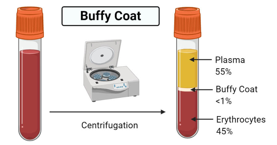
- In a test tube with whole blood, a thin grey-white layer of buffy coat is formed between the plasma and the hematocrit, consisting of leukocytes and platelets, both less dense than erythrocytes.
- Buffy coat accounts for about less than 1% of the total blood volume taken in a tube.
- Whole blood samples are generally fractioned as a pretreatment in order to separate the buffy coat from the plasma and erythrocytes.
- After centrifugation, the buffy coat remains between the plasma and the erythrocytes depending on its density.
- The color of the buffy coat usually remains between yellow to light brown, but there might be some variations in the color as the color depends on the concentration of neutrophils.
- The color is more greenish when the concentration of neutrophils is high, whereas it remains yellow if the concentration is low.

Buffy Coat Preparation
Buffy coat preparation in laboratories is usually performed for the concentration and observation of parasites in blood samples.
The following is the protocol for the preparation of buffy coat to concentrate parasites:
- A small narrow test tube or a bore plastic is taken and filed with an EDTA-treated anticoagulated blood sample with a Pasteur pipette.
- The blood sample is then centrifuged for a total of 15 minutes at the RCF of 1000g. The time might differ with laboratories as the time might range between 10 to 25 minutes.
- Following centrifugation, the supernatant containing the plasma above the buffy coat layer is removed onto a new tube.
- The buffy coat layer, along with some red cells present immediately below the buffy coat is transferred to one end of a slide. Buffy coat and the red cells are mixed with the tip of the pipette.
Note: In the case of some parasites like trypanosomes and microfilariae, a small amount of plasma is withdrawn together with the buffy coat as these parasites might be concentrated in the plasma. - A thin preparation is made on the slide with a smooth-edged spreader, and the slide is allowed to air dry.
- When dry, the preparation is fixed with absolute methanol or ethanol for 2 minutes.
- The slide is then stained using either the Field’s thin-film staining technique or with the Giemsa staining method.
- The stained preparation is examined first with a 40X objective and then with a 100X objective.
Buffy Coat Uses
- Buffy coats are important for DNA isolation from blood samples. Especially in the case of the mammalian blood sample with non-nucleated RBCs, DNA extraction is performed from white blood cells as leukocytes are about ten times more concentrated source of nucleated cells.
- The technique is also useful for the purification of large amounts of gDNA from relatively small sample sizes.
- Buffy coat preparation also allows the differentiation of white blood cells as they are more concentrated in the buddy coat than in the whole blood sample.
- The generation of buffy coat from the whole blood sample helps to concentrate large volumes of blood samples so that it decreases the downstream during cell separation and handling.
- The use of a buffy coat reduces donor variability as donor-specific soluble serum factors are eliminated with the rest of the discard.
- The platelet-rich buffy coats have become an alternative source for the platelet concentration method as the buffy coat preparation causes less platelet activation and damage.
- A quantitative buffy coat is a standard laboratory test to detect infection with malaria or other blood parasites like trypanosomes, Leishmania, and Histoplasma.
- Buffy coat preparation is a cheaper method of blood cell separation, and it is also the ideal method to meet the emergency requirement for platelets.
References and Sources
- Mescher AL (2016). Basic Histology. Fourteenth Edition. McGraw-Hill Education.
- 3% – https://microbeonline.com/buffy-coat-definition-composition-preparation-uses/
- 2% – https://accessmedicine.mhmedical.com/content.aspx?sectionid=190281178
- 1% – https://www.slideshare.net/kabitachatterjee/buffy-coat
- 1% – https://www.sciencedirect.com/science/article/pii/B9780124653306500051
- 1% – https://www.ncbi.nlm.nih.gov/pmc/articles/PMC3533502/
- 1% – https://quizlet.com/21450073/anatomy-blood-vocab-flash-cards/
- 1% – https://onlinelibrary.wiley.com/doi/pdf/10.1002/cyto.a.20133
- 1% – https://en.wikipedia.org/wiki/Buffy_coat
- <1% – https://www.researchgate.net/publication/5644132_Overnight_or_fresh_buffy_coat-derived_platelet_concentrates_prepared_with_various_platelet_pooling_systems
- <1% – https://stemcellsjournals.onlinelibrary.wiley.com/doi/full/10.1002/stem.2498
- <1% – https://quizlet.com/56416661/hemo-week-1-flash-cards/
- <1% – https://quizlet.com/115745962/chapter-4-microbiology-flash-cards/
- <1% – https://link.springer.com/referenceworkentry/10.1007/978-3-642-29613-0_38
