A fluorescence microscope is an optical microscope that uses fluorescence and phosphorescence instead of, or in addition to, reflection and absorption to study the properties of organic or inorganic substances.
Fluorescence is the emission of light by a substance that has absorbed light or other electromagnetic radiation while phosphorescence is a specific type of photoluminescence related to fluorescence.
Unlike fluorescence, a phosphorescent material does not immediately re-emit the radiation it absorbs. The fluorescence microscope was devised in the early part of the twentieth century by August Köhler, Carl Reichert, and Heinrich Lehmann, among others.
Interesting Science Videos
Fluorescence Microscope History
It was British scientist Sir George G. Stokes who first discovered fluorescence in 1852 although several researchers noted luminescence phenomena during the seventeenth and eighteenth century.
Following this, Stokes identified the Stokes shift, a wavelength shifts in emission spectra to longer values. Later at the beginning of the 20th century, fluorescence was first discovered in optical microscopy by August Köhler and Carl Reichert where they claimed fluorescence as an annoyance in UV microscopy.
German physicists Otto Heimstaedt and Heinrich Lehmann created the first fluorescence microscope with the help of which, autofluorescence in bacterial, animal, and plant tissues was seen.
Stanislav Von Provazek employed fluorescence microscopy to examine dye binding in both fixed tissues and live cells. The field of immunofluorescence didn’t come into existence until Albert Coons created a method for fluorescently tagging antibodies in the early 1940s.
- Light microscopes that use fluorescence microscopy have the ability to excite fluorophores and then detect the luminescence signal. Photoluminescence is a phenomenon that occurs when a molecule is excited by ultraviolet or visible light photons to produce luminescence. Depending on the electronic configuration of the excited state and the emission pathway, photoluminescence is divided into two categories: fluorescence and phosphorescence.
- When light excites or transfers an electron to a higher energy state, fluorescence is produced, which results in the generation of light after a brief time period (less than a microseconds) with a longer wavelength, lower energy, and a different hue from the first absorbed light. This time period is known as the fluorescence lifetime.
- Similar to fluorescence, phosphorescence happens in a similar way but has a significantly longer excited state lifespan.
- The pivotal component of the fluorescence microscopy is to ensure that the samples being analysed will need to be labelled, tagged or dyed, although some specimens contain intrinsic fluorescence (auto fluorescence) which don’t require any outside assistance to be fluorescent.
Several fluorescent dyes include:
Biological fluorescent stains
- Nucleic acid stains: Hoechst, DAPI, DRAQ5 and DRAQ7 stain the nucleus.
- Phalloidin: stain actin fibres of mammalian cells.
- Fluorochromes: Isothiocyanite, Alexa fluors or Dylight 488 stains the target sample.
Antibody labelled with fluorochrome is used to detect the antigen.
Principle of Fluorescence Microscope
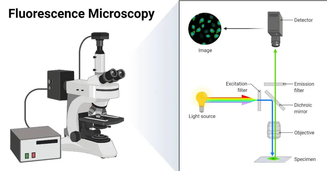
The initial step in the observation of the sample through a fluorescence microscope includes labelling the sample with fluorescent dyes. Then, light source which emits white light is allowed to fall onto the excitation filter This filter selects the light of a specific wavelength that can excite the fluorescent molecules tagged in the specimen and this excitation light incidents onto the dichroic mirror. The light after reflection from the dichroic mirror passes onto the specimen after emerging from the objective lens. This small wavelength light falls into the specimen stained with a fluorescent dye that results in emission of high wavelength light which passes again through the condenser lens and dichroic mirror. This allows green light in maximum along with some blue light to pass towards the emission filter. However, this filter only permits the longer wavelength green light to pass into the eyepiece and detector while at the same time rejecting the blue light completely. The detector detects the green light and permits it to fall back onto the specimen thereby, forming fluorescent green specimens against a dark background.
Parts of a Fluorescence Microscope
Some of the components of fluorescence microscope are as follows:
- Fluorophore: These are reactive fluorescent dyes which form a fluorescent image by generating highly contrasted visible green light after being activated by highly illuminating UV radiation.
- Light Source: Major light sources include xenon arc lamps, mercury-vapour lamps, lasers and high-power LEDs. A simple epifluorescent microscope makes use of a light source made up of xenon lamps, mercury lamps, and LEDs. On the other hand, laser light is mostly used by the advanced confocal fluorescence microscope.
- Excitation filter: The excitation filter is a bandpass filter that functions to narrow the wavelength of the light that illuminates the sample by blocking other sources of exciting light. Such light of shorter wavelength can be easily absorbed by the fluorescent dye.
- Dichroic Mirror: It is also known as dichromatic mirror or beam-splitter. It selectively reflects or transmits light with specific wavelengths.
- Objective/ Condenser lens: The light is then directed through a system of optics that serves as both a condenser, gathering the light into a narrow beam on the sample, and a focusing objective for the light emitted back by the specimen.
- Sample stage: The sample stage holds the specimen and contains x, y, and z-axis movements that can be controlled manually or by a computer.
- Emission filter: It is a bandpass filter that functions similarly to an excitation filter. The light emitted from the specimen includes light reflected from the sample, which will be the excitation wavelength as well as light emitted from the fluorescent components of the specimen. Since, certain samples may include different fluorescent material, light of different wavelengths may be emitted. It allows fluorophore light radiations to pass while blocking excitation light.
- Eyepieces and detector: The light after passing through an emission filter is directed toward a set of eyepieces to facilitate the user to see or to a camera. The fluorescence images can then be quantitatively analysed using a combination of digital imaging and image processing.
Types of Fluorescence Microscope
Wide-field Epifluorescence microscope
A parallel beam of light illuminates the entire specimen at once to excite the fluorophore. All of the resulting fluorescence of specimens can be viewed simultaneously, allowing for simple and quick imaging. All the fluorescence can be examined at once for multiple-prob specimens. Because all areas of the material may be examined at once, it enables for a rapid selection of fluorescent cells to scan.
The WF microscope does not acquire enough comprehensive information to allow 3D imaging because of its non-specific data collecting, including out-of-focus blur.
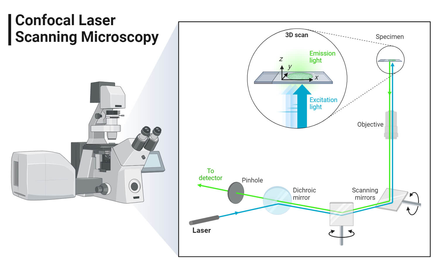
Confocal microscope
An additional set of optics is employed to ensure that only light from a small point in a narrow focal plane in the specimen reaches the observer, and any out-of-focus light is excluded. It facilitates observation of thick specimens with high resolution. Thus, it detects just what is in focus, and anything out of focus appears dark. This is accomplished by directing the light source, which is often a laser, to a specific place and detecting the image through a pinhole.
Confocal microscopes are classified into three types: laser scanning microscopes, which use a sharply focused laser to scan over the sample; spinning disk confocal microscopes, which use a disk with pinholes cut into it in the shape of a spiral; and programmable array microscopes (PAM), which work similarly to spinning disk microscopy but with the exception that the pinholes in the PAM can be opened and closed by the user.
It is quite expensive, which costs 2-7 times higher than a wide field microscope.
Multiphoton microscope
It is also known as non-linear or two-photon microscopy. In traditional fluorescence microscopy, a fluorophore is activated by absorbing a single photon of a specific wavelength. Two or three photons of a greater wavelength do the job of one when they hit the fluorophore at the same time (usually within few femtoseconds), resulting in fluorophore excitation and light emission. Photons combine their energy, allowing low-energy infrared photons to excite fluorophores. Infrared light penetrates tissue more deeply than the normal excitation light used in fluorescence microscopy. Because of its low intensity level, infrared light is less harmful and hence very beneficial when working with living samples.
Total internal reflection fluorescence microscope
It enables the imaging of fluorescent molecules near the glass/water (or glass/specimen) interface. This is accomplished by using an evanescent wave to excite the fluorophores rather than direct illumination from an arc lamp, LEDs, or lasers. The incident light is typically laser light, with the interface consisting of the glass of the coverslip and an aqueous solution film between the coverslip and adhering cells.
The optical phenomena, total internal reflection, follows Snell’s law. At the interface, reflected light generates an electromagnetic field, and an evanescent field forms through low refractive index material. The evanescent field can excite fluorophores near the interface that have the ability to electronic transition with or close to the wavelength of the laser beam.
This enables the study of membrane-associated processes such as cell adhesion, hormone binding, molecular transport, and exocytic and endocytic processes.
Operating Procedure of Fluorescence Microscope
- Choose microscope optics such as filters that are compatible with the fluorophore after deciding on a fluorophore and technique of tagging.
- Ensure that the microscope slide and coverslip are clean after first checking that these components are not auto-fluorescent in the desired wavelength range.
- Place a droplet of the sample on the slide, then gently cover it with a coverslip.
- Begin with a low magnification and adjust the focus, lighting, and x-y position as necessary.
- Continue to adjust focus and x-y position while increasing magnification to the desired level.
- Collect digital pictures of the section of interest from the sample specimen, making sure to identify the files with descriptive titles or making a note of the file names and descriptions for subsequent retrieval.
Applications of Fluorescence Microscope
- Neurotransmitters such as dopamine, serotonin, norepinephrine, and epinephrine can be detected using fluorescence microscopes even though they are much too small to be seen by a conventional light microscope.
- These are used by the food chemists in quality control by accessing the structural organisation and chemical processes of the food components and ensuring the safety of the consumers.
- It enables researchers to understand the cycles of protein synthesis and degradation as well as how proteins travel. The location of the mineral’s origin can also be determined more easily.
- It is employed in textile industries for the analysis of fibres and obtaining clear 3D images of fibres and yarns.
- It is used to examine the porosity of ceramics, enabling researchers to determine an object’s density, strength, and durability.
Advantages of Fluorescence Microscope
- Adaptable: Many conventional bright-field microscopy techniques can be replaced and enhanced by fluorescence microscopy techniques. If the instruments are outfitted with a few fluorescence attachments, the majority of compound microscopes can be utilized for fluorescence microscopy.
- Easy: Examining fluorescent specimens is easy due to the great contrast.
- Rapid: Specimens can be studied directly on a slide, frequently without any preliminary purification or concentration
- Reliable: The great sensitivity and specificity of fluorescence microscopy techniques are responsible for the good reliability and accuracy of microscopic analysis. Even when performed by technicians with less training, the likelihood of incorrect observations or discrepancies in test interpretation is decreased.
- Sensitive: Small things are simple to see because of the comparatively bright fluorescent picture on a dark background.
- Specific: High specificity for details is achieved by using modern fluorochromes and interference light filters.
- Universal: Numerous biomedical fields, including bacteriology, mycology, virology, parasitology, serology, immunology, autoimmunology, cytology, cell biology, and histochemistry, can benefit from the use of fluorescence microscopy.
Limitations of Fluorescence Microscope
- The addition of probes and dyes to a membrane system has the ability to alter the characteristics of the liposomal delivery system. However, it has been demonstrated that the physical characteristics of the membrane are only slightly affected by the application of low dye concentrations (1 mol%).
- The selection of the fluorescent dye is an important step since some dyes might produce significant changes in the host membrane and/or experimental errors, leading to incorrect data interpretation.
- Bleaching and a reduction in fluorescence intensity can also happen after prolonged exposure to fluorescent light.
Precautions
- Always keep track of the number of hours spent using a fluorescence microscope’s mercury lamp. Extending the lifespan of your fluorescent microscope may cause it to explode.
- When combining low power objectives with oil immersion objectives for your fluorescence microscope, exercise caution. When you swing the microscope’s nosepiece the wrong way, oil from your slide may contaminate your objectives. After using them, dab the lenses with lens paper to remove the oil. Wipe lightly.
- Frequently turning on and off a mercury lamp might shorten its life. If you expect someone else to use the fluorescent microscope in two hours, leave it on.
- Avoid gazing directly at mercury lamps since they emit extremely powerful and visible UV rays. Disassemble the lamp housing only if absolutely necessary. When changing filters, avoid looking through the eyepieces of the fluorescent microscope. Certain filters can directly reflect UV radiation into your eyes.
Examples of Fluorescence Microscope
Optical Microscope Dragonfly 600 (Andor Technology PLC)
- It includes SMLM, 3D super resolution module, B-TIRF, and Zoom Illumination.
- It produces exceptional multi-dimensional photos ranging from subcellular (nm) to whole organism (cm) while greatly increasing productivity.
- Its unique combination of speed and sensitivity enables researchers to find previously unknown dynamic events and image live organisms for days.
- It unravels detailed features such as dynamics of viral infection and the ultrastructure of chromatin or organelles.
Optical Microscope MSC- B107T (Infitek)
- The fluorescent achromatic objective lenses are inexpensive and help to meet the needs of daily fluorescence.
- It can be used to see the fine structure of flagellum and blood.
- Operators can obtain a high contrast image with a neutral background colour regardless of magnification. It is appropriate for seeing non-stained specimens.
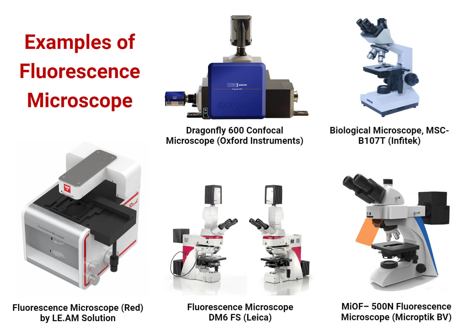
Fluorescence Microscope DM6 FS (Leica)
- It features equipment and accessories that employ leica optics which allow enough clearance around the specimen.
- It allows us to perform experiments with great mechanical and electronic stability.
- Specific techniques are added based on the Leica DM6 FS for Optogenetics and Cryo CLEM.
Optical Microscope MiOF-500N (Microptik BV)
- It provides research-grade performance as well as advanced features.
- It includes high-quality fluorescent plan objectives.
- It supports both fluorescence and bright field microscopy.
- It is also an excellent instrument for sedimentary rock analysis and impurity inspection in semiconductor companies.
References
- https://microscopeinternational.com/what-is-a-fluorescence-microscope-and-what-is-it-used-for/
- https://www.microscopyu.com/techniques/fluorescence/introduction-to-fluorescence-microscopy
- https://zeiss-campus.magnet.fsu.edu/articles/basics/fluorescence.html
- https://www.ncbi.nlm.nih.gov/pmc/articles/PMC5808202/
- https://applications.emro.who.int/dsaf/dsa281.pdf
- https://biologyreader.com/fluorescence-microscopy.html#Principle
- https://www.labmanager.com/lab-health-and-safety/how-to-use-a-fluorescence-microscope-19863
- https://www.researchgate.net/publication/353239749_Three_basic_types_of_fluorescence_microscopy_and_recent_improvement
- https://www.britannica.com/technology/microscope/Confocal-microscopes
- https://www.researchgate.net/publication/51469494_Functioning_Nanomachines_Seen_in_Real-Time_in_Living_Bacteria_Using_Single-Molecule_and_Super-Resolution_Fluorescence_Imaging
- https://ibidi.com/content/217-two-photon-microscopy
- https://www.medicalexpo.com/prod/andor-technology-plc/product-90363-1080669.html
- https://www.medicalexpo.com/prod/bioevopeak/product-301335-1057574.html
- https://www.medicalexpo.com/prod/leica-microsystems/product-70745-940613.html
- https://www.medicalexpo.com/prod/microptik-bv/product-112735-837749.html
it is Avery nice explanation
I’m happy to this page
Informative
Helpfull
Chala bagundi
Very nice