The re-emerging arbovirus, Zika Virus (ZIKV) was first detected in the rhesus monkey about 70 years ago in the Zika Forest in Uganda.
It is a single-stranded RNA virus belonging to the Flaviviridae family that is transmitted by the Aedes mosquito.
The two major geographically discrete lineages include the Asian and African lineages; the African lineage of virus affects the monkeys and apes as the main hosts and humans as occasional hosts whereas the Asian lineage affects humans as the main hosts.
Interesting Science Videos
Structure of Zika Virus

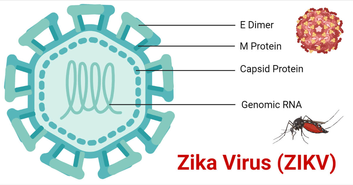
- The mature ZIKV resembles the shape of a golf ball with a smooth surface in which the major surface protein E lies parallel to the viral membrane.
- The virus is 50 nm in size with 180 copies of E and M protein present in its viral membrane.
- It consists of a positive-stranded RNA genome with three structural proteins and a lipid envelope.
- The E protein consists of 4 domains: stem transmembrane domain pair and 3 ectodomains I, II, and III.
- The E protein is dominant on the outer surface of the particle and approximately 50% of the protein is conserved in the ZIKV and dengue (DENV) virus strains.
- The smaller M protein lies under the larger E protein where the 180 E and M proteins are arranged in a raft configuration.
- The prM glycoprotein is connected to the viral membrane. The trimers of prM-E heterodimers form spiked projections arranged with icosahedral symmetry in immature ZIKV that assemble on the endoplasmic reticulum. The immature virion transit through the secretory pathway where the prM undergoes cleavage by host furin-like-protease.
- The mature virion has a relatively smooth surface made up of 90 E protein dimers oriented in a parallel plane with the viral membrane.
Genome Structure of Zika Virus

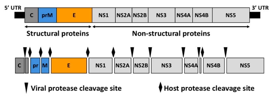
- The ZIKV consists of a single positive-stranded RNA genome of approximately 10.7kbp genome size which encodes a single open reading frame.
- It consists of two untranslated regions (UTRs) in each of its terminals.
- The single polyprotein codes for three structural proteins which include the capsid(C), pre-membrane (prM), and envelope (E), and seven non-structural proteins (NS1, NS2A, NS2B, NS3, NS4A, NS4B, and NS5).
- The structural proteins form the structure of the virus while the non-structural proteins are involved in genome replication, viral polyprotein processing, and regulation of host response.
Epidemiology of Zika Virus
- The Zika virus was first isolated in Uganda in 1947 during a study of yellow virus fever and has been associated to cause sporadic human infections in Africa and Asia.
- The first significant outbreak of the virus was identified in 2007 on the coast of Central Africa. Another outbreak was also identified the same year in Micronesia.
- Subsequent outbreaks were seen from 2013 to 2016 in French Polynesia and other Pacific islands, including New Caledonia, Easter Island, Cook Islands, Samoa, and American Samoa.
- The virus then rapidly spread through South and Central America. The United States detected its first locally transmitted Zika virus case in July of 2016.
- The actual incidence and prevalence of the virus can likely be underestimated due to the lack of routine laboratory testing for the pathogen.
Transmission of Zika Virus
The ZIKV transmission in humans mainly takes place via the bite of the infected Aedes aegypti mosquito.
Other species of Aedes like Aedes albopictus, A. africanus, A. luteocephalus, A. furcifer, and A. taylori are also seen to aid in the transmission of the virus.
Other routes of transmission of the virus include:
- Through sexual intercourse with the infected individual
- Through blood transfusions and organ transplantation
- Congenital infection from the mother to fetus as the virus is capable of crossing the placental barrier
- Through accidental exposure to laboratory workers
Replication of Zika Virus
The viral replication occurs in the cytoplasm, however, the detection of virus-specific antigen in the nuclei of cells infected with Zika virus has brought into question the role of the nucleus in viral replication. The mechanism of its replication includes:
- Attachment/Adsorption
The viral attachment occurs through the viral E proteins present in its envelope with the host receptors like the C-type lectin receptors (CLRs) expressed in myeloid cells, including monocytes, macrophages, and dendritic cells.
- Penetration
The virus is endocytosed through clathrin-mediated endocytosis aided by the mechanism of molecular mimicry.
- Uncoating
The clathrin-coated vesicle gets transported away from the plasma membrane, and the clathrin gets removed from it. The endocytic vesicle is then delivered to the early endosomes that mature into late endosomes. Due to the variation of the pH environment, the viral membrane then fuses with the endosomal membrane releasing the viral genome into the cytoplasm.
- Biosynthesis
The ssRNA genome is then translated to a single polyprotein that is cleaved into the structural and non-structural proteins. The viral replication occurs at the surface of the endoplasmic reticulum and a dsRNA genome is synthesized from the positive-stranded ssRNA. The dsRNA yields the viral mRNA and new ssRNA products through transcription and replication.
- Assembly
The viral assembly occurs at the endoplasmic reticulum from where it buds off and then enters the Golgi apparatus.
- Maturation
Viral maturation occurs in the Golgi apparatus where the prM protein of the immature virion gets cleaved and the mature viral particle is released into the cytoplasm.
- Release
The viral particle is released from the cell through the process of exocytosis.
Pathogenesis of Zika Virus
- There is a lack of sufficient data on the pathogenesis of the Zika virus.
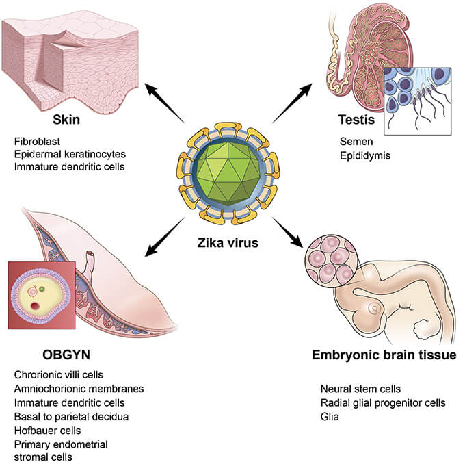
- The human dermal fibroblasts, epidermal keratinocytes, immature dendritic cells, monocytes, and macrophages act as receptor cells for the virus.
- The adhesion/entry factors like DC-SIGN, Tyro, AXL, and TIM-1 aid in the entry of the virus into the cells.
- The ZIKV replication induces the production of Type I interferons in the infected cells while also activating an antiviral immune response.
- From the skin, the virus spreads to the lymph nodes, replicates, and causes primary viremia. The virus spread to other visceral organs through the hematogenous circulation.
- The ZIKV RNA is detectable in the blood within 10 days from infection. Higher viral titers are observed during prolonged viremia.
- The viral shedding is seen to occur at high loads in urine, saliva, and other body fluids like semen, tears, and cervical mucus.
- Studies depicted the excretion of the virus in semen even for months after its clearance from the blood.
- Both the humoral and cell-mediated immune responses are induced following the infection.
- IgM antibodies produced against the virus are usually detectable for 2-3 months to over a year while IgG antibodies remain detectable for months or years and are reckoned to confer lifelong immunity.
Clinical Manifestations of Zika Virus
Zika virus infection may be asymptomatic, or if symptoms occur, are usually mild that arise 3-14 days after infection lasting for 2-7 days. The general symptoms include:
- Fever
- The appearance of maculopapular rash
- Joint pain
- Conjunctivitis
- Muscle pain
- Headache
- Pain behind the eye
- Vomiting

Other symptoms and complications include:
- Myalgia
- Asthenia
- Peripheral edema
- Gastrointestinal disturbances (abdominal pain, nausea, diarrhea)
- Congenital microcephaly
- Guillain-Barré syndrome
- Fetal losses in women infected during pregnancy
- Acute myelitis
- Meningoencephalitis
- Axillary and/or inguinal lymphadenopathy
- Leukopenia with monocytosis
- Thrombocytopenia
Diagnosis of Zika Virus
Culture
The virus can be cultured and isolated by inoculation in chicken embryo yolk sacs, allantoic sacs, and chorioallantoic membrane, as well as cell cultures in Vero, rhesus monkey kidney, and pig kidney cells. It can also be inoculated in suckling mice but shows less sensitivity than the cell cultures. The ZIKV has been successfully cultured from human blood, semen, and urine.
Molecular Detection of ZIKV RNA
The ZIKV RNA viruses can be detected by gene amplification in a two-step procedure through the reverse transcription of genomic RNA into a single-stranded DNA (cDNA), followed by the conversion to double-stranded DNA and the amplification of the DNA. Real-time PCR can be performed for a faster detection than conventional PCR.
Serological Diagnosis
The serological test for ZIKV is performed by ELISA and the test is confirmed by PRNT (Plaque Reduction Neutralization Test) according to the standard protocols. PRNT is considered the “gold standard” for anti-Flavivirus antibody differentiation. The IgM antibody response in primary Flavivirus was found to be specific for ZIKV with limited cross-reactivity with other flaviviruses. However, a high degree of serological cross-reactivity was observed during the secondary Flavivirus infection with other flaviviruses with both IgM ELISA and PRNT90. PRNT is, however, expensive and performed only in highly specialized laboratories.
Diagnosis of ZIKV in Endemic Countries
In countries with limited access to molecular testing methods, diagnosis is often performed by serological testing by IgM ELISA or rapid tests. A combined NS1 antigen and IgM antibody test increase the sensitivity and specificity of dengue fever diagnosis. If several patients test negative by DENV NS1 test, Zika and other Flavivirus infections are suspected.
Treatment of Zika Virus
- There is no specific treatment or antiviral drugs specific for the treatment of Zika virus infection.
- Acetaminophen can be given for fever and pain and an antihistamine for the rash.
- Drinking fluids and a nutritious diet are recommended for the patients.
- Due to the increased risk of hemorrhagic syndrome with other flaviviruses, it is recommended to avoid treatment with acetylsalicylic acid and nonsteroidal anti-inflammatory drugs.
- During the viremic phase of the infected patient, isolation of the patient is recommended to reduce the transmission of the virus through mosquito bites to other people.
Prevention and Control of Zika Virus
Although there are several vaccines in their development phase for the ZIKV infection, no vaccine is available for Zika fever as of now.
The preventive measures are the same for it as for other Aedes aegypti-borne diseases for which no vaccine is available, some of which include:
- Control of mosquito vectors by eliminating their breeding grounds through environmental modifications or the use of insecticides
- Use of mosquito repellants, coils, vaporizers, and oils for the prevention of mosquito bites
- Minimizing skin exposures in areas where the virus remains endemic
- Countries at high risk for ZIKV infection should develop basic virologic and serologic laboratories for better diagnosis of the ZIKV
References
- Musso, D., & Gubler, D. J. (2016). Zika Virus. Clinical Microbiology Reviews, 29(3), 487–524. https://doi.org/10.1128/CMR.00072-15
- Rawal G, Yadav S, Kumar R. Zika virus: An overview. J Family Med Prim Care 2016;5:523-7
- Pierson, T. C., & Diamond, M. S. (2018). The emergence of Zika virus and its new clinical syndromes. Nature, 560(7720), 573–581. https://doi.org/10.1038/s41586-018-0446-y
- Wolford, R. W., & Schaefer, T. J. (2019, September 20). Zika Virus. Nih.gov; StatPearls Publishing. https://www.ncbi.nlm.nih.gov/books/NBK430981/
- Australia, H. (2019, December 3). Zika virus. Www.healthdirect.gov.au. https://www.healthdirect.gov.au/zika-virus
- Zika virus. (2018, July 20). Www.who.int. https://www.who.int/news-room/fact-sheets/detail/zika-virus
- CDC. (2014, November 5). Zika Virus. CDC. https://www.cdc.gov/zika/index.html
- Mayo Clinic. (2018). Zika virus – Symptoms and causes. Mayo Clinic. https://www.mayoclinic.org/diseases-conditions/zika-virus/symptoms-causes/syc-20353639
- NHS Choices. (2019). Zika virus. NHS. https://www.nhs.uk/conditions/zika/
- Symptoms. (2014, November 5). CDC. https://www.cdc.gov/zika/symptoms/symptoms.html
- Zika Virus Infection – Infections. (n.d.). MSD Manual Consumer Version. https://www.msdmanuals.com/home/infections/arboviruses-arenaviruses-filoviruses/zika-virus-infection
- Ávila-Pérez, G.; Nogales, A.; Martín, V.; Almazán, F.; Martínez-Sobrido, L. Reverse Genetic Approaches for the Generation of Recombinant Zika Virus. Viruses 2018, 10, 597.
- Zika virus: Symptoms and treatment. (2017, July 31). Www.medicalnewstoday.com. https://www.medicalnewstoday.com/articles/305163#diagnosis
- Sirohi, D., & Kuhn, R. J. (2017). Zika Virus Structure, Maturation, and Receptors. The Journal of Infectious Diseases, 216(suppl_10), S935–S944. https://doi.org/10.1093/infdis/jix515
