- The term hypersensitivity denotes a condition in which an exaggerated immune response of a host results in inappropriate reactions that leads to destruction of host tissues.
- According to the most widely accepted classification of Gell and Coombs, there are four main types of hypersensitivity reactions as type I, II, III and IV. Type I, II and III are immunoglobulin-mediated (immediate) hypersensitivity reactions while type IV reaction is lymphoid cell-mediated or simply cell mediated hypersensitivity (delayed-type).
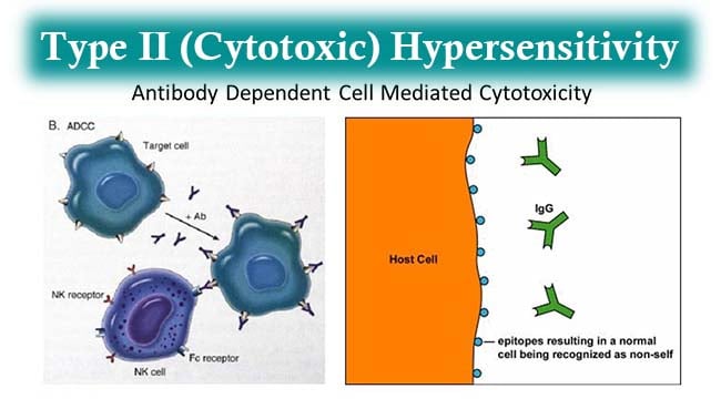
- Type II hypersensitivity reaction also known as cytotoxic hypersensitivity is the antibody mediated destruction of healthy cells.

- It is primarily mediated by antibodies of the IgG or IgM classes which are one or more tissue specific. Complement, phagocytes and K cells also play a role.
- The reaction time is minutes to hours and thus considered as immediate hypersensitivity.
- Type II hypersensitivity as in other cases of hypersensitivity occur when the mechanism of self tolerance is breached and some self reactive immune cells are activated to produce antibodies that attach to antigens on host cells.
- The antigens are cell bound surface antigens and can either be intrinsic (endogenous) or extrinsic (exogenous). Intrinsic antigens are those that are made by the host tissue itself while extrinsic antigens refer to a foreign substance lodged onto a host tissue such as a drug.
Interesting Science Videos
Mechanism of Type II Hypersensitivity Reactions
- The reaction is completed in two phases – sensitization phase and effector phase.
- A sensitization phase leads to production of antibodies that recognize substances or metabolites that accumulate in cellular membrane structures. In the effector phase, target cells become coated with antibodies which lead to cellular destruction.
- Antibody bound to a surface antigen can induce the death of the antibody-bound cell by three distinct mechanisms – by activation of the complement system, cell destruction by antibody dependent cell mediated cytotoxicity (ADCC) or by the process of opsonization.
- First, IgG or IgM antibodies coating target cells can bind to Fc receptors present on cells such as macrophages and neutrophils and mediate phagocytosis.
- IgG or IgM antibodies can also activate complement via the classical pathway. This leads to deposition of C3b, which can mediate phagocytosis. Complement activation also leads to production of the membrane attack complex (MAC), which forms pores in the cellular membrane resulting in cytolysis.
- Finally, IgG antibodies can bind FcγRIII on NK cells and macrophages, thus mediating release of granzymes and perforin and resulting in cell death by apoptosis (ADCC).
- Alternatively, the antigen-antibody complex thus formed may be non- cytolytic but impair the normal functioning of the cell in which the antibody interrupt the cell receptor function. Such antibodies are referred to as” antireceptor antibodies”.
Examples of Type II Hypersensitivity
-
Rhesus incompatibility (Rh hemolytic disease)
During subsequent pregnancies, when Rh –ve mother conceive Rh +ve fetus, small numbers of fetal erythrocytes that pass across the placenta stimulate a memory response which results in IgG antibodies destroying the fetal erythrocytes (hemolytic disease of the newborn).
-
Transfusion Reactions
Natural antibodies to major blood group antigens (A, B) bind to transfused erythrocytes carrying the target antigens resulting in massive hemolysis.
-
Cell Destruction due to Autoantigens
Antibodies to a variety of self antigens such as basement membranes of lung and kidney (Goodpasture’s Syndrome), the acetylcholine receptor (Myasthenia Gravis) and erythrocytes (Autoimmune Hemolytic Anemia) can result in tissue damaging reactions.
-
Drug Induced Hemolytic Anemia
Drugs such as penicillin, cephalosporin and streptomycin can absorb non-specifically to surface proteins on erythrocytes and cause IgG-mediated damage to such red cells.
References
- Beenhouwer,D.(2009). Molecular Pathology. Burlington: Academic Press.
- Brooks, G. F., Jawetz, E., Melnick, J. L., & Adelberg, E. A. (2010). Jawetz, Melnick, & Adelberg’s medical microbiology. New York: McGraw Hill Medical.
- Kristine, K. (2009, September 23). Real examples of hypersensitivity reactions. Retrieved from https://www.pathologystudent.com/real-examples-of-hypersensitivity-reactions
- Lydyard, P.M., Whelan,A.,& Fanger,M.W. (2005).Immunology (2 ed.).London: BIOS Scientific Publishers.
- Owen, J. A., Punt, J., & Stranford, S. A. (2013). Kuby Immunology (7 ed.). New York: W.H. Freeman and Company.
- Playfair, J., & Chain, B. (2001). Immunology at a Glance. London: Blackwell Publishing.
