Sudan stain is a special stain used for staining of fats and fat droplets using several Sudan dyes, which include Sudan II, Sudan III, Sudan IV, Oil Red O, and Sudan Black B.
- These Sudan groups of dyes are defined as lysochrome dyes which are soluble in fats and lipids and also lipid solvents.
- Structurally they are diazo dyes i.e dyes that are commercially used in coloring textiles, garments, and plastic.
- Sudan dyes are synthetic organic compounds used to stain sudanophilic biological samples like fats and lipids.
- They stain fats and lipids red but the color also depends on the type of dye used such as Sudan Black B stains fats and lipids blue-black.
- Sudan I, Sudan III, Oil Red O, are lysochrome i.e fat-soluble dye, which are used in histochemistry to stain lipids, lipoproteins, and triglycerides, from frozen sections or paraffin sections depending on the specimen. The stain appears as a red powder with a melting point of 156–158 °C.
- Sudan II has industrial functions whereby it is used to color nonpolar substances such as wax, grease, fats, oils, hydrocarbons, and acrylic emulsions.
- Sudan III is a lysochrome and diazole dye used for staining lipid
- Sudan Black B which is the most commonly known and used Sudan dyes for the staining of a wide range of Lipids including phospholipids, strokes, and neutral triglycerides. It is nonfluorescent hence it can be used in an environment with light. It is also a thermally stable dye.
- The Sudan Black B is not specific for lipids like the other Sudan dyes, hence it can also be used to stain chromosomes, Golgi bodies, and leukocyte granules.
Interesting Science Videos
Principle of Sudan Staining: Sudan Black B dye
Sudan stain uses frozen tissue sections that are fixed using formalin solution or paraffinized sections. Sudan Black B dye is the most commonly used dye from the Sudan dye groups. Sudan Black B is a slightly basic dye that combines with the acidic groups in the lipid compounds, hence staining the phospholipids, lipoproteins, and triglycerides found in the staining specimen.
Reagents and reagent preparation
1. Propylene Glycolin two Coplin jars
2. 85% Propylene Glycol
- Propylene glycol 85.0 ml
- Distilled water 15.0 ml
3. Hematoxylin
4. Glycerin Jelly
5. Sudan Black B/Propylene
- Sudan Black B 0.7 gm
- Propylene glycol 100.0 ml
- Prepare it by dissolving Sudan Black in propylene glycol, slowly, while stirring. Heat it to 100°C for a few minutes, while stirring constantly. Filter the solution using the Whatman 2 filter paper and cool it. Filter the solution again using a frittered glass filter with medium porosity. Store the solution at 60°C in an oven, and the solution can remain stable for 1 year. Solution stable for 1 year.
Procedure for Sudan Black B Staining
Specimen: Frozen sections of the sample
- Frozen sections are fixed on a clean glass slide using fresh 10% formalin.
- Wash the fixed section with distilled water and tap off the excess.
- Add propylene glycol in two changes, each of 5 minutes.
- Add Sudan black solution and leave it at 7 minutes for agitation.
- Add 85% propylene glycol and leave for 3 minutes.
- Rinse the stain in distilled water.
- Add nuclear Fast Red and leave for 3 minutes.
- Wash in tap water, twice, and rinse in distilled.
- Mount with aqueous mounting jelly, that is glycerin jelly.
Results and Interpretation of Sudan Black B Staining
- Sudan Black B dye stains fats, lipids, triglycerides, lipoproteins blue-black.
- The nuclei stain Red.

Limitations
- Some of the Sudan dyes used are carcinogenic and poisonous such as Sudan III, hence wearing protective clothing while performing the Sudan Staining technique.
Applications of Sudan Staining
- To stain fats, hence it has been used to demonstrate triglycerides, lipids, and lipoproteins.
- To determine the level of fecal fat for the diagnosis of steatorrhea.
- To stain chromosomes, Golgi apparatus, and leukocytes.
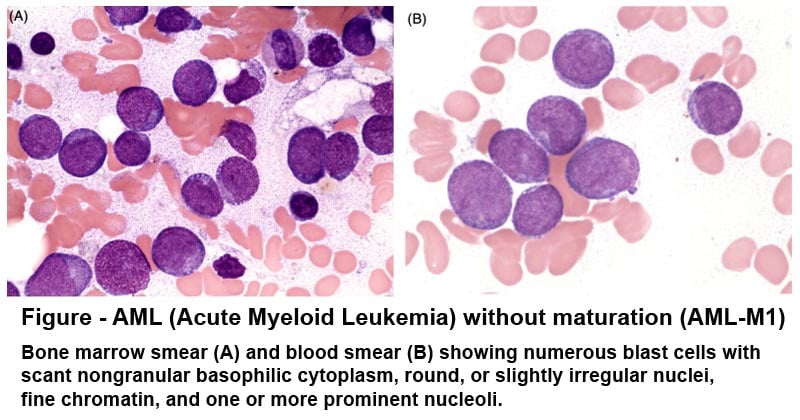
- To study hematological pathologies such as defects in the myeloblasts.
- It can also detect defects associated with abnormal mitochondria.
- It is used to stain granulocytes (eosinophils, mast cells), promyelocytes, and myeloblasts.
- Oil Red O is used in forensic pathology for fingerprinting.
- Sudan staining dyes are used for printing fabrics and garments
References and Sources
- https://www.clinisciences.com/en/buy/cat-sudan-black-b-stain-3954.html
- https://www.clinisciences.com/en/buy/cat-sudan-black-b-stain-3954.html
- https://webpath.med.utah.edu/HISTHTML/MANUALS/SUDANF.PDF
- https://www.pulsus.com/scholarly-articles/sudanese-traditional-stains-for-staining-some-biological-samples.html
- https://www.urmc.rochester.edu/urmc-labs/pathology/stainsmanual/index.html?SUDANBLACKBFORPHOSPHOLIPIDSINPARAFFINSECTIONSANDFATSINFROZENSECTIONS
- https://en.wikipedia.org/wiki/Sudan_II
- https://en.wikipedia.org/wiki/Sudan_III
- https://en.wikipedia.org/wiki/Oil_Red_O
- https://en.wikipedia.org/wiki/Sudan_stain

God bless you