Interesting Science Videos
Spermatogenesis
Spermatogenesis is the process of formation of mature sperm cells through a series of mitotic and meiotic divisions along with metamorphic changes in the immature sperm cell.
- It is the male version of gametogenesis which results in the formation of mature male gametes.
- In mammals, this takes place in the seminiferous tubules of the male reproductive system. Spermatogenesis requires optimal conditions to occur and is essential for sexual reproduction.
- The complete process of spermatogenesis occurs in different stages that take place in different structures within the male reproductive system.
- It begins in the seminiferous tubules within testes and then continues into the epididymis where maturation of the male gamete occurs, and they are further stored under ejaculation.
- The location of the testes/scrotum is critical as a lower temperature (usually 1-8°C lower than the average human body temperature) is essential for the process of spermatogenesis.
- Spermatogenesis begins in male after puberty and it is continued throughout life. Even though sperms are continuously being formed in the testes, not all areas of the testes can form sperm at the same time.
- It takes as long as 74 days for an immature germ cell to develop into a mature male gamete, and during that time, there are many intermittent resting stages.

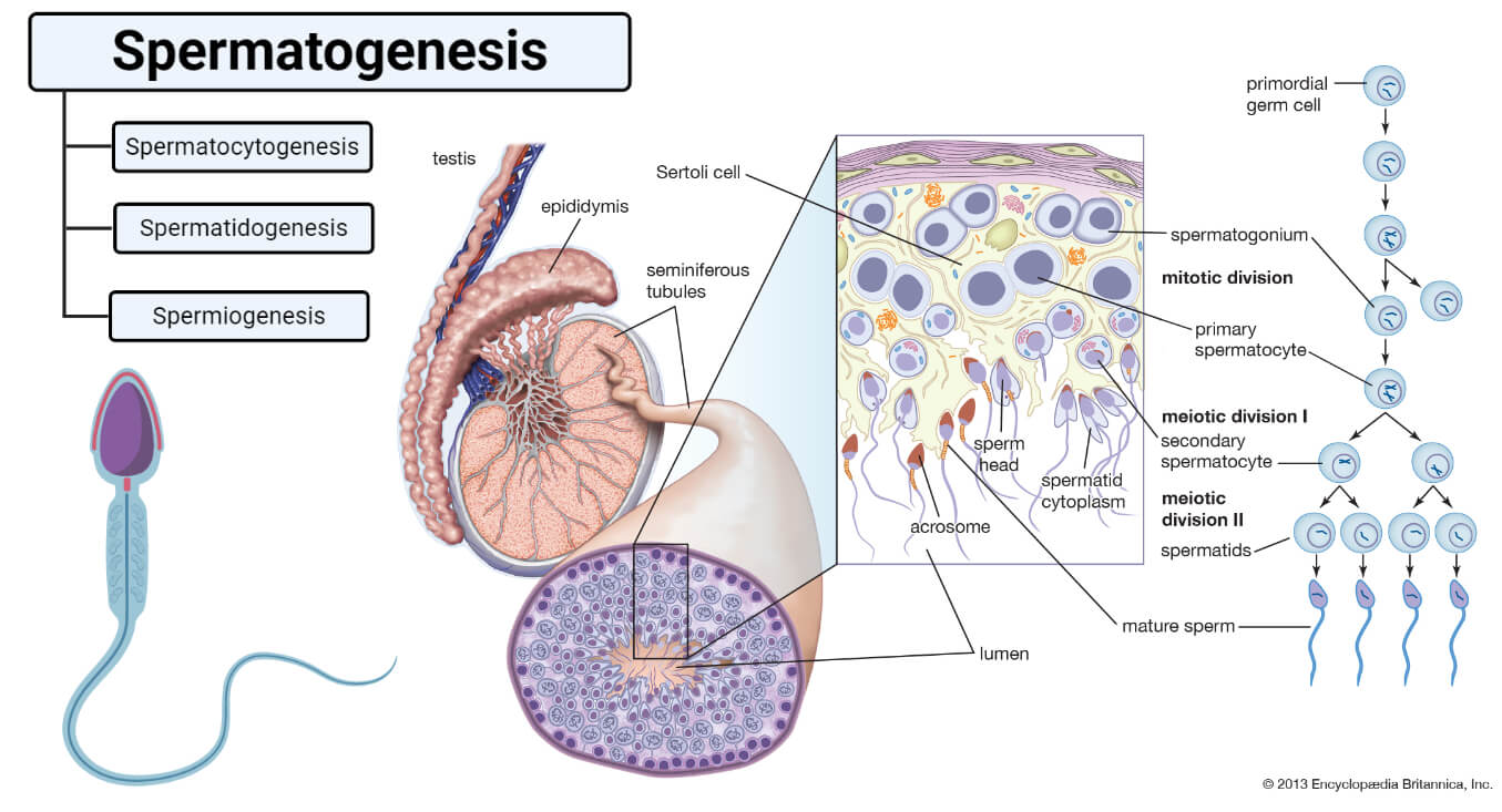
Image Source: Britannica, Created with BioRender.com.
The process of spermatogenesis can be broken into the following stages:
A. Spermatocytogenesis
- Spermatocytogenesis is the first stage of spermatogenesis which involves the division of single diploid cells into four haploid spermatocytes.
- The testis is composed of numerous tightly coiled tubules called seminiferous tubules which are lined with stem cells. The immature cells called spermatogonia are formed from these stem cells.
- The stem cells divide mitotically of which, the first half develop to form sperm cells, whereas the rest remain as stem cells to provide a continuous flow of stem cells in the tubules.
- These cells then migrate towards Sertoli cells. Sertoli cells are the cells that are randomly scattered throughout the seminiferous tubules and provide nutrients to the developing spermatogonia.
- Spermatogonia that cross the barrier to the Sertoli cells enlarge to form large primary spermatocytes.
- After a resting period, these primary spermatocytes move towards the lumen of seminiferous tubes and undergo meiotic division I to produce two haploid secondary spermatocytes.
- The number of chromosomes thus reduces from 46 to 23 in each spermatocyte. The genetic diversity observed in sexual reproduction is sourced through meiotic divisions.
B. Spermatidogenesis
- Secondary spermatocytes rapidly enter meiosis II to produce haploid spermatids. As a result, four haploid spermatids are formed from single diploid spermatogonia.
- Spermatidogenesis lasts for a brief period and is barely seen in histological studies.
- Out of the 23 pairs of chromosomes in the spermatogonia, one pair is of sex chromosomes is composed of one X chromosome, which is the female chromosome, and one Y chromosome, which is the male chromosome.
- During meiotic division, the X chromosome enters into one spermatid and the Y chromosome enters into another spermatid. The sex of the zygote is then determined based on which one of these two spermatids fertilize the ovum.
C. Spermiogenesis
- Spermiogenesis is the last stage of spermatogenesis where the spermatids undergo changes in the shape and structure to form a mature sperm cell.
- The spermatids retain the structure of epithelioid cells for a short time but soon change into an elongated structure called spermatozoa.
- A spermatozoon consists of a head and a tail. The tail is formed of microtubules which collectively form an axoneme and a large number of mitochondria.
- The genetic material within the spermatozoon becomes highly condensed and is packed within the head. About two-thirds of the head is surrounded by a thick cap called the acrosome.
- The acrosome is formed mainly of Golgi Body and contains enzymes like hyaluronidase and other powerful proteolytic enzymes that later help the sperm to fertilize the ovum.
- Under the influence of testosterone, spermatozoa gain maturity. The excess cytoplasm and other organelles are phagocytosed by the Sertoli cells.
- The mature spermatozoa are now released from the Sertoli cells into the lumen of the seminiferous tubules, but they still lack motility.
- The non-motile spermatozoa then enter the epididymis with the help of testicular fluid secreted by the Sertoli cells and peristaltic contraction.
- In the epididymis, mature spermatozoa gain motility and are then stored until the next ejaculation.
Read Also: 18 Differences Between Spermatogenesis and Oogenesis
References
- Hall JE and Guyton AC. (2011) Textbook of Medical Physiology. Twelfth Edition. Elsevier Saunders.
- Waugh A and Grant A. (2004) Anatomy and Physiology. Ninth Edition. Churchill Livingstone.
- Marieb EN and Hoehn K. (2013) Human Anatomy and Physiology. Ninth Edition. Pearson Education, Inc.
- Yasuzumi, G., Tanaka, H., & Tezuka, O. (1960). Spermatogenesis in animals as revealed by electron microscopy. VIII. Relation between the nutritive cells and the developing spermatids in a pond snail, Cipangopaludina malleata Reeve. The Journal of biophysical and biochemical cytology, 7(3), 499–504. https://doi.org/10.1083/jcb.7.3.499
- https://www.britannica.com/science/spermatogenesis
- https://en.wikipedia.org/wiki/Spermatogenesis
