Rhizopus spp are saprophytic fungi found on plants and it is parasitic on animals.
- They are multicellular fungi, with about 8 species.
- They are responsible for causing an opportunistic infection known as invasive mucormycosis in humans and animals.
- Some species are used for industrial importance such as Rhizopus stolonifer which caused bread mold.
- Rhizopus infections may also be a complication of ketoacidosis.
- Phylogenic characterization of Rhizopus categorizes eight species Rhizopus schipperae, Rhizopus delemar, Rhizopus microsporus, Rhizopus caespitosus, Rhizopus arrhizus (Rhizopus oryzae), Rhizopus reflexus, Rhizopus homothallicus, and Rhizopus stolonifer.
- Medically important groups are Rhizopus oryzae and Rhizopus microsporus.

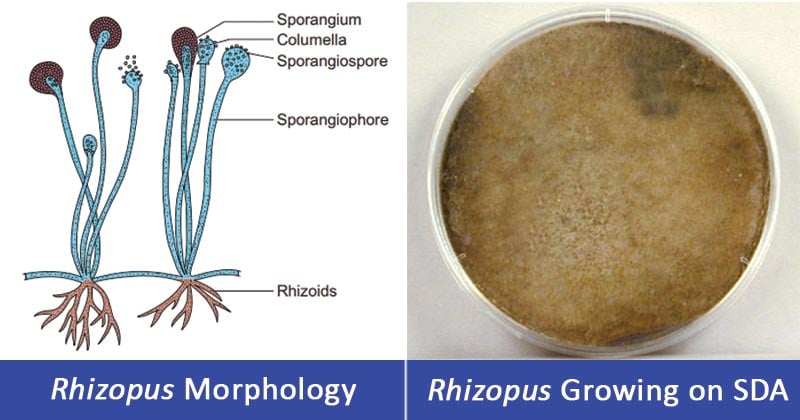
Image Source: BrainKart and Dr. Gary E. Kaiser.
Interesting Science Videos
Habitat of Rhizopus spp
- This species is a cosmopolitan group of fungi.
- It is found on a wide variety of surfaces, on organic substances like soil, decaying fruits, grown fruits and vegetables, jellies, tobacco, peanuts, leather, bread, and syrups.
- They are also found in animal feces
Morphological and Cultural Characteristics of Rhizopus spp
- Rhizopus species grow as filamentous, with branching hyphae which are coenocytic (multinucleated).
- The branched hyphae are of three types: stolons, pigmented rhizoids, and unbranched sporangiophores.

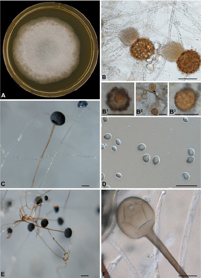
Figure: Macroscopic and microscopic morphology of Rhizopus microsporus CBS 700.68. A: Colony on MEA after 2 days incubation at 30 °C; B: Zygospores with unequal suspensors; B1–B3. Azygospore from CBS 344.29. C: Columella; D: Sporangia; E: Rhizoids; F: Columella with a collar. Scale bar = 10 μm. Image Source: Springer Nature.
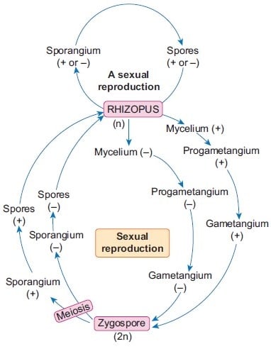
- They reproduce both sexually and asexually.
- Asexual reproduction produces sporangiospores which are formed in a spherical structure known as a sporangium.
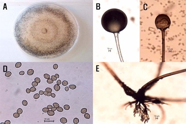
- The sporangiospores are globose to ovoid, single-celled, hyaline to brown, and striate.
- The sporangia are produced in large numbers, which are dark, and are formed on a long stalk conidiophore known as a sporangiophore.
- The sporangiophores arise from root-like rhizoids, and they rounded, responsible for producing numerous multinucleated spores.
- Sexual reproduction produces dark zygospores formed when two compatible mycelia fuse.
- Zygospores germinate forming genetically different offsprings from the parents.
- Colonies are fast-growing, covering the surface of the agar.
- Rapidly growing colonies fade from white to dark during sporulation.
- The colonies have a dense cottony growth or candy flossy or fairly floss in texture.

Figure: Life cycle of Rhizopus. Image Source: BrainKart
Pathogenesis of Rhizopus spp
- Rhizopus spp causes infections in animals, humans, and plants.
- Human infection by Rhizopus spp is known as mucormycosis also known as zygomycosis.
- Plant diseases are collectively known as rots affecting harvested fruits (strawberries), tomatoes, sweet potatoes, tobacco, papayas, and stone fruits.
Pathogenesis in human and animal host: Murcomycosis/Zygomycosis
- The infection is caused by Rhizopus arrhizus, Rhizopus oryzae and Rhizopus microsporus.
- The fungus is transmitted into the host via air spore inhalation which deposits in the paranasal sinuses and the lung.
- Risk factors include diabetes mellitus, malnutrition, malignancies (lymphomas and leukemias), renal failure, organ transplant, burns, immunosuppressive therapy, cirrhosis, and AIDS.
- Patients with diabetic ketoacidosis and patients on dialysis who receive treatment with iron chelator deferoxamine are also more susceptible to mucormycosis.
- The most common form of mucormycosis is rhinocerebral mucormycosis.
Clinical manifestation of mucormycosis/zygomycosis
- The infection is characterized by the invasion of blood vessels in humans and animals.
- The infection then progresses to other body areas including the lungs and the brains.
- Generally, these infections are broken down into five presentations: rhinocerebellar, pulmonary, cutaneous, gastrointestinal, and disseminated.
Manifestations include:
- Sinus infection (sinusitis) characterized by fever, headaches, nasal congestion, nasal discharge, and sinus pain.
- Dissemination to out part of the sinus characterized by necrosis of the mouth palate, bone and cartilage disintegration causing division of the nostril septum, swelling of the nose area, erythema of the skin and eye socket.
- Bluing of the sinus and eye sockets due to lack of oxygen (cyanosis)
- Blurred vision and extended necrosis which can damage the facial structure.
- Dissemination to brain characterized by lethargy, seizures, slurred speech, partial paralysis, abnormalities of the nerves of the face and eyes (cranial neuropathies), a brain abscess, altered consciousness, and coma.
- Paralysis of eye muscles, known as ophthalmoplegia may also occur.
- The involvement of the sinus and the brain is known as rhinocerebral mucormycosis.
- Pulmonary mucormycosis is a progressive infection of the respiratory system.
- It is associated with the involvement of the respiratory system characterized by fever and an unproductive cough with spitting or hemoptysis, chest pain, and difficulty breathing (dyspnea).
- Cutaneous mucormycosis is characterized by skin inflammation and hardening involving underlying tissues. Skin redness, with painful swelling, also occurs.
- Ulceration also occurs with blistering and tissue necrosis causing the skin to turn black.
- Cutaneous mucormycosis can be severe and fulminant.
- Gastrointestinal infection is uncommon but not rare.
- It is characterized by abdominal pain and vomiting of blood (hematemesis).
- Lesions form in the stomach or intestines.
- Inflammation of the peritonitis, abdominal lining, and abdominal organs can occur.
- Severe bowel pain due to lack of blood flow (bowel infarctions) and hemorrhagic shock.
- Disseminated mucormycosis occurs in immuno-compromised.
- It is rare but when it occurs, it causes a severe infection that spreads to other body parts affecting the brain, heart, spleen, skin, and other organs.
- Rarely it affects the kidney, heart chambers, and valves and bones.
Laboratory Diagnosis of Mucormycosis (Rhizopus spp)
Specimen: Sputum, pus/tissue biopsies, blood/serum, transbronchial biopsy, sinuses biopsy, gastrointestinal tissue biopsy, skin biopsy
Microscopic Examination
- 10-20% KOH wet mount to observe nonseptate or sparsely septate broad hyphae, sporangiophores, root-like hyphae (rhizoids), sporangia, sporangiospores.
- Observe brown colored unbranched sporangiospores occurring in clusters at the tips of the hyphal tubes.
- Singly occurring spores may also be observed which are ovoid-shaped, hyaline to brown colored.
- The spores are rounded with flattened bases.

Figure: Morphological characteristics of Rhizopus oryzae isolated from orange. A: Colony on PDA after 7 days of inoculation, B: Sporangium and sporangiophore, C: Columellum, D: Sporangiospores, E: Rhizoid. Image Source: Korea Institute of Science and Technology Information.
Cultural Examination
- Sabouraud Dextrose Agar (SDA) with cycloheximide at 37°C produces a characteristic dense cottony-fluffy growth with a flossy texture.
- Macroscopic observation of the color of the colonies is initially white and turns grey to yellowish-brown as culture matures, Reverse pigmentation is white to pale
Histological examination
- Using Hematoxylin and eosin stain (H&E stain or HE stain) to observe for wide, thin-walled, hyaline septate or nonseptate hyphae in tissue samples. The hyphae are also observed to be non-parallel and branched
Molecular Diagnosis
- PCR and genomic sequencing for phylogenic identification and differentiation of the Rhizopus specific species.
Differential diagnosis
- Differentiating Pulmonary mucormycosis from other fungal infections such as aspergillosis can also assist in the identification of causative agents.
- This is done during the physical and symptomatic examination of patients inclusive with laboratory diagnosis.
Treatment of Mucormycosis
- Mucormycosis treatment involves combined antifungal treatment along with the surgical procedure of the involved tissues.
- Intravenous administration of amphotericin B is the drug of choice.
- Posaconazole or isavuconazole can be used as second line of tretament after amphotericin B.
- Adjunctive treatment with hyperbaric oxygen is given in combination with primary therapy.
- Other antifungal drugs include voriconazole, fluconazole, and flucytosine can be used depending o the causative agent of mucormycosis.
- Treatments for underlying conditions are mandatory with close observation of patients for contraindications.
Selected species of Rhizopus and industrial uses
There are 8 known species of Rhizopus of which 3 are of industrial and medical importance and they include:
- Rhizopus microsporus var. oligosporus
- Rhizopus oryzae
- Rhizopus stononifer
Rhizopus microsporus var oligosporus
- It is used to produce fumaric acid, lactic acid, and cortisone.
- It is used to make fermented soybeans known as tempeh.
Rhizopus oryzae or Rhizopus arrhizus
- It is used in the production of lactic acid and cortisone.
- It is also used for alcoholic fermentation
- Its also used for the biosorption of heavy metals which is the passive adsorption of chemical contaminants by organisms.
Rhizopus stononifer
- It is also known as black bread mold.
- It causes fruit rot on strawberry, tomato, and sweet potato (soft rot).
- It is used in the commercial production of fumaric acid, lactic acid, and cortisone.
References and sources
- https://mycology.adelaide.edu.au/descriptions/zygomycetes/rhizopus/
- https://www.britannica.com/science/Rhizopus
- drfungus/knowledgebase/rhizopus
- https://en.wikipedia.org/wiki/Rhizopus
- https://www.uptodate.com/contents/mucormycosis-zygomycosis/print
- https://www.ncbi.nlm.nih.gov/books/NBK544364/
- https://www.sciencedirect.com/science/article/pii/B012227055X011299

can i get the complete physiology and morphological characteristics of rhizopus stolonifer specifically