Interesting Science Videos
What is phagocytosis?
- Phagocytosis is a process by which cells ingest large particles (> 0.5 micrometers) into membrane-bound vesicles called phagosomes, which are then targeted to the lysosomes for enzymatic degradation.
- Phagocytosis is greatly enhanced by the opsonization of the bacteria.
- Attachment of a specific IgG or complement fragment, C3, can greatly enhance the efficiency of phagocytosis, although phagocytosis can occur without opsonization.
- Complement receptors, CR1 and CR3, are the primary receptors for opsonization by the complement.
- The Polymorphonuclear Leukocytes (PMNs) also express receptors for IgG fragment Fc (FcγRs) that facilitates phagocytosis.
- The two most prominently expressed Fcγ receptors on circulating PMNs are known as FcγRII (CD32) and FcγIII (CD16), and binding to these receptors triggers the oxidative burst in PMNs.
- The binding of antibody and complement receptors at the PMNs surface activates the phagocytic process.

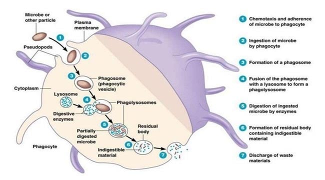
Image Source: SlideShare, Credit: Pearson Inc.
Mechanism of Phagocytosis
Destruction of the Microbes
The following are important factors that help destroy microorganisms within a phagolysosome:
Oxygen Radicals: A complex of proteins called phagocyte oxidase in the membrane of a phagolysosome generates oxygen radicals in the phagosome. A single electron is taken from NADPH and added to oxygen, partially reducing it. The resulting highly reactive molecules react with proteins, lipids and other biological molecules.
Nitric Oxide: Nitric oxide synthase synthesizes nitric oxide, a reactive substance that reacts with superoxide to create further molecules that damage various biological molecules.
Anti-Microbial Proteins: Lysosomes contain several proteases, including a broad spectrum enzyme, elastase, which is important or even essential for killing various bacteria. Another anti-microbial protein is lysozyme, which attacks the cell walls of certain (gram-positive) bacteria.
Anti-Microbial Peptides: Defensins and certain other peptides attack bacterial cell membranes. Similar molecules are found throughout much of the animal kingdom.
Binding Proteins: Lactoferrin binds iron ions, which are necessary for the growth of bacteria. Another protein binds vitamin B12.
Hydrogen Ion Transport: Transporters for hydrogen ions (a second role of the oxidase) acidify the phagolysosome, which kills various microorganisms and is important for the action of the proteases described above.
In addition to destroying the microorganism, phagocytes also release molecules that diffuse to other cells and help coordinate the overall response to an infection.
Regulatory molecules that regulate an immune response are called cytokines. Most are small proteins and are mainly released by white blood cells and their relatives, such as macrophages.
Several types of cells in the immune system engulf microorganisms via phagocytosis.
Neutrophils: Neutrophils are abundant in the blood, quickly enter tissues, and phagocytize pathogens in acute inflammation.
Macrophages: Macrophages are closely related to monocytes in the blood. These longer-lived cells predominate in chronic inflammation. They also release some important inflammatory paracrine.
Dendritic Cells: Phagocytosis in these cells is important for the elaboration of a specific immune response rather than for directly destroying the pathogens.
B Lymphocytes: A small amount of phagocytosis in these cells is often necessary in order for them to develop into cells that release antibodies.
- The PMNs have two broad types of killing mechanisms.
- One is oxygen-dependent and the other is oxygen-independent.
- Phagocytosis of microbes stimulates the production of superoxide radicals and other reactive oxygen species.
- These are potent microbicidal agents and include, among others, hydrogen peroxide and chloramines.
- The enzyme NADPH oxidase is found in the cell membrane of the PMNs and generates the superoxide (called the respiratory burst).
- The superoxide is unstable and quickly dismutates to hydrogen peroxide and other substances
that are microbicidal. - These reactions take place inside the phagolysosome (also called phagosome).
Steps in the phagocytosis of a bacterium
- Bacterium becomes attached to membrane evaginations called pseudopodia.
- The bacterium is ingested, forming a phagosome.
- Phagosome fuses with a lysosome.
- The bacterium is killed and then digested by lysosomal enzymes.
- Digestion products are released from a cell.
Example: Macrophage phagocytizing a virus.
- The virus and the cell need to come into contact with each other.
- The virus binds to the cell surface receptors on the macrophage.
- The macrophage starts to invaginate around the virus, engulfing it into a pocket.
- The invaginated virus becomes completely enclosed in a bubble-like structure, called a “phagosome”, within the cytoplasm.
- The phagosome fuses with a lysosome, becoming a “phagolysosome”.
- Phagolysosome lowers the pH to break down its contents.
- Once the contents have been neutralized, the phagolysosome forms a residual body that contains the waste products from the phagolysosome.
- The residual body is eventually discharged from the cell.
