Opsonization definition
- The term opsonization refers to the capacity of antibodies and complement components (as well as other proteins) to coat dangerous antigens that can then be recognized by antibodies or complement receptors on phagocytic cells.
- Opsonization is the molecular mechanism whereby molecules, microbes, or apoptotic cells are chemically modified to have stronger interactions with cell surface receptors on phagocytes and antibodies.
- This is the mechanism of identifying invading particles (antigens) by the use of specific components called opsonins.
- The opsonins act as markers or tags that allow recognition by the immune system of the body.
- An opsonin is any molecule that enhances phagocytosis by marking an antigen for an immune response or marking dead cells for recycling.
- The purpose of opsonization is to make the antigens palatable to the antibody or the phagocytic cells.
Mechanism of Opsonization
- Opsonization of pathogens can occur via antibodies or the complement system.
Antibody-mediated Opsonization
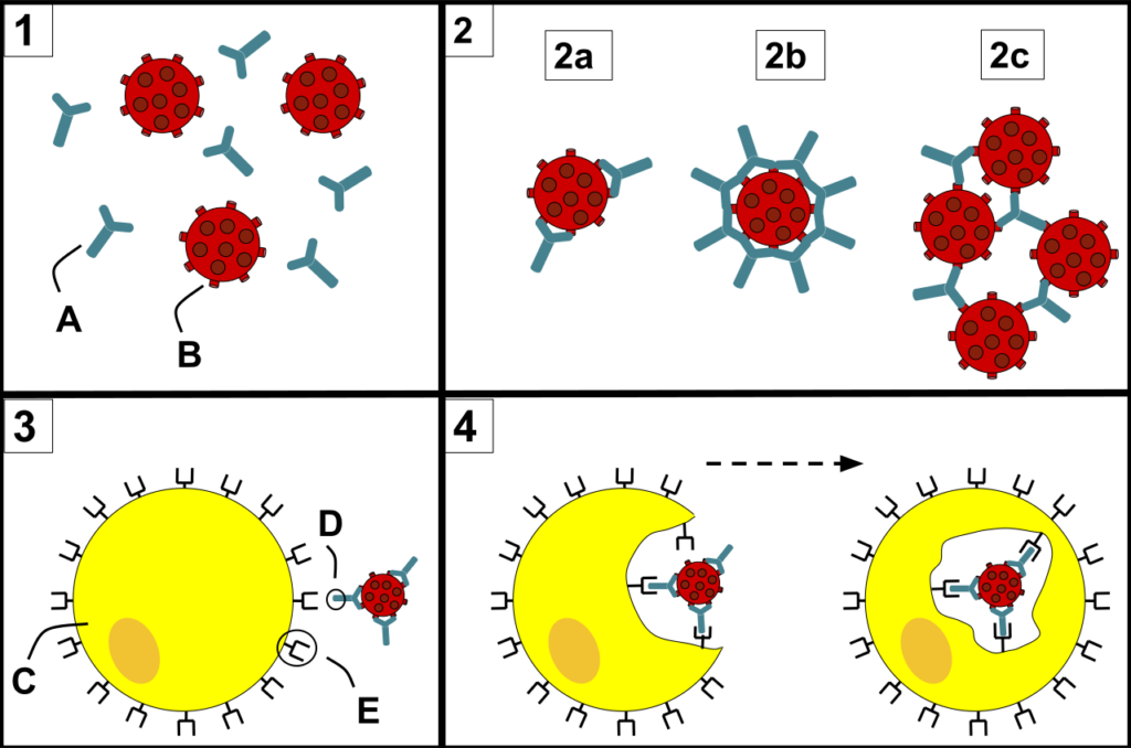
Figure: 1) Antibodies (A) and pathogens (B) free roam in the blood. 2) The antibodies bind to pathogens and can do so in different formations such as opsonization (2a), neutralization (2b), and agglutination (2c). 3) A phagocyte (C) approaches the pathogen, and the Fc region (D) of the antibody binds to one of the Fc receptors (E) on the phagocyte. 4) Phagocytosis occurs as the pathogen is ingested. Source: Wikipedia.
- The mechanism of opsonization is employed by antibodies in order to inhibit and clear infection.
- Antibody-mediated opsonization by antibodies involves the coating of pathogens with antibodies so that they are recognized and phagocytosed by innate immune cells.
- Encapsulated bacteria that resist phagocytosis become extremely attractive to neutrophils and macrophages when coated with antibody and their rate of clearance from the bloodstream is strikingly enhanced.
- As the structure of immunoglobulins was deciphered, it was apparent that IgG is the heat-stable serum factor responsible for antigen-specific opsonization.
- IgG combines by its two antigen-binding pieces, Fab, with antigenic determinants on the surface of the microorganism (or another particle).
- Upon combination with antigen, the IgG molecule undergoes specific conformational and configurational changes in the F(ab)2 hinge region.
- Phagocytic cells of all types have receptors for the IgG molecule on their plasma membranes.
- The number of these receptors on each mouse peritoneal and alveolar has been estimated at 1-2 million. These receptors are resistant to tryptic proteolysis and mediate binding of IgG-coated particles at 4°C as well as at 37°C and in the absence of divalent cations.
- Even though all four subclasses of human IgG bind to the antigen, only IgG1, and IgG3 are capable of binding to receptors on phagocytic cells.
- The phagocytic cell’s receptors for IgG bind only to the Fc portion of the molecule, and are therefore known as Fc receptors.
- In the case of antibody-mediated opsonization, binding of a pathogen (antigen)-antibody complexes to an Fc receptor on phagocytes will induce internalization of the complex and internal digestion of the pathogen in lysosomes.
- Generally, multiple antibodies bind to various sites on the antigen, increasing the chance and efficiency in which the pathogen is engulfed in the phagosome and destroyed by lysosomes.
Complement-mediated Opsonization
- The complement system is composed of over 30 proteins that improve the ability of antibodies and phagocytic cells to fight invading organisms.
- It initiates phagocytosis by opsonizing antigens. This system is also responsible for enhancing inflammation and cytolysis.
- The most critical heat-labile opsonin, and perhaps the most essential opsonin of all, is C3b (C3b is the fragment of C3 that binds to particles when C3 is cleaved by a C3-convertase).
- Either through the classical pathway (initiated with binding of IgG or IgM molecules to antigen, which results in binding and activation of the C 1 complex) or the alternate pathway (initiated by the presence of lipid-carbohydrate complexes found in the cell wall of bacteria ), C3 is cleaved into C3a and C3b.
- It is C3b that binds to the surface of the particle and serves as an opsonin.
- Once a particle is coated with C3b, it must then be recognized by and bound to, the surface of a phagocytic cell before it can be ingested.
- The mechanisms by which these events occur have been partially characterized. All mononuclear phagocytes and polymorphonuclear leukocytes thus far studied have receptors on their plasma membranes for C3b.
- However, the binding of C3b-coated particles to C3b receptors of some cells requires the presence of divalent cations in the medium.
- The activated macrophages then ingest particles coated with C3b.
- Besides, in microorganisms like Hemophilus influenza, C3b cleaves the aromatic dipeptides present in the neutrophils as C3b has an enzymatic activity for aromatic dipeptides.
- The cleavage of aromatic dipeptides on the neutrophil’s plasma membrane is considered as a means by which C3b mediates phagocytosis of particles to which it is bound.
Opsonins and Types
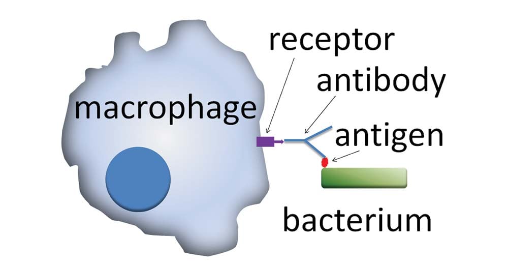
Figure: Action of opsonins: A phagocytic cell recognizes the opsonin on the surface of an antigen. Source: Wikipedia.
- An opsonin is any molecule that enhances phagocytosis by marking an antigen for an immune response or marking dead cells for recycling.
- Opsonin molecules usually bind, on one end, to the receptors present in the antigen and, on the other end, to the receptors on the phagocytes.
- This binding results in different mechanisms that ultimately lead to the destruction or removal of the particular antigen.
- Opsonin molecules ensure that the binding of the antigen to the immune cells is greatly enhanced.
- Opsonins have important roles in the immune system like marking of dead and dying cells for clearance by macrophages and neutrophils.
- Besides, opsonins also aid in activating the complement proteins and destruction of cells by natural killer (NK) cells.
- The mechanisms utilized by opsonins to enhance the kinetics of phagocytosis involve favoring the interaction between opsonin and cell surface receptors on the immune cells.
- These molecules override the negative charges on the cell membrane, which make it difficult for two cells to come close together.
Types
- Opsonins involved in the immune system include the following molecules:
Antibodies
- Antibodies are the molecules of the adaptive immunity that are released by B cells as an immune response.
- In the case of IgG antibodies, the presence of the Fc domain allows the binding of receptors on the phagocytes to the Fc domain while the Fab domain of the antibody binds to the antigen.
- IgM antibodies, however, lack the Fc receptors and thus, are ineffective in enhancing phagocytosis. But they are highly effective in activating the complement system and are considered as an opsonin.
- The binding of antibodies to the antigen and the immune cells results in the release of lysis products from the effector cells.
Complement proteins
- Among the various complement proteins, C3b, C4b, and C1q are the common proteins that also serve as opsonins.
- C3b is by far the most effective opsonin that initiates phagocytosis as it can be recognized by phagocyte receptors.
- One added advantage with complement proteins acting as opsonin is that Complement receptor 1 is expressed in all phagocytes and recognizes several complement proteins like C3b and C4b.
- C1q is a part of the C1 complex which interacts with the Fc region of antibodies and performs as an opsonin.
Circulating proteins
- A number of circulating proteins like pentraxins, collectins, and ficolins also serve as opsonins.
- These proteins are the PRRs (pattern recognizing receptors) capable of coating the microbes as an opsonin, which enhances the activity of neutrophils by several mechanisms.
- A significant property of these proteins is the ability to bind in a Ca‐dependent fashion, as a pattern recognition molecule, to several microorganisms that contain phosphorylcholine in their membranes which then activates the complement system.
- Collectins like Mannose Binding Lectin (MBL) is the basis for the lectin pathway of complement activation.
- Ficolins, in turn, typically recognize N‐acetylglucosamine residues in complex‐type carbohydrates in addition to other ligands on different antigens.
Examples
The common opsonins are:
- IgM antibodies
- IgG antibodies
- C3b proteins
- C4b proteins
- C1q proteins
- Pentraxins
- Collectins
- Ficolins
- Mannose-binding lectin (MBL)
References
- Peter J. Delves, Seamus J. Martin, Dennis R. Burton, and Ivan M. Roitt(2017). Roitt’s Essential Immunology, Thirteenth Edition. John Wiley & Sons, Ltd.
- Judith A. Owen, Jenni Punt, Sharon A. Stranford (2013). Kuby Immunology. Seventh Edition. H. Freeman and Company
- Thau L, Mahajan K. Physiology, Opsonization. [Updated 2020 Mar 25]. In: StatPearls [Internet]. Treasure Island (FL): StatPearls Publishing; 2020 Jan-. Available from: https://www.ncbi.nlm.nih.gov/books/NBK534215/
- Griffin F.M. (1977) Opsonization. In: Day N.K., Good R.A. (eds) Biological Amplification Systems in Immunology. Comprehensive Immunology, vol 2. Springer, Boston, MA
Sources
- 4% – https://www.ncbi.nlm.nih.gov/books/NBK534215/
- 2% – https://patents.google.com/patent/US20040013673A1/en
- 2% – https://makkypedia.blogspot.com/2016/06/opsonization.html
- 1% – https://www.sciencedirect.com/topics/immunology-and-microbiology/immunoglobulin-g
- 1% – https://www.sciencedirect.com/book/9780120442201/methods-for-studying-mononuclear-phagocytes
- 1% – https://www.ncbi.nlm.nih.gov/pmc/articles/PMC4139653/
- 1% – https://quizlet.com/93116857/immunology-chapters-2-and-3-flash-cards/
- 1% – https://quizlet.com/74073384/ap-ii-chpt-21-the-immune-system-flash-cards/
- <1% – https://www.sciencedirect.com/topics/immunology-and-microbiology/opsonization
- <1% – https://www.sciencedirect.com/science/article/pii/S0006349500766021
- <1% – https://www.ncbi.nlm.nih.gov/pmc/articles/PMC4559511/
- <1% – https://www.ncbi.nlm.nih.gov/pmc/articles/PMC4019044/
- <1% – https://www.ncbi.nlm.nih.gov/pmc/articles/PMC344229/
- <1% – https://quizlet.com/187777219/lectin-pathway-classical-and-rest-of-chapter-3-flash-cards/
- <1% – https://deepblue.lib.umich.edu/bitstream/handle/2027.42/25474/0000014.pdf?sequence=1


