Mucosa, or mucous membrane, is a thick, soft tissue lining that forms a protective layer for internal organs of the body, body canals including orifices such as nostrils, ears, lips, urethral opening, anus, etc.
They are mainly associated with the secretion of a fluid-like, thick, lubricating substance known as mucus, a mucopolysaccharide consisting of mucin, which is secreted by specialized mucus-secreting cells in the body.
Interesting Science Videos
Structure of Mucous Membrane
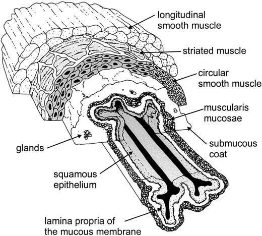
A mucosal membrane is characterized by the presence of several layers of cells and tissue varying with location but exhibits a general configuration applicable to most of the locations in the body.
The typical structure of mucosal membrane includes the following three basic structural sub-layers:

Epithelial Layers
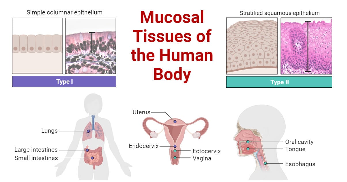
It is the outermost non-keratinized covering of mucous layer, consisting of, stratified or even simple squamous epithelial cells varying with location. It serves as a protective barrier against pathogens, irritants, and foreign particles. Their arrangement may vary with the location of the body. Some epithelial cells are equipped with cilia responsible for the removal of dust and other foreign particles, the process known as mucociliary clearance.
Lamina Propria
Lamina propria, a thin layer of connective tissue is located immediately beneath the epithelial layers, comprising blood vessels, lymphatic vessels, and mucus-secreting cells. It is responsible for structural integrity and offers a wide range of immune cells for the destruction of invading pathogens.
Muscularis Mucosae
It is a smooth muscle layer situated beneath lamina propria responsible for the movements of the mucosal layer, aiding in secretion and the process of absorption.
Along with three major layers, goblet cells, special secretory cells are also present in the mucosal layer, protecting and lubricating the mucous membrane. They are also responsible for trapping and washing away particulate matter such as dust particles and other foreign substances.
Locations in the Body
They are found lining the body cavities, which majorly include:
- Respiratory system:
It includes nasal, olfactory, and bronchial mucosa. They are abundant in regions of the nostrils and throat.
- Gastrointestinal tract:
It includes oral, esophageal, gastric, and intestinal mucosa found in the mouth, tongue, esophagus, and intestinal region.
- Urogenital system:
It includes urethral and vaginal mucosa found in the genitals.
- Ocular mucosa:
It is found in the region of the eyes.

Functions of Mucous Membrane
General functions include:
- Secretion
- Protection
- Absorption
- Primary line of defense: The primary and foremost function of the mucosal layer is to protect our body as it forms the first line of defense of our immune system. They form a barrier against foreign particles clearing them out by trapping them in the sticky secretion. The lubrication prevents any mechanical injury. Also, immune cells and naturally present antibiotics defend against invading pathogens.
- Oral mucosa: It forms a lining over the soft palate and floor of the mouth. It also provides a surface for chewing.
- Esophageal mucosa: It lines the inner cavity of the esophagus preventing it from abrasion.
- Gastric mucosa: It produces mucus, digestive enzymes, and the cells that stimulate acid production to break down food in the stomach. It also protects the stomach and bladder from the abrasive effects of highly acidic (in the stomach) or alkaline environments.
- Intestinal mucosa: It forms a gut lining aiding in the absorption of nutrients and water from foods.
Mucosal Immunity
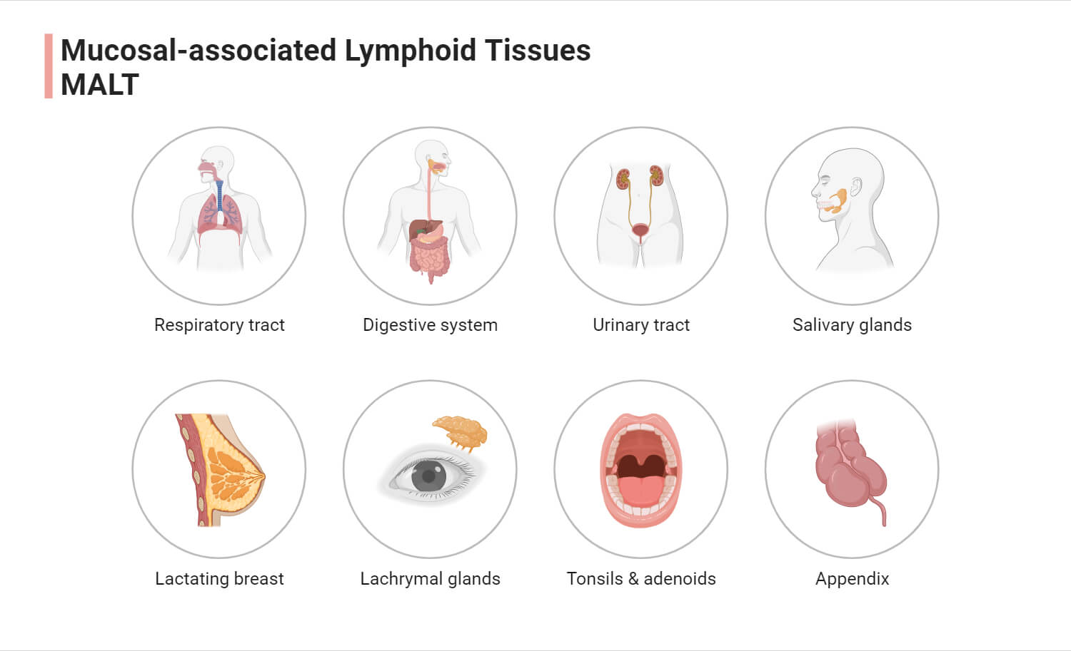
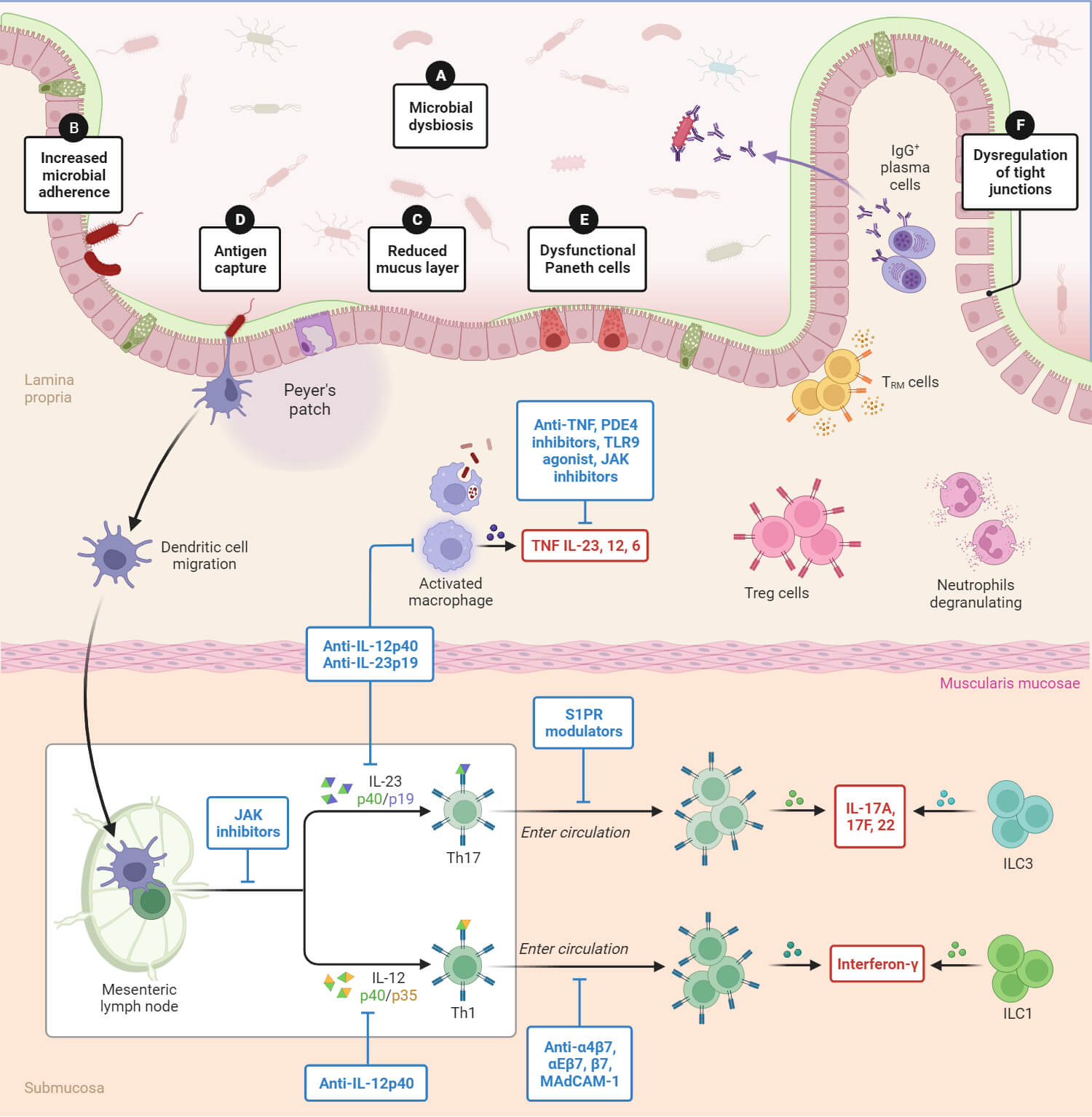
Mucosal immunity, also known as mucosal-associated lymphoid tissue (MALT), is a part of the immune system of the body that provides defense at the surface of the mucous membrane protecting the host from potentially harmful pathogens in the environment. It is associated with mucosal sites such as gut mucosa comprising Peyer’s Patches (PPs), intestinal lamina propria, cryptopatches, and lymphoid follicles. It can be broadly categorized based on location as gut-associated lymphoid tissue (GALT), nasopharynx-associated lymphoid tissue (NALT), bronchus-associated lymphoid tissue (NALT), conjunctiva-associated lymphoid tissue (CALT), lachrymal duct associated (LDALT). IgA cells are the most common B cells involved in the immunity exhibited by the mucosal layer. MALT majorly functions to produce and secrete these IgA antibodies across the surface of the mucosa. Also, the T cells of thymic origin play a role in immunity majorly associated with the regulation of differentiation of IgA precursors. IgA can neutralize food antigens along with the inhibition of commensal and bacterial pathogens.

Common disorders affecting mucous membranes
- Oral and genital herpes
- oral thrusts and genital yeast infections
- Genital and oral lichen planus
- Oral bullous pemphigoid
- Oral pemphigus
- Mouth sores
- Mononucleosis
- Oropharyngeal HPV infection
- Hand, foot and mouth disease
- H. pylori infection
- Infections and inflammations (e.g., gingivitis, bronchitis)
- Allergic reactions involving mucous membrane
- Autoimmune diseases affect mucosa.
Mucosal Health
Since the mucosal membrane is responsible for providing you with the first line of defense against various types of infection, mucosal health is highly essential to maintaining the healthy status of the body. It can be well-maintained with proper hydration and nutrition and avoiding potential irritants.

Comparisons with Other Tissues
With serous membranes
| Character | Mucous membrane | Serous membrane |
| Location | It lines the cavities and structure that open directly to the environment | It lines the closed cavities in the body that do not open directly to the outside environment |
| Epithelial layer | Stratified or simple squamous epithelium | Usually, simple squamous epithelium |
| Secretions | Goblet cells secrete mucus | Epithelium secretes serous fluid |
| Function | It acts as a protective layer against pathogens, irritants, and foreign particles; it is also responsible for mucosal immunity | Serous fluid helps in lubrication of the membrane lowering friction and abrasion during the movement of organs in the abdominal or thoracic cavity. |
With cutaneous (skin) tissues
| Character | Mucous membrane | Cutaneous membrane |
| Location | It lines cavities that are open to the outside environment. | It forms the outer surface of the body forming skin. |
| Epithelial layer | Stratified or squamous epithelium depending upon the location | Stratified squamous epithelium in the epidermis; apical surface keratinized; and a layer of connective tissue in the dermis layer of skin |
| Secretions | Mucus secretion by goblet cells | The sweat gland and sebaceous gland secrete sweat and sebum respectively |
| Function | Protection, immunity | Physical barrier, sensory receptors responsible for sensations of touch, pressure, temperature, pain |
References
- Biologydictionary.net Editors. (2017, January 31). Mucous Membrane. Retrieved from https://biologydictionary.net/mucous-membrane/. Accessed on 12/27/2023.
- Britannica. (2023, November 22). Mucous membrane. Retrieved from https://www.britannica.com/science/mucous-membrane. Accessed on 12/27/2023.
- Cesta, M. F. (2006). Normal structure, function, and histology of mucosa-associated lymphoid tissue. Toxicologic pathology, 34(5), 599-608. https://doi.org/10.1080/01926230600865531. Accessed on 12/28/2023.
- Cleveland Clinic. (2022, July 24). Mucosa. Retrieved from https://my.clevelandclinic.org/health/body/23930-mucosa. Accessed on 12/27/2023.
- Fundamentals of Anatomy and Physiology. Types of Tissues. University of Southern Queensland Pressbooks. Retrieved from https://usq.pressbooks.pub/anatomy/chapter/types-of-tissues/
- Liao, D., Zhao, J., & Gregersen, H. (2017). Esophagus. Biomechanics of Living Organs (pp. 147-167). Academic Press. https://doi.org/10.1016/B978-0-12-804009-6.00007-9
- López M.C. (2014) Mucosal immunity. Reference Module in Biomedical Sciences. Retrieved from https://doi.org/10.1016/B978-0-12-801238-3.01992-9. Accessed on 12/31/2023.
- Medicine LibreTexts. 4.3 A: Epithelial Membranes Retrieved from https://med.libretexts.org/Bookshelves/Anatomy_and_Physiology/Anatomy_and_Physiology_2e_(OpenStax)/01%3A_Levels_of_Organization/04%3A_The_Tissue_Level_of_Organization/4.03%3A_Epithelial_Tissue Accessed on 12/28/2023.
- SEER Cancer Statistics. Membranes. National Cancer Institute. Retrieved from https://training.seer.cancer.gov/anatomy/cells_tissues_membranes/membranes.html. Accessed on 12/30/2023.
- Squier, C. A., & Kremer, M. J. (2001). Biology of oral mucosa and esophagus. JNCI Monographs, 2001(29), 7-15. Retrieved from https://doi.org/10.1093/oxfordjournals.jncimonographs.a003443
