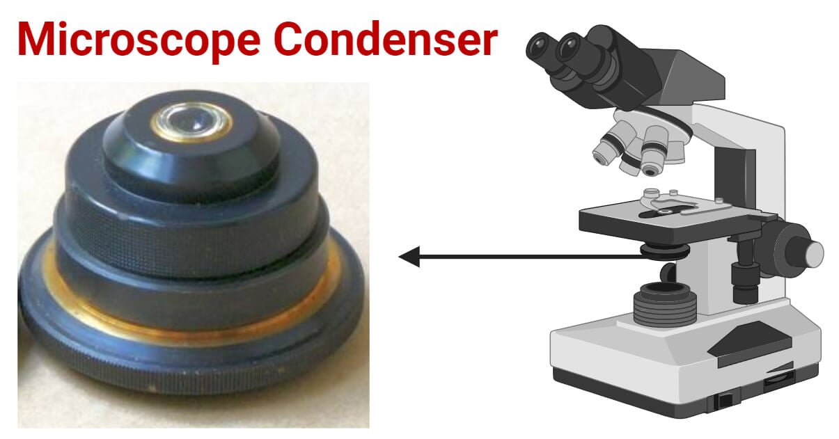
A microscope condenser is an optical component that focuses and directs light onto the specimen for observation. It contributes to the illumination and clarity of the microscopic image.
- Microscope condensers are adjustable and allow for customized height and aperture settings based on the specimen and microscopy technique for optimal lighting conditions.
- Condensers contribute to enhanced resolution and contrast and reveal finer details of the microscopic specimen.
- Condensers concentrate and direct light onto the specimen and enhance visibility and clarity in microscopic observations.

Interesting Science Videos
Basics of Microscope Condenser
The microscope condenser, a fundamental component of microscopy, is pivotal in shaping the quality of microscopic observations.
A condenser is an optical lens system that focuses and controls the illumination that passes through the specimen.
Positioned beneath the microscope stage, the condenser is crucial in directing light onto the specimen. It contributes to the overall brightness and clarity of the image.
Structure of Microscope Condenser
The condenser, a pivotal component of microscopes, exhibits a distinct physical structure that influences its functionality. Typically characterized by a cylindrical or prism-like shape, it is encased in a housing made of metal or plastic.
This protective enclosure encompasses essential adjustment features, such as knobs and screws. It enables users to fine-tune the condenser’s position and settings.
Within the condenser lies a complex lens system composed of multiple lenses, each assigned specific functions.
The intricacies of this lens system vary depending on the condenser type and microscope model, accommodating diverse microscopy techniques.
Furthermore, the condenser has essential adjustment mechanisms and controls, including height adjustment controls that regulate the distance between the condenser and the specimen.
Aperture controls modify the size of the condenser aperture, allowing for customization tailored to specific imaging requirements.
Functions of Microscope Condenser
- Concentration and Focusing of Light
- Enhancement of Image Contrast
- Improvement of Resolution
- Regulation of Numerical Aperture
- Adaptation to Microscopy Techniques
Types of Microscope Condensers
Microscope condensers are like magic lenses, each with its own tricks to make tiny worlds clearer.
Let’s uncover their powers and see how these optical marvels bring hidden details into focus!
Abbe Condenser
The Abbe condenser is a fundamental type of microscope condenser known for its simple and effective design. Typically consisting of a lens system, the Abbe condenser is positioned below the microscope stage.
It often incorporates an iris diaphragm for controlling the aperture size and allows users to adjust the amount of light reaching the specimen.
The Abbe condenser is commonly utilized in brightfield microscopy, the standard technique for observing stained or naturally pigmented specimens.
Its design aids in producing a well-illuminated, evenly lit field to enhance the contrast and visibility of specimen details.
Darkfield Condenser
The Darkfield condenser is specialized for darkfield microscopy, a technique that utilizes oblique illumination to create a bright image of the specimen against a dark background.
The Darkfield condenser achieves this by preventing direct light from entering the objective, only allowing scattered light to reach the specimen.
The condenser enhances contrast and reveals minute details in translucent or unstained specimens. It’s beneficial for observing live, unstained biological specimens.
Phase Contrast Condenser
Phase contrast microscopy is a technique designed to visualize transparent, unstained specimens that would be practically invisible in brightfield microscopy.
This method exploits the phase differences of light passing through different specimen parts.
In phase contrast microscopy, the condenser plays a critical role in transforming subtle phase differences in the specimen into visible contrast.
It achieves this by manipulating the amplitude and phase of the light passing through the specimen. It enhances the visibility and detail of transparent structures without staining or other contrast-enhancing techniques.
Adjusting and Using the Microscope Condenser
Adjusting and using the condenser in microscopy involves a meticulous process to ensure optimal imaging conditions.
Proper alignment and centering, the initial steps in this procedure, are crucial for maximizing light transmission through the specimen.
Using centering screws and alignment tools, users can fine-tune the condenser’s position under the objective.
Subsequently, adjusting the condenser height and aperture plays a pivotal role in image quality. Controlling the condenser’s height dictates the distance between the condenser and the specimen.
Simultaneously, adjusting the aperture regulates the amount and angle of light reaching the specimen, directly influencing contrast and resolution. Users can employ specific tips for optimizing condenser settings tailored to different microscopy techniques to refine microscopy outcomes further.
These adjustments, troubleshooting measures, and practical considerations empower microscopists to achieve superior imaging results across diverse applications.
Video on How to use a Microscope Condenser
Conclusion
The condenser is crucial to microscopy, shaping image quality and precision. Its structure, resembling a cylinder or prism, is strengthened by adjustment mechanisms. It forms the basis for fine-tuning optics.
The complex lens system guides and focuses light, enhancing specimen illumination. Together, these features play a pivotal role in ensuring clear and detailed observations in microscopy.
References
- https://www.ncbi.nlm.nih.gov/pmc/articles/mid/NIHMS502860/
- https://bolioptics.com/n-a-1-25-4-100x-microscope-abbe-condenser/
- http://www.columbia.edu/itc/hs/medical/sbpm_histology_old/microscope.html
- https://www2.hawaii.edu/~johnb/micro/m140/syllabus/week/handouts/m140.2.4.html]
- https://micro.magnet.fsu.edu/primer/pdfs/basicsandbeyond.pdf
- https://onlinelibrary.wiley.com/doi/full/10.1111/jmi.12181
- https://www2.mrc-lmb.cam.ac.uk/microscopes4schools/microscopes2.php

