Interesting Science Videos
History of Microscope
- In the 1st Century AD, the Romans invented the glass and used them to magnify objects.
- In the early 14th Century AD, eyeglasses were made by Italian spectacle makers.
- In 1590, two Dutch spectacle makers, Hans, and Zacharias Jansen created the first microscope. It was a simple tube with 2 lenses system and had 9X magnification.
- In 1670, Robert Hooke, an English Chemist, Mathematician, Physicist, and Inventor, improvise the microscope of that time and developed the compound microscope. He first developed the 3 lenses microscope.
- In 1675, Anton Van Leeuwenhoek ground a glass ball into a convex lens and used it to make a single-lens microscope with 270X magnification. Using this microscope, he first observed the bacterial cells.
- In 1729, Chester Moore Hall first presented and used achromatic lenses in the microscope.
- In 1830, Joseph Jackson Lister suggested the use of multiple low-power lenses to achieve clear magnification.
- In 1878, Ernst Abbe, a German Physicist and Optical Scientist, developed a mathematical theory relating wavelength with image resolution. He was the first to develop and use water and oil immersion lenses.
- In 1903, Richard Zsigmondy invented the ultramicroscope. This could view objects smaller than the wavelength of light.
- In 1932, Frits Zernike invented the phase-contrast microscope.
- In 1938, Max Knoll and Ernst Ruska invented the first electron microscope. It was a transmission electron microscope (TEM). It used a beam of an electron instead of light to make an enlarged image.
- In early 1940, Russian physicist Sergey Y. Sokolov developed concept of ultrasound microscope. But, only in 1970, its working model was developed in America.
- In 1942, Ernst Ruska improved TEM into a scanning electron microscope (SEM). In this type, electron beams are passed across the specimen instead of passing through it as in TEM.
- In 1951, British physicists William Nixon and Ellis Coslett invented X-ray microscope.
- In 1981, Gerd Binnig and Heinrich Rohrer invented the scanning tunneling microscope. This allowed us to get the 3-D image of an object.
What is Microscopy?
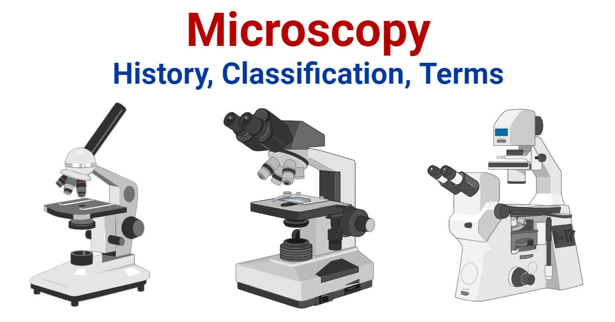
Microscopy can simply be understood as the ‘use of microscope’. Microscopy can be defined as the scientific discipline of using microscopes for getting a magnified view of objects that can’t be viewed by naked eyes.
It is a very important tool in biology and nanotechnology. In microbiology, it is one of the most important tools used in observing microbial cells. Medical sectors, pathology, histology, molecular biology, and cytology are in great debt of microscopy.
Microscopy Classification
A. Optical Microscopy (Light Microscopy)
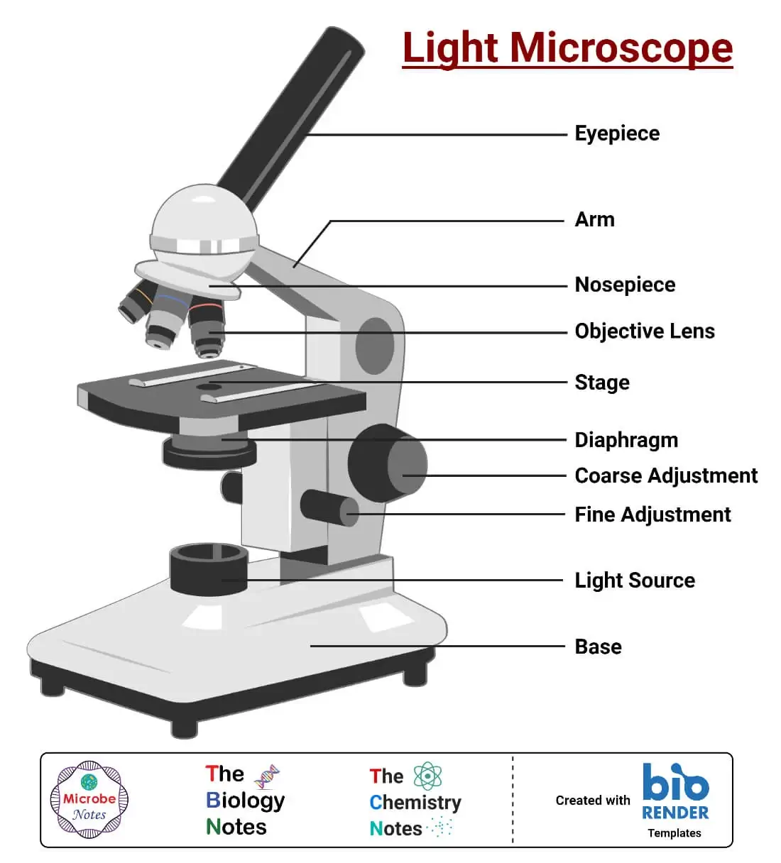
Optical Microscopy (Light Microscopy) is the microscopy technique that uses transmitted visible light, either natural or artificial, for developing the image of an object. It is the most common type of microscopy. It is further classified into several groups;
1 Bright Field Microscopy
Bright Field Microscopy is the simplest and the most common type. A simple or compound light microscope is used in this technique. It uses transmitted visible light to develop magnified images.
It has a low contrasting capacity, low optical resolution, requires staining and has a limited magnification of around 1300X.
It is simple and can be used to observe living cells and microorganisms.
2 Dark Field Microscopy
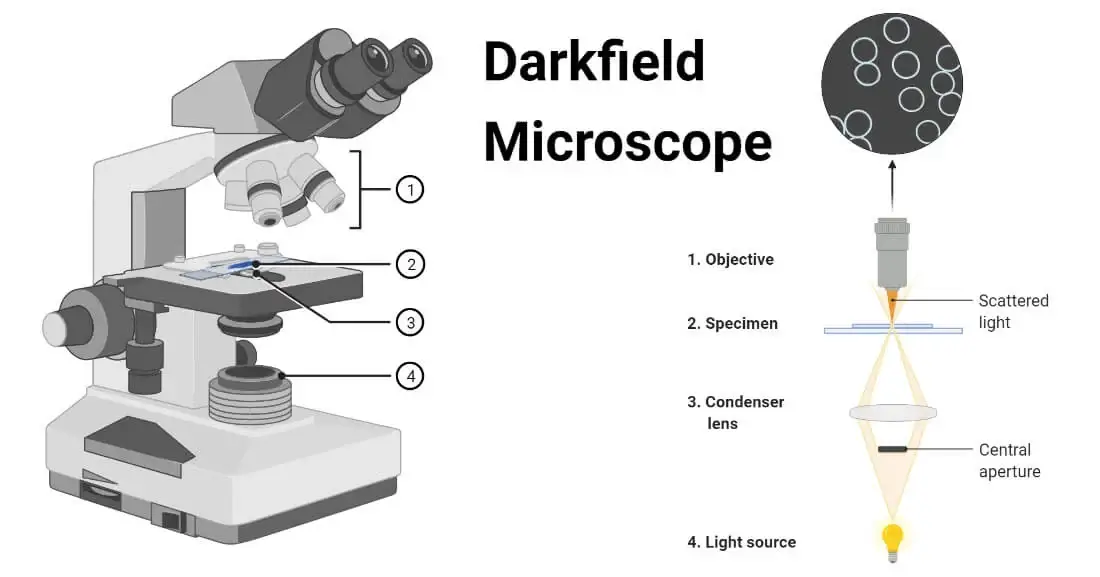
Dark Field Microscopy uses dark-ground microscopes. The reflected light is used, instead of transmitted light, to form a magnified image.
It is used to observe very thin specimens and motility of flagella and cilia.
3 Phase Contrast Microscopy
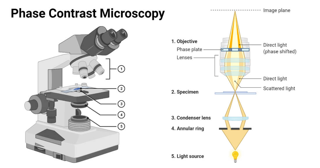
In Phase Contrast Microscopy, the phase-contrast microscope is used. It converts phase shifts into differences in intensity of light-producing more contrast images.
It is used for observing living cells and cellular structures.
4 Fluorescence Microscopy
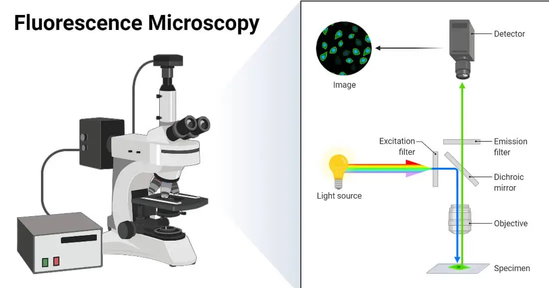
Fluorescence Microscopy is a microscopy technique that uses a fluorescent microscope with a UV light source. It is widely used in detecting antigens, antibodies, and other macromolecules.
5. Confocal Microscopy
Confocal Microscopy is a newer microscopy technique that uses a focused laser beam. It is used to get high-resolution 3-D images of biological samples.
6. Differential Interference Contrast Microscopy
Differential Interference Contrast Microscopy is a newer microscopy technique used in unstained and transparent samples to enhance the contrast of their image.
B. Electron Microscopy
Electron Microscopy is a microscopy technique that uses a beam of electrons to develop a highly magnified image of microscopic samples.
In this technique, electron microscopes are used.
C. Scanning Probe Microscopy
Scanning Probe Microscopy is a microscopy technique that uses a physical probe to scan specimens and form magnified images. This measures the surface features of the specimen.
D. X-ray Microscopy
X-ray Microscopy is a microscopy technique that uses soft X-ray radiation to produce a magnified image of the specimen.
Microscopy Terms
What is Magnification?
Magnification is the process of producing an enlarged image of a specimen by using a lens system. In a microscope, magnification can be computed by calculating the product of the magnification power of the eyepiece by the magnification power of the objective in use.
What is Magnifying Power?
Magnifying Power is defined as the ratio of the angle subtended by the image at the eye to the angle subtended by the object at the eye when placed at a minimum distance of distinct vision.
It can be mathematically defined as;
M=1+ D/F
where,
M = magnifying power
D = least distance of distinct vision
F = Focal length of a convex lens
What is Refractive Index?
Refractive Index can be defined as velocity of light in a vacuum to velocity of light in a medium (substance). Simply it is the measure of bending of a light ray when passing from one medium to another.
Mathematically it can be defined as;
n = c/v
where,
n = refractive index
c = speed of light in vacuum
v = velocity of light in a medium
What is Resolution?
Resolution can be defined as the shortest distance between two points on a specimen that can be distinguished by a microscope in its image. It is the ability of a microscope to distinguish details on a specimen.
Mathematically it is given as;
r = ⋋/2NA
where,
r = resolution
⋋ = imaging wavelength
NA = numerical aperture
References
- Images created using biorender.com
- Microscopy: Intro to microscopes & how they work (article) | Khan Academy
- What is Microscopy? (with pictures) (infobloom.com)
- Microscopy and Types of Microscopy (brainkart.com)
- What is Microscopy? | The University of Edinburgh
- Microscope Glossary of Terms: Microscope A-Z – Microscope and Laboratory Equipment Reviews (microscopespot.com)
- Resolution | Nikon’s MicroscopyU
- What is Microscope Resolution? – Microscope Clarity
- How does a microscope work? – Explain that Stuff
- Types of Microscopes: Definition, Working Principle, Diagram, Applications, FAQs (byjus.com)
- Fluorescence Microscopy – Principle, Components, Mechanism (noteshippo.com)
- https://www.olympus-lifescience.com/en/microscope-resource/primer/techniques/fluorescence/anatomy/fluoromicroanatomy/
- What is confocal microscopy ? | Cherry Biotech
- Fish K. N. (2009). Total internal reflection fluorescence (TIRF) microscopy. Current protocols in cytometry, Chapter 12, Unit12.18. https://doi.org/10.1002/0471142956.cy1218s50
- Dark-field Microscopy: Principle and Uses • Microbe Online
- What is Dark Field Microscopy – Microscope Clarity
- Acoustic Microscopy – Benefits, Limitations and Uses (microscopemaster.com)
- X-Ray Spectroscopy Principle, Instrumentation and Applications (microbiologynote.com)
- Jacobsen, C. (2019). Index. In X-ray Microscopy (Advances in Microscopy and Microanalysis, pp. 573-579). Cambridge: Cambridge University Press.
- Principle, Instrumentation, Types and Applications of XRD (14impressions.in)
- Molecular Expressions Microscopy Primer: Specialized Microscopy Techniques – Polarized Light Microscopy – Compensators and Retardation Plates (fsu.edu)

Superb notes
Good