Interesting Science Videos
Measles Virus

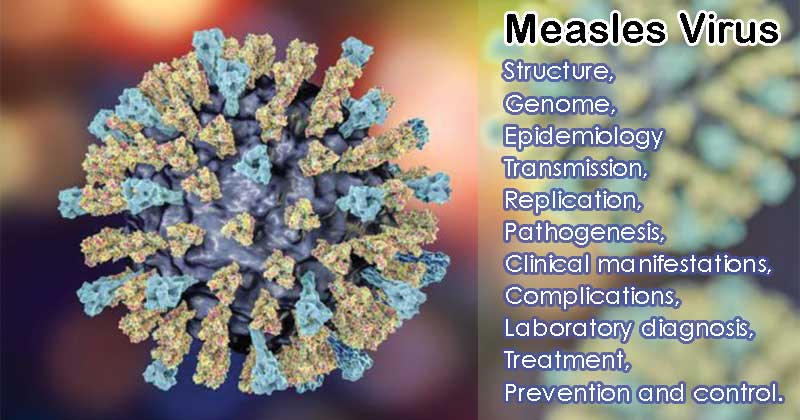
Image Source: BBC
Structure
- Measles virus is the prototypic member of the Morbillivirus genus of the family Paramyxoviridae.
- Measles virus is spherical with a diameter ranging from 100 to 300 nm and has two major structural components: one is the helical ribonucleoprotein (RNP) core formed by the association of the nucleoprotein (N), phosphoprotein (P) and large protein (L) with the viral genome, the other is the cellular membrane-derived lipid envelope surrounding the RNP core.
- The measles virus has two glycoproteins spikes that play a significant role in pathogenesis: F (fusion) protein and the H (haemagglutinin) protein.
- Fusion protein are responsible for the fusion of the virus with host cell membranes and viral penetration.
- Haemagglutinin protein are responsible for the binding of the virus to cells and it is antigen against which neutralizing antibodies are formed.
- Large protein (L) and nucleoprotein (N) together form the nucleocapsid that covers the viral RNA.
- The viral genome is non-segmented, negative-sense single-stranded RNA.
Genome
- The measles virus genome is non-segmented, negative-sense single-stranded RNA.
- The RNA genome consists of approximately 16,000 nucleotides and is enclosed in a lipid-containing envelope derived from the host cell.
- The genome begins with a 52 nucleotides non-coding region known as the leader and ends with a 37 nucleotides non-coding region known as the trailer, both of which are essential for the transcription and replication of the genome.
- The organization of the MeV genome comprises of six genes coding for eight viral proteins arranged as 3′-N,P,V,C,M,F,H,L-5′, each flanked by gene-end and gene-start sequences.
- The genome encodes eight proteins, two of which (V and C) are nonstructural proteins and are transcribed from the phosphoprotein (P) gene.
- Of the six structural proteins, large protein (L), and nucleoprotein (N) form the nucleocapsid housing the viral RNA.
- The hemagglutinin protein (H), fusion protein (F), and matrix protein (M), together with lipids from the host cell membrane, form the viral envelope.
Epidemiology and transmission
- Measles virus is primarily transmitted by respiratory droplets over short distances and, less commonly, by small-particle aerosols that remain suspended in the air for long periods of time.
- In the Northern Hemisphere, the incidence tends to rise in the winter.
- In tropical regions, epidemics are less marked.
- In the pre-vaccination era, the maximum incidence was seen in children aged 5 – 9 years. By the age of 20, approximately 99% of subjects have been exposed to the virus.
- Measles epidemics occur every 2 years in developed countries in the absence of widespread use of the vaccine.
- With the introduction of the vaccine, measles infection has shifted to teens in countries with an efficient program.
- In contrast, in third world countries, measles infection has its greatest incidence in children under 2 years of age.
- Malnutrition is one of the main underlying causes of this excess mortality.
- In general measles, mortality is highest in children < 2 years and in adults.
Replication
- The initial binding of MeV to the cell surface is mediated by the tetrameric H protein via interaction with cell surface receptors, which triggers the conformational change of the trimeric F protein and then, the membrane fusion and the delivery of the viral RNP core into the cytoplasm.
- The two identified cell surface receptors are CD46 and CD150 (SLAM).
- SLAM, an acronym for signaling lymphocyte activation molecule, is expressed on activated T and B lymphocytes and antigen-presenting cells.
- Following cell entry, the genomic RNPs are released into the cytosol and the encapsidated viral RNA serves as a template of the RdRP complex for both transcription and replication.
- Transcription begins at the 3′ end of the genome and viral genes are transcribed in the 3′ to 5′direction with a sequential “stop-start” mechanism.
- Newly synthesized viral mRNAs are translated to viral proteins by using the host translation machinery.
- The negative-strand genome is also used to synthesize a positive-strand anti-genome, which is a complimentary copy of the entire genome that produces more genomes via the same viral RNA polymerase.
- During replication, the newly synthesized genomic RNA is tightly wrapped with the N protein to provide a helical template for viral transcription and replication.
- The F and H proteins assemble intracellularly prior to receptor binding and are co-transported to the plasma membrane.
- The M protein associates with the RNP complex in the cytoplasm and then carries it to the plasma membrane, where the assembly with F and H proteins occurs and the virus is released via budding.
Pathogenesis
- Measles virus is transmitted by respiratory droplets over short distances and, less commonly, by small-particle aerosols that remain suspended in the air for long periods of time.
- The virus replicates in the respiratory tract and then spreads to the local lymphatic tissues.
- Amplification of a virus in the lymph nodes produces a primary viremia that results in the spread of the virus to multiple lymphoid tissues and the skin, kidney, gastrointestinal tract, and liver.
- Viral dissemination is predominantly mediated by the cell-to-cell transmission of the virus.
- In these organs, the virus replicates in endothelial cells, epithelial cells, and monocytes and macrophages.
- The measles virus can be detected in peripheral blood leukocytes at the time of onset of prodromal symptoms and for several days after the onset of the rash.
- The fusion of the measles virus‐infected cells leads to the formation of multinucleated giant cells, a pathological hallmark of this infection.
- MeV spreads systemically to other organs and tissues through infected circulating CD150 immune cells and, in some rare cases, infects endothelial cells, neurons, astrocytes and oligodendrocytes in vivo.
- Although most measles cases resolve without complications, the measles virus is capable of persisting in neurons as a defective variant that spreads from neuron to neuron directly, without passage through the extracellular environment.
- In this way, the virus avoids detection and elimination by circulating high titers of measles virus-specific neutralizing antibody.
- The persistence of measles virus in the CNS can result in progressive fatal encephalitis called SSPE developing in immunocompetent individuals several months to years after infection and recovery from acute measles virus
- Although the induction of this autoimmune response is poorly understood, “molecular mimicry” based on structural similarities between MeV proteins and myelin has been suggested as a pathogenic mechanism.
- The disease is hallmarked by demyelination, which results in ataxia, motor, and sensory loss and mental status changes and can result in death.
Clinical manifestations
- The time from infection to clinical disease is approximately 10 days to the onset of fever and 14 days to the onset of rash.
- After an incubation period of 10 – 11 days, the patient enters the prodromal stage with fever, malaise, sneezing, rhinitis, congestion, conjunctivitis and cough.
- Koplik’s spots, which are pathognomonic of measles, appear on the buccal and lower labial mucosa opposite the lower molars.
- The distinctive maculopapular rash appears about 4 days after exposure and starts behind the ears and on the forehead.
- From here the rash spreads to involve the whole body.
- Atypical measles infection may be seen in people who have been incompletely vaccinated and are characterized by a sudden onset of high fever with a headache, abdominal pain, and myalgia.
- In contrast to acute measles, the rash develops on the distal extremities and spreads centripetally and the majority of cases develop pneumonia.
Complications
Secondary bacterial infection – otitis media, bronchitis, and pneumonia.
Measles Pneumonia– This is giant cell pneumonia which occurs mainly in people with immunocompromised patients.
Acute measles encephalitis
- Acute encephalitis is a severe complication with a frequency of around 1 in 1000-5000 with the mortality rate around 15%.
- Encephalitis usually develops when exanthem is still present within a period of 8 days after the onset of measles.
- CSF findings in measles encephalitis consist usually of mild pleocytosis and the absence of measles antibodies.
Subacute measles encephalitis
- It is most common in children with leukemia undergoing axial radiation therapy.
- The incubation period ranges from 5 to 6 months. The condition commences with focal convulsions, other signs include hemiplegia, coma, and this condition is frequently confused with SSPE.
Subacute sclerosing panencephalitis (SSPE)
- SSPE is a rare slowly progressing fatal degeneration of the brain.
- It is seen in children and young adults and occurs 6 – 8 years after the initial attack of measles.
- The incidence is of the order of 1 in 100,000 cases of acute measles.
- The course of SSPE is highly variable but usually starts with generalized intellectual deterioration or psychological disturbance.
- In 75% of cases, the retina, a chorioretinitis develops leading to blindness.
- The CSF characteristically has high levels of antibodies against the measles virus as well as elevated levels of gammaglobulin.
Myocarditis– ECG abnormalities have been reported in up to 20% of children with uncomplicated measles but frank measles myopericarditis is rare.
Thrombocytopenic purpura– this is a rare complication of measles
Measles in pregnancy
- Measles in pregnancy results in a high rate of spontaneous abortion and premature delivery.
- There is some evidence that measles may be transmitted transplacentally as infants delivered during the mother’s incubation often develops a rash simultaneously with the mother.
- While some infants with perinatally acquired measles have mild illnesses, others develop severe disease with pneumonia.
Laboratory diagnosis
- Demonstration of clinical form i.e koplik’s spots.
Microscopy – demonstration of multinucleated giant cells measuring 100 nm in diameter obtained from nasopharyngeal secretion and stained with Giemsa stain
Virus isolation
- The measles virus can be isolated from a variety of sources, e.g. throat or conjunctival washings, sputum, urinary sediment cells and lymphocytes.
- The primary human kidney cell line can be used for the isolation of the virus.
- A continuous cell line like a Vero cell line can be used.
- The cytopathic effect can be seen in between 2 -15 days and consists of either a broad syncytium or a stellate form with inclusion bodies.
Antigen detection– Measles virus antigen detection is done by direct and indirect immunofluorescence from NPS specimens.
Antibody detection
- Detection of antibody titers which rises by 4 fold between the acute and the convalescent phase or detection of measles-specific IgM.
- Antibody detection is done by HAI, CF, neutralization and ELISA tests.
- In the case of SSPE, the presence of measles specific antibodies in the CSF is the most reliable means of laboratory diagnosis.
Molecular diagnosis – It accounts for the identification of measles virus RNA from a clinical specimen by PCR.
Treatment
- There is no prescribed medication for measles, however acetaminophen to relieve fever and muscle aches, vitamin A supplements are given to patients and are suggested to drink plenty of water.
- Administration of human anti-measles gammaglobulin is recommended.
Prevention and control
- The measles vaccine is most commonly administered as part of a combination of live attenuated vaccines that include measles, mumps, rubella or measles, mumps, rubella and varicella (MMR or MMRV).
- The MMR vaccine is a three-in-one vaccination that protects individuals from measles, mumps, and rubella.
- Children 12 months of age or older should have 2 doses, separated by at least 28 days.
- Two doses of MMR (measles, mumps & rubella) vaccine is nearly 100% effective at preventing measles.
- Practicing hygiene and cleanliness such as washing hands with soap and water or sanitizer, covering mouth and nose with a tissue when coughing or sneezing and avoiding close contact, such as kissing, hugging, or sharing eating utensils or cups, with people who are sick.
Sources
- 6% – http://www.virology-online.com/viruses/MEASLES.htm
- 4% – https://www.sciencedirect.com/topics/neuroscience/measles-virus
- 4% – https://www.mdpi.com/1999-4915/8/11/308/pdf
- 3% – https://www.sciencedirect.com/topics/immunology-and-microbiology/measles-virus
- 2% – https://www.slideshare.net/doctorrao/measles-1371951
- 2% – https://www.ncbi.nlm.nih.gov/pmc/articles/PMC4997572/
- 2% – https://pdfs.semanticscholar.org/bb37/7560951a1d99d11fed7b95659fb53eb82e14.pdf
- 1% – https://www.virology-online.com/viruses/MEASLES.htm
- 1% – https://www.slideshare.net/doctorrao/measles-update
- 1% – https://www.sciencedirect.com/topics/veterinary-science-and-veterinary-medicine/measles-virus
- 1% – https://www.sciencedirect.com/topics/nursing-and-health-professions/measles
- 1% – https://www.researchgate.net/publication/5821354_Structure_of_the_measles_virus_hemagglutinin
- 1% – https://www.powershow.com/viewht/547405-ZGQ5Z/Epidemiology_20of_20Measles_powerpoint_ppt_presentation
- 1% – https://www.ncbi.nlm.nih.gov/pubmed/25122787
- 1% – https://www.ncbi.nlm.nih.gov/pmc/articles/PMC5127022/
- 1% – https://www.ecdc.europa.eu/en/measles/facts/factsheet
- 1% – https://www.centrallakesclinic.biz/mosaic-virus/cns.html
- 1% – https://quizlet.com/184125003/measles-mumps-rubella-herpes-varicella-flash-cards/
- 1% – https://academic.oup.com/jid/article/170/Supplement_1/S15/936339
- 1% – http://www.vdh.virginia.gov/epidemiology/epidemiology-fact-sheets/pediatric-autoimmune-neuropsychiatric-disorders-associated-with-streptococcal-infections-pandas/
- 1% – http://www.authorstream.com/Presentation/drdpraveen-356561-measles-measles4-education-ppt-powerpoint/
- 1% – http://www.authorstream.com/Presentation/aSGuest139977-1479297-ashry-epidemiology-measles/
- 1% – http://myistm.istm.org/HigherLogic/System/DownloadDocumentFile.ashx?DocumentFileKey=eabd91fe-2ee8-4adf-a616-bb1067c6870f

Very interesting. Thank so much for the sharing.
Julio Santiago