Interesting Science Videos
Flow Cytometry Definition
- Flow cytometry is a standard laser-based technology that is used in the detection and measurement of physical and chemical characteristics of cells or particles in a heterogeneous fluid mixture.
- The use of flow cytometry has increased over the years as it provides a rapid analysis of multiple characteristics (both qualitative and quantitative) of the cells.
- The properties that can be measured by this process include a particle’s size, granularity or internal complexity, and fluorescence intensity.
- These characteristics are determined using an optical-to-electronic coupling system that detects the cells based on laser scattered by the cells.
- A flow cytometer, despite its name, does not necessarily deal with cells; it deals with cells quite often, but it can also deal with chromosomes or molecules or many other particles that can be suspended in a fluid.
Flow Cytometry Principle
The basic principle of flow cytometry is based on the measurement of light scattered by particles, and the fluorescence observed when these particles are passed in a stream through a laser beam.

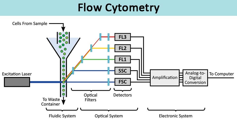
Figure: Schematic of a common flow cytometer, illustrating the fluidic, optical, and electronic systems. Image Source: AAT Bioquest, Inc.
Light Scattering
- Light scattering results when a particle deflects incident laser light. The extent to which this happens depends on the physical properties of a particle, namely its size and internal complexity.
- Forward-scattered light (FSC) is proportional to the cell-surface area or size of the cell. It is a measurement of mostly diffracted light and detects rays that are just off the axis of the incident laser beam dispersed in the forward direction by a photodiode.
- Side-scattered light (SSC) indicates the cell granularity or internal complexity of the cells. SSC is a measurement of mostly refracted and reflected light that occurs at any interface within the cell where there is a change in the refractive index.
- The measurements of FSC and SSC are used for the differentiation of cell types in a heterogeneous cell population.
Fluorescence
- Fluorescent markers used to detect the expression of cellular molecules such as proteins or nucleic acids in a system.
- The fluorescent compound absorbs light energy over a range of wavelengths that is characteristic of that compound.
- This absorption of light causes an electron in the fluorescent compound to be raised to a higher energy level.
- The excited electron quickly decays to its ground state, emitting the excess energy in the form of fluorescence which is then collected by detectors.
- In a mixed population of cells, different fluorochromes can be used to distinguish separate subpopulations.
- The fluorescence pattern of each subpopulation, combined with FSC and SSC data, can be used to identify which cells are present in a sample and to count their relative percentages.
- The electronics system then converts the detected light signals into electronic signals that can be processed by the computer.
Instrumentation/Parts of Flow Cytometry
A flow cytometer is made up of three main systems: fluidics, optics system, and electronics system.
Fluidics
- The purpose of the fluidics system is to transport particles in a fluid stream to the laser beam. To accomplish this, the sample is injected into a stream of sheath fluid (usually a buffered saline solution) within the flow chamber.
- The design of the flow chamber allows the sample core to be focused in the center of the sheath fluid where the laser beam then interacts with the particles.
- Focusing is achieved by injecting the sample suspension into the center of a sheath liquid stream. The flow of the sheath fluid moves the particles and restricts them to the center of the sample core.
Optics System
- The optical system of the cytometer consists of excitation optics and collection optics.
- The excitation optics consists of the laser and lenses that are used to shape and focus the laser beam to the flow of the sample.
- The collection optics consist of a collection lens to collect light emitted after the particle interacts with the laser beam and a system of optical mirrors that divert the specified wavelengths of the collected light to designated optical detectors.
- After a cell or particle passes through the laser light, the rays emitted on the side and the fluorescence signals are directed to the photomultiplier tubes (PMTs), and a photodiode collects the signals.
- To achieve the specificity of a detector for a particular fluorescent dye, a filter is placed in front of the tubes, which allows only a narrow range of wavelengths to reach the detector.
Electronics system
- The electronic system converts the signals from the detectors into digital signals that can be read by a computer.
- Once the light signals strike one side of the PMT or the photodiode, they are converted into a relative number of electrons that are multiplied to create a more significant electrical current.
- The electrical current moves to the amplifier and is converted to a voltage pulse.
- The highest point of the pulse is achieved when the particle strikes the center of the beam, in which case the maximum amount of scatter or fluorescence is achieved.
- The Analog-to-Digital Converter (ADC) then converts the pulse to a digital number.
Protocol/Procedure/Process/Steps of Flow Cytometry
The process of flow cytometry consists of the following:
Sample Preparation
- Before running in the flow cytometers, the cells under analysis must be in a single-cell suspension.
- Clumped cultured cells or cells present in solid organs should first be converted into a single cell suspension before the analysis by using enzymatic digestion or mechanical dissociation of the tissue, respectively.
- It is then followed by mechanical filtration should to avoid unwanted instrument clogs and obtain higher quality flow data.
- The resulting cells are then incubated in test tubes or microtiter plates with unlabeled or fluorescently conjugated antibodies and analyzed through the flow cytometer machine.
Antibody Staining
- Once the sample is prepared, the cells are coated with fluorochrome-conjugated antibodies specific for the surface markers present on different cells. This can be done either by direct, indirect, or intracellular staining.
- Indirect staining, cells are incubated with an antibody directly conjugated to a fluorophore.
- In indirect staining, the fluorophore-conjugated secondary antibody detects the primary antibody
- The intracellular staining procedure allows direct measurement of antigens presents inside the cell cytoplasm or nucleus. For this, the cells are first made permeable and then are stained with antibodies in the permeabilization buffer.
Running Samples
- At first, control samples are run to adjust the voltages in the detectors.
- The flow rates in the cytometer are set and the sample is run.
Types of Flow Cytometry
There are different types of flow cytometers based on the purpose and precision of the process:
-
Traditional flow cytometers
- The traditional cytometers are the common cytometer using sheath fluid for focusing the sample stream.
- The most common lasers used in traditional flow cytometers are 488 nm (blue), 405nm (violet), 532nm (green), 552nm (green), 561 nm (green-yellow), 640 nm (red) and 355 nm (ultraviolet).
-
Acoustic Focusing Cytometers
- In these cytometers, ultrasonic waves are used to focus the cells for analysis.
- This prevents sample clogging and also allows higher sample inputs.
-
Cell sorters
- Cell sorters are a category of traditional flow cytometers which allows the user to collect samples after processing.
- The cells that are positive for the desired parameter can be separated from those that are negative for the parameters.
-
Imaging flow cytometer
- Imaging cytometers are traditional cytometers combined with fluorescence microscopy.
- Imaging cytometer allows for rapid analysis of a sample for morphology and multi-parameter fluorescence at both a single cell and population level.
Applications/Uses
Flow Cytometry is used in several fields including molecular biology, pathology, immunology, virology, plant biology, and marine biology. Some of the common application include:
- It is used in clinical labs for the detection of malignancy in bodily fluids like leukemia.
- Cytometers like cell sorters can be used to separate the cells of interest in separate collection tubes physically.
- It can be used for the detection of the content of DNA by using fluorescent markers.
- Flow cytometers allow the analysis of replication cells by using fluorescent dye for four different stages of the cell cycle.
- Acoustic flow cytometers are used in the study of multi-drug resistant bacteria in the blood and other samples.
- The different stages of cell death, apoptosis, and necrosis can be detected by flow cytometers based on the differences in the morphological and biochemical changes.
Limitations
- This process doesn’t provide information on the intracellular location or distribution of proteins.
- Over time, debris is aggregated, which might result in false results.
- The pre-treatment associated with sample preparation and staining is a time-consuming process.
- Flow cytometry is an expensive process that requires highly qualified technicians.
References
- Biotech, M. (2018). Flow cytometry instrumentation – an overview. Current Protocols in Cytometry, e52. DOI: 10.1002/cpcy.52
- McKinnon K. M. (2018). Flow Cytometry: An Overview. Current protocols in immunology, 120, 5.1.1–5.1.11. https://doi.org/10.1002/cpim.40
- Dean, P.N. and Hoffman, R.A. (2007), Overview of Flow Cytometry Instrumentation. Current Protocols in Cytometry, 39: 1.1.1-1.1.8. DOI:1002/0471142956.cy0101s39
- https://enquirebio.com/flow-cytometry
Sources
- 7% – https://www.bosterbio.com/protocol-and-troubleshooting/flow-cytometry-principle
- 5% – http://www.bu.edu/flow-cytometry/files/2010/10/BD-Flow-Cytom-Learning-Guide.pdf
- 3% – https://www.ncbi.nlm.nih.gov/pmc/articles/PMC5939936/
- 2% – https://www.slideshare.net/Raghuveer19/flow-cytometry-73042192
- 2% – https://www.slideshare.net/PradeepNarwat/flow-cytometry-152440593
- 2% – https://www.amrepflow.org.au/public/documents/SyllabusAMREPFlowCoreFlowCytometry160120.pdf
- 2% – https://docs.abcam.com/pdf/protocols/Introduction_to_flow_cytometry_May_10.pdf
- 2% – http://www.rsc.org/suppdata/c7/sc/c7sc00260b/c7sc00260b1.pdf
- 2% – http://site.iugaza.edu.ps/hesawwaf/files/2016/09/Lab.5-immunofluorescence1.pptx
- 1% – https://www.usbio.net/promos/flow-cytometry
- 1% – https://www.thefreelibrary.com/Flow+cytometry%3a+Principles+and+clinical+applications+in+hematology.-a0209536695
- 1% – http://nikitavsurpatne.yolasite.com/optics.php
- <1% – https://www.sciencedirect.com/science/article/pii/S0169409X04001838
- <1% – https://www.ncbi.nlm.nih.gov/pmc/articles/PMC4856776/
- <1% – https://www.ncbi.nlm.nih.gov/pmc/articles/PMC100149/
- <1% – https://www.nature.com/articles/s41467-020-14929-2
- <1% – https://www.labome.com/method/Flow-Cytometry-A-Survey-and-the-Basics.html
- <1% – https://www.bdbiosciences.com/en-us/instruments/research-instruments/research-cell-sorters/facsaria-iii
- <1% – https://answers.yahoo.com/question/index?qid=20120419080608AAzfZea
- <1% – http://sgpwe.izt.uam.mx/files/users/uami/cobe/Adan-Flow_cytometry-basic_principles_and_applications.pdf

What is you mention in daigram fl1 fl2 fl 3
could you please upload more notes on Msc biotechnology