Interesting Science Videos
Epithelial Tissue definition
Epithelial Tissue is one of the four types of tissue (epithelial, muscular, connective, and nervous) in animals which consists of closely aggregated polyhedral cells adhering firmly to one another, forming cellular sheets that line the interior of hollow organs and cover the body surface. An epithelial tissue or epithelium (plural is epithelia) consists of cells arranged in continuous sheets, in either single or multiple layers.
Characteristics of Epithelial Tissue
- Even though epithelial tissue present in different parts of the body might differ in structure and function, they all have some common characteristics.
- Some of these characteristics are given below:
- Shape and Size
- The shapes and sizes of epithelial cells are variable, ranging from tall columnar to cuboidal to low squamous.
- The cell’s size and morphology are generally based on their function.
- Polarity
- Epithelial cells generally show polarity, with organelles and membrane proteins distributed unevenly within the cell.
- The apical (free) surface of an epithelial cell is present towards the body surface, the body cavity, the lumen of an internal organ, or a gland duct that receives cell secretions. Apical surfaces may contain cilia or microvilli.
- The lateral surfaces of an epithelial cell, facing the adjacent cells on either side, may contain intercellular adhesion and other junctions.
- The basal surface of an epithelial cell adheres to extracellular materials such as the basement membrane, which is an inert connective tissue made by the epithelial cells themselves.
- Basement Membrane
- The basement membrane is a thin extracellular layer that commonly consists of two layers, the basal lamina, and the reticular lamina.
- The basal lamina is closer to and secreted by the epithelial cells and contains proteins like laminin and collagen as well as some glycoproteins and proteoglycans.
- The reticular lamina is closer to the connective tissue, present underneath, and contains collagen protein produced by connective tissue cells called fibroblasts.
- Intercellular Adhesion and Other Junctions
- Several membrane-associated structures provide adhesion and communication between cells.
- Tight junctions, also called zonulae occludens, are the most apical of the junctions that form a band completely encircling each cell.
- The second type of junction is the adherens junction or zonula adherens, which also encircles the epithelial cell, usually immediately below the tight junction.
- Another anchoring junction is the desmosome or macula adherens which are disc-shaped structures at the surface of one cell that matches with identical structures at an adjacent cell surface.
- Gap junctions mediate intercellular communication rather than adhesion or occlusion between cells.
- Avascular
- Epithelial tissue is avascular, relying on the blood vessels of the adjacent connective tissue to bring nutrients and remove wastes.
- The exchange of substances between epithelial tissue and connective tissue occurs by diffusion.
- Innervated
- Epithelial tissue is innervated; that is, it has its own nerve supply.
- Renew and Repair
- Epithelial cells have a high rate of cell division which allows the epithelial tissue to continually renew and repair itself by sloughing off dead or injured cells and replacing them with new ones.
Functions of Epithelial Tissue
Based on the location, epithelial tissue performs a bunch of functions. Some of which are:
Protection
- One of the most critical functions of epithelial tissue is protection. It protects the cells present below against radiation, desiccation, invasion by pathogens, toxins, and physical trauma.
- The absence of blood vessels in the epithelial tissue thus prevents bleeding in the tissue during abrasion.
Transportation
- Epithelial tissue also functions in the transportation of different molecules in and out of the cells with different pumps present in the epithelial tissue.
- Besides, in the digestive, respiratory, and urinary system, it allows the exchange of molecules between the underlying cells and the body cavity, capillaries, and ducts.
Secretion
- Glandular epithelium secretes various macromolecules like hormones responsible for multiple bodily functions.
- Many endocrine and exocrine glands also help maintain the body surfaces (skin) as well as support the functions of various organs (digestive system).
Absorption
- By the function of various specialized structures like cilia and microvilli on the surface of cells, epithelial tissue also aids in the absorption of multiple molecules by increasing the surface area.
- In the digestive system, columnar cells of the small intestine help in the absorption of water and various other nutrients.
Receptor function
- Some cells in the epithelial tissue are specialized to perform sensory functions that can detect the sensory information and convert them into neural signals.
- Cells in epithelial tissue like the pseudostratified columnar epithelium of the olfactory mucosa contain apical cilia that allow the sensation of odor.
Types / Classification with examples and location
Epithelial tissue is divided into two types:
- Covering and lining epithelium, also called the surface epithelium, that forms the outer covering of the skin and some internal organs and also forms the inner lining of blood vessels, ducts, body cavities, and the inner lining of the respiratory, digestive, urinary, and reproductive systems.
- Glandular epithelium that makes up the secreting portion of glands such as the thyroid gland, adrenal glands, sweat glands, and digestive glands.
Further, types of covering and lining epithelial tissue are classified according to the arrangement of cells and the shapes of those cells.
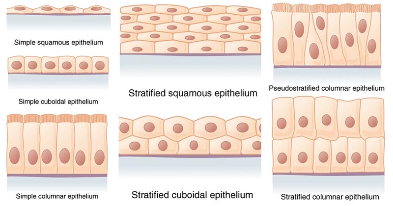
Image Source: Rice University (OpenStax)
Simple epithelium
- Simple epithelium is made up of a single layer of identical cells, which are usually found on secretory and absorptive surfaces, where the single layer enhances these processes.
- Simple epithelium is divided into three main types, and these are named according to the shape of the cells, which differ based on their functions.
a. Simple squamous epithelium
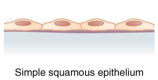
Image Source: Rice University (OpenStax)
- The simple squamous epithelium consists of a single layer of flat cells that resembles the tiles on a floor when viewed from the apical surface with a centrally located nucleus that is flattened and oval or spherical.
- This epithelium most commonly lines the cardiovascular and lymphatic system (heart, blood vessels, lymphatic vessels), where it is known as endothelium and forms the epithelial layer of serous membranes (peritoneum, pleura, pericardium), where it is called
- It is also found in air sacs of lungs, glomerular (Bowman’s) capsule of kidneys, and the inner surface of the tympanic membrane (eardrum).
b. Simple cuboidal epithelium
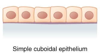
Image Source: Rice University (OpenStax)
- Simple cuboidal epithelium is a single layer of cube-shaped cells that are round and have a centrally located nucleus.
- It covers the surface of the ovary, lines the anterior surface of the capsule of the lens of the eye, forms pigmented epithelium at the posterior surface of retina of the eye, lines kidney tubules and smaller ducts of various glands, makes up secreting portion of some glands like the thyroid gland and ducts of some glands such as the pancreas.
c. Simple columnar epithelium
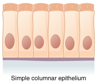
Image Source: Rice University (OpenStax)
- The columnar epithelium is made by a single layer of cells, rectangular in shape, on a basement membrane.
- This epithelium lines many organs and is often derived to make it well suited for a specific function.
- Columnar epithelium lines the stomach without any surface structures. However, the free surface of the columnar epithelium lining the small intestine is covered with microvilli, which provide a vast surface area for the absorption of nutrients from the small intestine.
- In the trachea, the columnar epithelium is ciliated. Also, it contains goblet cells that secrete mucus, and in the uterine tubes, ova are propelled along by ciliary action towards the uterus.
Stratified epithelium
- A stratified epithelium consists of several layers of cells of various shapes, and basement membranes are usually absent.
- As basal cells divide, daughter cells arising from cell divisions are pushed older cells upward toward the apical layer.
- As they move toward the surface and away from blood supply in underlying connective tissue, they become dehydrated and less metabolically active.
- Tough proteins predominate as cytoplasm is reduced, and cells become tough, hard structures that eventually die.
- At the apical layer, after dead cells lose cell junctions, they are sloughed off, but they are continuously replaced as new cells emerge from basal cells.
- There are two main types of stratified epithelium: stratified squamous, stratified cuboidal, and stratified columnar epithelium.
a. Stratified squamous epithelium
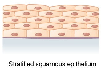
Image Source: Rice University (OpenStax)
The stratified squamous epithelium has two or more layers of cells. The cells in the apical layer and several layers deep to it are squamous while the cells in deeper layers vary from cuboidal to columnar.
i. Keratinized stratified squamous epithelium
- This epithelium develops a tough layer of keratin in the apical segment of cells and several layers deep to it
- The relative amount of keratin increases in cells as they move away from the nutritive blood supply and the organelles eventually die.
- The keratin forms a tough, relatively waterproof protective layer that prevents drying of the live cells present underneath.
- Keratinized stratified squamous epithelium forms a superficial layer of skin.
ii. Non-keratinized stratified squamous epithelium
- This epithelium does not contain large amounts of keratin in the apical layer, and several layers deep and is moistened continuously by mucus from salivary and mucous glands.
- Nonkeratinized stratified squamous epithelium lines wet surfaces (lining of mouth, esophagus, part of the epiglottis, part of the pharynx, and vagina) and covers the tongue.
b. Stratified cuboidal epithelium
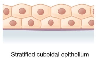
Image Source: Rice University (OpenStax)
- Stratified cuboidal epithelium has multiple layers of cells in which the apical layer is made up of cuboidal cells while the deeper layer can be either cuboidal or columnar.
- Stratified cuboidal epithelium is seen in the excretory ducts of salivary and sweat glands.
c. Stratified columnar epithelium
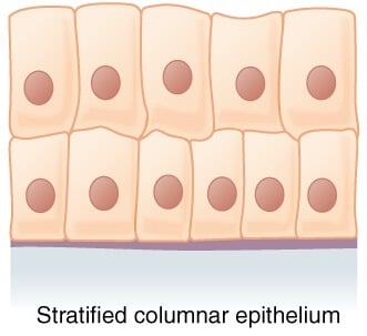
Image Source: Rice University (OpenStax)
- The stratified columnar epithelium has multiple layers of cells in which the apical layer is made up of columnar cells while the deeper layer can be either cuboidal or columnar.
- This type of epithelium is present in the conjunctiva of the eyes, parts of the urethra, and the small area in the anal mucosa.
Pseudostratified columnar epithelium
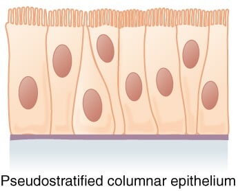
Image Source: Rice University (OpenStax)
- Pseudostratified epithelium appears to have several layers because the nuclei of the cells are present at various levels.
- Although all the cells are attached to the basement membrane in a single layer, some cells do not reach the apical surface.
- As a result of these features, it appears as a multilayered tissue, but in fact, is the simple epithelium.
- This epithelium lines epididymis, larger ducts of many glands, and parts of male urethra and airways of most of the upper respiratory tract.
Transitional epithelium tissue
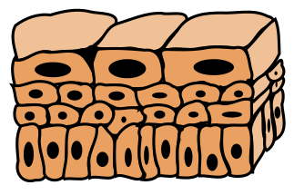
Figure: Transitional epithelium tissue. Image Source: Wikipedia.
- Transitional epithelium (urothelium) has a variable appearance (transitional).
- In a relaxed or unstretched state, looks like stratified cuboidal epithelium, except apical layer cells tend to be broad and rounded.
- As tissue is stretched, cells become flatter, giving the appearance of stratified squamous epithelium. Multiple layers and elasticity make it ideal for lining hollow structures (urinary bladder) subject to expansion from within.
Glandular Epithelium
- Epithelial cells that function mainly to produce and secrete various macromolecules may occur in epithelia with other significant functions or comprise specialized organs called glands.
- Scattered secretory cells, sometimes called unicellular glands, are common in simple cuboidal, simple columnar, and pseudostratified epithelia.
- Glands develop from covering epithelia in the fetus by cell proliferation and growth into the underlying connective tissue, followed by further differentiation.
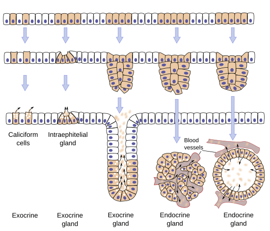
Figure: Main types of glands. Glands differentiate from epithelial tissues during embryo development. Arrows point to the released substances. Image Source: University of Vigo.
Endocrine glands
- The secretions of endocrine glands, called hormones, enter the interstitial fluid and then diffuse into the bloodstream without flowing through a duct.
- Endocrine secretions have far-reaching effects because they are distributed throughout the body by the bloodstream.
- Examples of endocrine glands include pituitary gland at the base of the brain, the pineal gland in the brain, thyroid and parathyroid glands near larynx (voice box), adrenal glands superior to kidneys, pancreas near the stomach, ovaries in the pelvic cavity, testes in the scrotum, thymus in the thoracic cavity.
Exocrine glands
- Exocrine glands secrete their products into ducts that release the secretions onto the surface of organs such as the skin surface or the lumen of a hollow organ.
- The effects of exocrine gland secretions are limited, and some of them would be harmful if they entered the bloodstream.
- Sweat, oil, and earwax glands of the skin, digestive glands such as salivary glands (secrete into mouth cavity) and pancreas (secretes into the small intestine) are the examples of exocrine glands.
References
- Mescher AL (2016). Basic Histology. Fourteenth Edition. McGraw-Hill Education.
- Tortora GJ and Derrickson B (2017). Principles of Physiology and Anatomy. Fifteenth Edition. John Wiley & Sons, Inc.
- Waugh A and Grant A. (2004) Anatomy and Physiology. Ninth Edition. Churchill Livingstone.
- https://www.kenhub.com/en/library/anatomy/overview-and-types-of-epithelial-tissue
Internet Sources
- 3% – https://quizlet.com/25768290/chapter-3-tissues-flash-cards/
- 2% – https://quizlet.com/47458913/chapter-4-flash-cards/
- 2% – https://quizlet.com/26692499/chapt-4-epithelial-tissue-flash-cards/
- 2% – https://quizlet.com/25669314/anatomy-ch-4-reading-flash-cards/
- 2% – https://doctor2017.jumedicine.com/wp-content/uploads/sites/7/2018/01/Surface-epithelium.pdf
- 1% – https://www.youtube.com/watch?v=0NEV-Rd7OgA
- 1% – https://www.thoughtco.com/animal-anatomy-epithelial-tissue-373206
- 1% – https://www.thenursepage.com/epithelial-tissues-types-of-epithelial-tissues/
- 1% – https://www.cram.com/flashcards/ap-i-ch4-6264721
- 1% – https://www.coursehero.com/file/p5d8jsc/Epithelial-tissue-may-be-divided-into-two-types-1-Covering-and-lining/
- 1% – https://study.com/academy/lesson/simple-cuboidal-epithelium-location-structure-function.html
- 1% – https://quizlet.com/gb/340412792/histology-epithelia-flash-cards/
- 1% – https://quizlet.com/ca/310892532/chapter-4-bio-235-flash-cards/
- 1% – https://quizlet.com/95543301/epithelial-tissue-covering-and-lining-epithelium-flash-cards/
- 1% – https://quizlet.com/72533810/anatomy-and-physiology-tissue-level-of-organization-flash-cards/
- 1% – https://quizlet.com/6320460/anatom-ch3-terms2-flash-cards/
- 1% – https://quizlet.com/50190298/44-epithelial-tissue-quiz-1-flash-cards/
- 1% – https://quizlet.com/326765543/apchapter-4-the-tissue-level-of-organization-flash-cards/
- 1% – https://quizlet.com/26142932/anatomy-lecture-3-tissues-flash-cards/
- 1% – https://quizlet.com/25583191/anatomy-ch-4-epithelial-tissue-flash-cards/
- 1% – https://quizlet.com/225671385/final-exam-specific-objectives-chapter-4-tissues-flash-cards/
- 1% – https://quizlet.com/211826315/chapter-4-the-tissue-level-of-organization-flash-cards/
- 1% – https://quizlet.com/20156045/the-tissue-level-of-organisation-flash-cards/
- 1% – https://quizlet.com/17285188/biomed-module-7-flash-cards/
- 1% – https://quizlet.com/14302876/epithelial-tissues-flash-cards/
- 1% – https://quizlet.com/13823304/classification-of-tissues-part-1-flash-cards/
- 1% – https://healthjade.net/basement-membrane/
- 1% – https://doctor2019.jumedicine.com/wp-content/uploads/sites/10/2020/01/Glandular-Epithelium.pdf.pdf
- <1% – https://www.studymode.com/essays/Outline-The-Structure-Of-The-Main-1455732.html
- <1% – https://www.lab.anhb.uwa.edu.au/mb140/CorePages/Epithelia/epithel.htm
- <1% – https://www.kenhub.com/en/library/anatomy/overview-and-types-of-epithelial-tissue
- <1% – https://www.differencebetween.com/difference-between-simple-stratified-and-pseudostratified-epithelial-tissue/
- <1% – https://www.coursehero.com/file/p502iic/630x-LM-Mark-Nielsen-Sectional-view-of-keratinized-stratified-squamous/
- <1% – https://www.britannica.com/science/microvilli
- <1% – https://quizlet.com/99144165/epithelium-tissue-functions-and-locations-flash-cards/
- <1% – https://quizlet.com/9814963/chapter-5-tissues-flash-cards/
- <1% – https://quizlet.com/24665603/body-tissues-flash-cards/
- <1% – https://quizlet.com/18641983/chapter-3-cells-and-tissues-flash-cards/
- <1% – https://oerpub.github.io/epubjs-demo-book/content/m46048.xhtml
- <1% – https://micro.magnet.fsu.edu/primer/anatomy/brightfieldgallery/index.html
- <1% – https://en.wikipedia.org/wiki/Stratified_cuboidal_epithelium
- <1% – https://en.m.wikipedia.org/wiki/Urethra
- <1% – https://courses.lumenlearning.com/boundless-ap/chapter/epithelial-tissue/
- <1% – https://courses.lumenlearning.com/boundless-ap/chapter/absorption/
- <1% – https://anatomyandphysiologyi.com/epithelial-tissue/
- <1% – http://www.histology.leeds.ac.uk/tissue_types/epithelia/epi_specialisations.php
- <1% – http://www.differencebetween.net/science/health/difference-between-exocrine-and-endocrine/
- <1% – http://medcell.med.yale.edu/systems_cell_biology/epithelium_lab.php
