Interesting Science Videos
The Human Digestive System Definition
- The human digestive system is the collective name used to describe the alimentary canal, some accessory organs, and a variety of digestive processes that take place at different levels in the canal to prepare food eaten in the diet for absorption.
- It has a general structure that is modified at different levels to provide for the processes occurring at each level.
- The complex of digestive processes gradually breaks down the foods eaten until they are in a form suitable for absorption.
- After absorption, nutrients are used to synthesize body constituents.
- They provide the raw materials for the manufacture of new cells, hormones and enzymes, and the energy needed for these and other processes and for the disposal of waste materials.
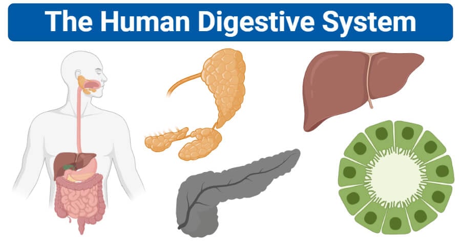
The human digestive system consists of the alimentary tract and accessory organs.
Alimentary Tract of The Human Digestive System
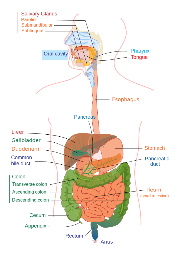
Image Source: Mariana Ruiz.
The alimentary canal begins at the mouth, passes through the thorax, abdomen, and pelvis and ends at the anus. It is thus a long tube through which food passes. It has various parts that are structurally remarkably similar. The parts include:
- Mouth
- Pharynx
- Oesophagus
- Stomach
- Small intestine
- Large intestine
- Rectum and Anal canal.
A. Mouth
- The mouth or oral cavity is bounded by muscles and bones: anteriorly —by the lips, posteriorly — it is continuous with the oropharynx, laterally —by the muscles of the cheeks, superiorly —by the bony hard palate and muscular soft palate, inferiorly —by the muscular tongue and the soft tissues of the floor of the mouth.
- It is lined throughout with mucous membrane, consisting of stratified squamous epithelium containing small mucus-secreting glands.
- The palate forms the roof of the mouth and is divided into the anterior hard palate and the posterior soft palate. The soft palate is muscular, curves downwards from the posterior end of the hard palate, and blends with the walls of the pharynx at the sides.
- The uvula is a curved fold of muscle covered with mucous membrane, hanging down from the middle of the free border of the soft palate.
- It consists of the following important parts:
The Tongue
- The tongue is a voluntary muscular structure that occupies the floor of the mouth.
- It is attached by its base to the hyoid bone and by a fold of its mucous membrane covering, called the frenulum, to the floor of the mouth.
- The superior surface consists of stratified squamous epithelium, with numerous papillae (little projections), containing nerve endings of the sense of taste, sometimes called the taste buds.
- The tongue plays an important part in:
- mastication (chewing)
- deglutition (swallowing)
- speech
- taste
The Teeth
- The teeth are embedded in the alveoli or sockets of the alveolar ridges of the mandible and the maxilla.
- Each individual has two sets, the temporary or deciduous teeth, and the permanent teeth.
- At birth, the teeth of both dentitions are present in an immature form in the mandible and maxilla.
- There are 20 temporary teeth, 10 in each jaw. They begin to erupt when the child is about 6 months old, and should all be present after 24 months.
- The permanent teeth begin to replace the deciduous teeth in the 6th year of age and this dentition, consisting of 32 teeth, is usually complete by the 24th year.
Types and Functions of the teeth
The incisor and canine teeth are the cutting teeth and are used for biting off pieces of food, whereas the premolar and molar teeth, with broad, flat surfaces, are used for grinding or chewing food.

Image Source: Scientific Animations.
B. The Pharynx
- Food passes from the oral cavity into the pharynx then to the esophagus below, with which it is continuous.
- The pharynx is divided for descriptive purposes into three parts, the nasopharynx, oropharynx, and laryngopharynx.
- The nasopharynx is important in respiration. The oropharynx and laryngopharynx are passages common to both the respiratory and the digestive systems.
Function of Pharynx
The pharynx has roles in both the respiratory and digestive systems and can be thought of as the point where these systems diverge.
For the digestive system, its muscular walls function in the process of swallowing, and it serves as a pathway for the movement of food from the mouth to the esophagus.
- The constrictive circular muscles of the pharynx’s outer layer play a big role in peristalsis. A series of contractions will help propel ingested food and drink down the intestinal tract safely.
- The inner layer’s longitudinal muscles, on the other hand, will widen the pharynx laterally and lift it upward, thus allowing the swallowing of ingested food and drink.
C. The Oesophagus
- The esophagus is about 25 cm long and about 2 cm in diameter and lies in the median plane in the thorax in front of the vertebral column behind the trachea and the heart.
- It is continuous with the pharynx above and just below the diaphragm it joins the stomach.
- The upper and lower ends of the esophagus are closed by sphincter muscles.
- The upper cricopharyngeal sphincter prevents air from passing into the esophagus during inspiration and the aspiration of oesophageal contents.
- The cardiac or lower oesophageal sphincter prevents the reflux of acid gastric contents into the esophagus.
Functions of Oesophagus
- The esophagus serves to pass food and liquids from the mouth down to the stomach. This is accomplished by periodic contractions (peristalsis).
- The esophagus is an important connection to the digestive system through the thoracic cavity, which protects the heart and lungs.
- Two sphincters on either side of the esophagus separate food into small units known as a bolus.
D. The Stomach
- The stomach is a J-shaped dilated portion of the alimentary tract situated in the epigastric, umbilical, and left hypochondriac regions of the abdominal cavity.
- The stomach is continuous with the esophagus at the cardiac sphincter and with the duodenum at the pyloric sphincter.
- It has two curvatures. The lesser curvature is short, lies on the posterior surface of the stomach, and is the downwards continuation of the posterior wall of the esophagus. Just before the pyloric sphincter, it curves upwards to complete the J shape.
- Where the esophagus joins the stomach the anterior region angles acutely upwards, curves downwards forming the greater curvature then slightly upwards towards the pyloric sphincter.
- The stomach is divided into three regions: the fundus, the body, and the antrum.
- At the distal end of the pyloric antrum is the pyloric sphincter, guarding the opening between the stomach and the duodenum.
- Stomach size varies with the volume of food it contains, which may be 1.5 liters or more in an adult.
- In the stomach, gastric muscle contraction consists of a churning movement that breaks down the bolus and mixes it with gastric juice and peristaltic waves that propel the stomach contents towards the pylorus.
- About 2 liters of gastric juice are secreted daily by special secretory glands in the mucosa.
- It consists of Water, mineral salts, mucus secreted by goblet cells in the glands and on the stomach surface, hydrochloric acid, Intrinsic factor, inactive enzyme precursors, etc.
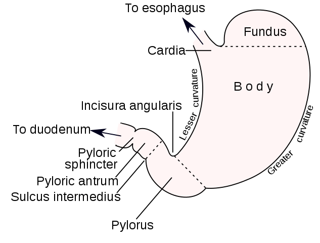
Image Source: Henry Vandyke Carter / Mysid.
Functions of the Stomach
- Temporary storage allowing time for the digestive enzymes, pepsins, to act.
- Chemical digestion — pepsins convert proteins to polypeptides.
- Mechanical breakdown — the three smooth muscle layers enable the stomach to act as a churn, gastric juice is added and the contents are liquefied to chime.
- Performs limited absorption of water, alcohol and some lipid-soluble drugs
- Non-specific defense against microbes — provided by hydrochloric acid in gastric juice.
- Preparation of iron for absorption further along the track — the acid environment of the stomach solubilizes iron salts, which is required before iron can be absorbed
- Production of intrinsic factor needed for absorption of vitamin B12 in the terminal ileum
- Regulation of the passage of gastric contents into the duodenum. When the chyme is sufficiently acidified and liquefied, the pyloric antrum forces small jets of gastric contents through the pyloric sphincter into the duodenum.
E. The Small Intestine
- The small intestine is continuous with the stomach at the pyloric sphincter and leads into the large intestine at the ileocaecal valve.
- It is a little over 5 meters long and lies in the abdominal cavity surrounded by the large intestine.
- In the small intestine, the chemical digestion of food is completed and most of the absorption of nutrients takes place.
- The small intestine comprises three main sections continuous with each other:
- The duodenum: It is about 25 cm long and curves around the head of the pancreas. Secretions from the gall bladder and pancreas are released into the duodenum through a common structure, the hepatopancreatic ampulla, and the opening into the duodenum is guarded by the hepatopancreatic sphincter (of Oddi).
- The jejunum: It is the middle section of the small intestine and is about 2 meters long.
- The ileum, or terminal section, is about 3 meters long and ends at the ileocaecal valve, which controls the flow of material from the ileum to the caecum, the first part of the large intestine, and prevents regurgitation.
- The surface area of the small intestine mucosa is greatly increased by permanent circular folds, villi, and microvilli.
- The villi are tiny finger-like projections of the mucosal layer into the intestinal lumen, about 0.5 to 1 mm long.
- Their walls consist of columnar epithelial cells, or enterocytes, with tiny microvilli (1 μm long) on their free border.
Functions of the small intestine
- The small intestine is the part of the intestines where 90% of the digestion and absorption of food occurs, the other 10% taking place in the stomach and large intestine.
- The main function of the small intestine is the absorption of nutrients and minerals from food.
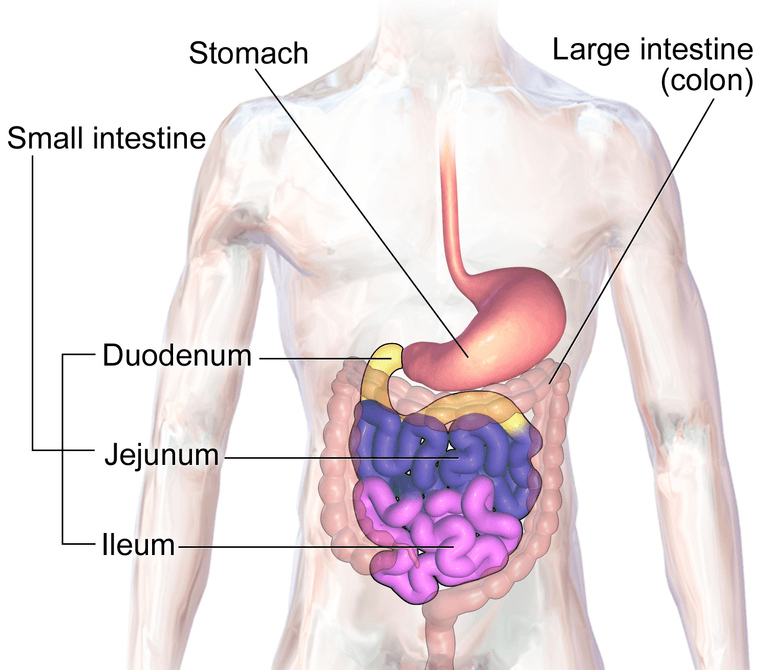
Image Source: BruceBlaus.
F. The Large Intestine
- It is about 1.5 meters long, beginning at the caecum in the right iliac fossa and terminating at the rectum and anal canal deep in the pelvis.
- Its lumen is larger than that of the small intestine. It forms an arch around the coiled-up small intestine.
- The colon is divided into the caecum, ascending colon, transverse colon, descending colon, sigmoid colon rectum, and anal canal.
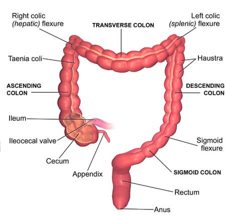
Image Source: BruceBlaus.
The caecum
- This is the first part of the colon. It is a dilated region which has a blind end inferiorly and is continuous with the ascending colon superiorly.
- Just below the junction of the two, the ileocaecal valve opens from the ileum.
- The vermiform appendix is a fine tube, closed at one end, which leads from the caecum. It is usually about 13 cm long and has the same structure as the walls of the colon but contains more lymphoid tissue.
The ascending colon
- This passes upwards from the caecum to the level of the liver where it curves acutely to the left at the hepatic flexure to become the transverse colon.
The transverse colon
- This is a loop of the colon which extends across the abdominal cavity in front of the duodenum and the stomach to the area of the spleen where it forms the splenic flexure and curves acutely downwards to become the descending colon.
The descending colon
- This passes down the left side of the abdominal cavity then curves towards the midline. After it enters the true pelvis it is known as the sigmoid colon.
The sigmoid colon
- This part describes an S-shaped curve in the pelvis then continues downwards to become the rectum.
G. The Rectum and the Anal Canal
- It is a slightly dilated section of the colon about 13 cm long. It leads from the sigmoid colon and terminates in the anal canal.
- The anal canal is a short passage about 3.8 cm long in the adult and leads from the rectum to the exterior.
- Two sphincter muscles control the anus; the internal sphincter, consisting of smooth muscle fibers, is under the control of the autonomic nervous system and the external sphincter, formed by skeletal muscle, is under voluntary control.
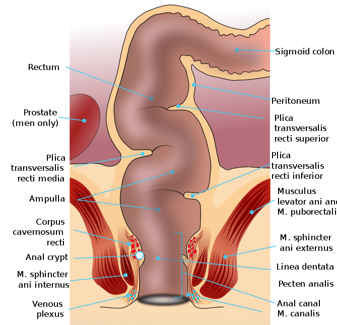
Image Source: Armin Kübelbeck.
Functions of the large intestine, rectum and anal canal
Absorption
- The contents of the ileum which pass through the ileocaecal valve into the caecum are fluid, even though some water has been absorbed in the small intestine.
- In the large intestine absorption of water continues until the familiar semisolid consistency of feces is achieved.
- Mineral salts, vitamins, and some drugs are also absorbed into the blood capillaries from the large intestine.
Microbial activity
- The large intestine is heavily colonized by certain types of bacteria, which synthesize vitamin K and folic acid. They include Escherichia coli, Enterobacter aerogenes, Streptococcus faecalis, and Clostridium perfringens (welchii).
Defaecation
- Usually, the rectum is empty, but when a mass movement forces the contents of the sigmoid colon into the rectum the nerve endings in its walls are stimulated by a stretch.
- Defaecation involves involuntary contraction of the muscle of the rectum and relaxation of the internal anal sphincter.
- Contraction of the abdominal muscles and lowering of the diaphragm increases the intra-abdominal pressure (Valsalva’s maneuver) and so assists the process of defaecation.
Video Animation: Human digestive system – How it works! (Thomas Schwenke)

Accessory Organs of The Human Digestive System
- Various secretions are poured into the alimentary tract, some by glands in the lining membrane of the organs, e.g. gastric juice secreted by glands in the lining of the stomach, and some by glands situated outside the tract.
- The latter are the accessory organs of digestion and their secretions pass through ducts to enter the tract. They consist of:
- 3 pairs of salivary glands
- Pancreas
- Liver and the biliary tract.
- The organs and glands are linked physiologically as well as anatomically.
A. The Salivary glands
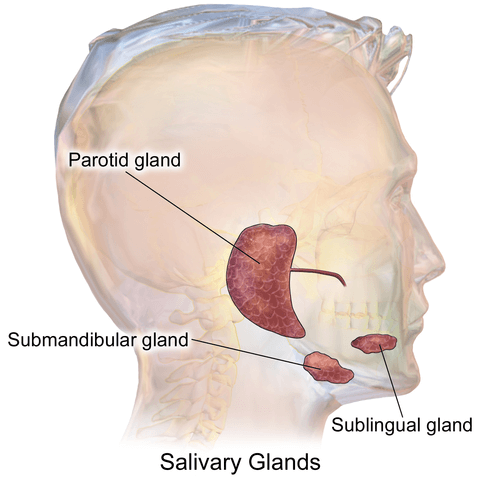
Image Source: BruceBlaus.
- Salivary glands are present in the oral cavity and pour their secretions into the mouth.
Saliva is the combined secretions from the salivary glands and the small mucus-secreting glands of the lining of the oral cavity. About 1.5 liters of saliva is produced daily and it consists of:
- water
- mineral salts
- enzyme: salivary amylase
- mucus
- lysozyme
- immunoglobulins
- blood-clotting factors.
- There are three pairs: the parotid glands, the submandibular glands, and the sublingual glands.
Parotid glands
- These are situated one on each side of the face just below the external acoustic meatus.
- Each gland has a parotid duct opening into the mouth at the level of the second upper molar tooth.
Submandibular glands
- These lie one on each side of the face under the angle of the jaw.
- The two submandibular ducts open on the floor of the mouth, one on each side of the frenulum of the tongue.
Sublingual glands
- These glands lie under the mucous membrane of the floor of the mouth in front of the submandibular glands.
- They have numerous small ducts that open into the floor of the mouth.
Functions of Salivary Glands and Saliva
- Chemical digestion of polysaccharides. Saliva contains the enzyme amylase that begins the breakdown of complex sugars, reducing them to the disaccharide maltose.
- Lubrication of food. Dry food entering the mouth is moistened and lubricated by saliva before it can be made into a bolus ready for swallowing.
- Cleansing and lubricating. An adequate flow of saliva is necessary to cleanse the mouth and keep its tissues soft, moist and pliable. It helps to prevent damage to the mucous membrane by rough or abrasive foodstuffs.
- Non-specific defense. Lysozyme, immunoglobulins and clotting factors combat invading microbes.
- The taste buds are stimulated only by chemical substances in solution. Dry foods stimulate the sense of taste only after thorough mixing with saliva.
B. The Pancreas
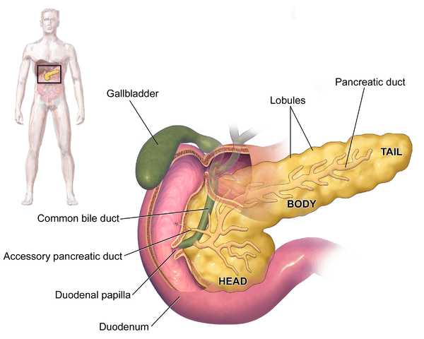
Image Source: BruceBlaus.
- The pancreas is a pale grey gland weighing about 60 grams.
- It is about 12 to 15 cm long and is situated in the epigastric and left hypochondriac regions of the
- abdominal cavity.
- It consists of a broad head, a body, and a narrow tail. The head lies in the curve of the duodenum, the body behind the stomach, and the tail lies in front of the left kidney and just reaches the spleen.
- The pancreas is both an exocrine and an endocrine gland.
The exocrine pancreas
- It consists of a large number of lobules made up of small alveoli, the walls of which consist of secretory cells.
- Each lobule is drained by a tiny duct and these unite eventually to form the pancreatic duct, which extends the whole length of the gland and opens into the duodenum.
- Just before entering the duodenum, the pancreatic duct joins the common bile duct to form the hepatopancreatic ampulla. The duodenal opening of the ampulla is controlled by the hepatopancreatic sphincter (of Oddi).
- The function of the exocrine pancreas is to produce pancreatic juice containing enzymes that digest carbohydrates, proteins, and fats.
The endocrine pancreas
- Distributed throughout the gland are groups of specialized cells called the pancreatic islets (of Langerhans).
- The islets have no ducts so the hormones diffuse directly into the blood.
- The function of the endocrine pancreas is to secrete the hormones insulin and glucagon, which are principally concerned with control of blood glucose levels.
Functions of the Pancreas
As part of the exocrine system, the pancreas secretes enzymes that work in tandem with bile from the liver and gallbladder to help break down substances for proper digestion and absorption.
Enzymes produced by the pancreas for digestion include:
- lipase to digest fats
- amylase to digest carbohydrates
- chymotrypsin and trypsin for digesting proteins
- The pancreas produces enzymes as soon as food reaches the stomach.
- These enzymes travel through a series of ducts until they reach the main pancreatic duct.
- The main pancreatic duct meets the common bile duct, which carries bile from the gallbladder and liver towards the duodenum. This meeting point is called the ampulla of Vater.
- Bile from the gallbladder and enzymes from the pancreas are released into the duodenum to help digest fats, carbohydrates, and proteins so they can be absorbed by the digestive system.
Endocrine Function
As part of the endocrine system, the pancreas secretes two main hormones that are vital to regulating glucose (also known as blood sugar) level:
Insulin. The pancreas secretes this hormone to lower blood glucose when levels get too high.
Glucagon: The pancreas secretes this hormone to increase blood glucose when levels get too low.
Balanced blood glucose levels play a significant role in the liver, kidneys, and even brain. Proper secretion of these hormones is important to many bodily systems, such as your nervous system and cardiovascular system.
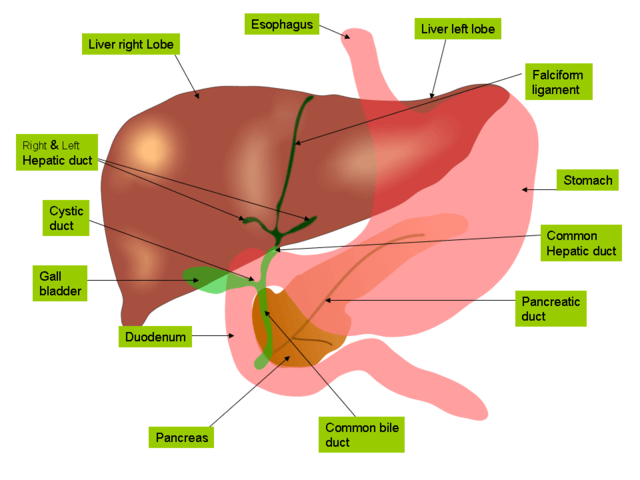
Image Source: Jiju Kurian Punnoose.
C. The Liver
- The liver is the largest gland in the body, weighing between 1 and 2.3 kg.
- It is situated in the upper part of the abdominal cavity occupying the greater part of the right hypochondriac region, part of the epigastric region, and extending into the left hypochondriac region.
- Its upper and anterior surfaces are smooth and curved to fit the undersurface of the diaphragm; its posterior surface is irregular in outline.
- The liver is enclosed in a thin inelastic capsule and incompletely covered by a layer of peritoneum. Folds of peritoneum form supporting ligaments attaching the liver to the inferior surface of the diaphragm. It is held in position partly by these ligaments and partly by the pressure of the organs in the abdominal cavity.
- The liver has four lobes. The two most obvious are the large right lobe and the smaller, wedge-shaped, left lobe. The other two, the caudate and quadrate lobes, are areas on the posterior surface.
- The lobes of the liver are made up of tiny lobules just visible to the naked eye.
- These lobules are hexagonal in outline and are formed by cubical-shaped cells, the hepatocytes, arranged in pairs of columns radiating from a central vein.
- Between two pairs of columns of cells, there are sinusoids (blood vessels with incomplete walls) containing a mixture of blood from the tiny branches of the portal vein and hepatic artery.
- Amongst the cells lining the sinusoids are hepatic macrophages (Kupffer cells) whose function is to ingest and destroy any foreign particles present in the blood flowing through the liver.
- Blood drains from the sinusoids into central or centrilobular veins. These then join with veins from other lobules, forming larger veins, until eventually, they become the hepatic veins that leave the liver and empty the inferior vena cava just below the diaphragm.
Functions of the liver
- Secretion of bile. The hepatocytes synthesize the constituents of bile from the mixed arterial and venous blood in the sinusoids. These include bile salts, bile pigments, and cholesterol.
- Carbohydrate metabolism. Conversion of glucose to glycogen in the presence of insulin, and converting liver glycogen back to glucose in the presence of glucagon. These changes are important regulators of the blood glucose level.
- Fat metabolism. Desaturation of fat i.e. converts stored fat to a form in which it can be used by the tissues to provide energy.
- Protein metabolism. Deamination of amino acids removes the nitrogenous portion from the amino acids not required for the formation of new protein; urea is formed from this nitrogenous portion which is excreted in the urine.
- It also breaks down the genetic material of worn-out cells of the body to form uric acid which is excreted in the urine.
- Transamination — removes the nitrogenous portion of amino acids and attaches it to other carbohydrate molecules forming new non-essential amino acids.
- Synthesis of plasma proteins and most of the blood clotting factors from the available amino acids occur in the liver.
- Breakdown of erythrocytes and defense against microbes. This is carried out by phagocytic Kupffer cells (hepatic macrophages) in the sinusoids.
- Detoxification of drugs and noxious substances. These include ethanol (alcohol) and toxins produced by microbes.
- Metabolism of ethanol.
- Inactivation of hormones. These include insulin, glucagon, cortisol, aldosterone, thyroid, and sex hormones.
- Synthesis of vitamin A from carotene. (Carotene is the provitamin found in some plants, e.g. carrots and green leaves of vegetables).
- Production of heat. The liver uses a considerable amount of energy, has a high metabolic rate, and produces a great deal of heat. It is the main heat-producing organ of the body.
- It is involved in the storage of:
- fat-soluble vitamins: A, D, E, K
- iron, copper
- some water-soluble vitamins, e.g. riboflavin, niacin,
- pyridoxine, folic acid, and vitamin B12.
References
- Waugh, A., & Grant, A. (2009). Ross and Wilson: Anatomy and Physiology in Health and Illness. (11th edition). Churchill Livingstone
- Hall, J. E. 1. (2016). Guyton and Hall textbook of medical physiology (13th edition.). Philadelphia, PA: Elsevier.
- Chung, K. W., Chung, H. M., & Halliday, N. L. (2015). BRS Gross anatomy (Eighth edition.). Philadelphia: Wolters Kluwer Health.
- Marieb, Elaine Nicpon,Hoehn, Katja. (2012) Human anatomy & physiology /Boston : Pearson
Internet Sources
- 3% – https://www.slideshare.net/harshraman1989/anatomy-and-physiology-of-gi-system-and-diagnostic-techniques
- 3% – https://www.healthline.com/health/what-does-the-pancreas-do
- 3% – http://www.radiation-therapy-review.com/Oesophagus_Stomach_Small_and_Large_Bowels_Rectum_and_Anus.html
- 2% – https://www.slideshare.net/svchavan71/13-digestive-system-75383549
- 1% – https://www.slideshare.net/zeeshanazmi069/the-digestive-system-127337389
- 1% – https://www.slideshare.net/VictorEkpo2/anatomy-of-the-digestive-system-76148303
- 1% – https://www.slideshare.net/brissomathewarackal/the-pancreas-66618516
- 1% – https://www.news-medical.net/health/What-Does-the-Small-Intestine-Do.aspx
- 1% – https://www.homeobook.com/applied-anatomy-of-mouth/
- 1% – https://quizlet.com/28175702/digestive-system-flash-cards/
- 1% – https://mybiblioteka.su/10-38611.html
- 1% – https://medical-dictionary.thefreedictionary.com/Receptors%2c+pancreatic+hormone
- 1% – https://brainly.com/question/13903300
- 1% – https://biologydictionary.net/esophagus/
- 1% – https://basicmedicalkey.com/the-digestive-system/
- 1% – https://answers.yahoo.com/question/index?qid=1006041807300
- 1% – http://www.anaphy.com/mouth/
- 1% – http://shodhganga.inflibnet.ac.in/bitstream/10603/29138/9/09_chapter1.pdf
- 1% – http://pharmasy.weebly.com/uploads/3/7/3/0/37303361/stomach.pptx
- <1% – https://www.youtube.com/watch?v=PNQpktQhxJc
- <1% – https://www.sciencedirect.com/topics/medicine-and-dentistry/oral-cavity
- <1% – https://www.sciencedirect.com/topics/medicine-and-dentistry/pharynx
- <1% – https://www.sciencedirect.com/topics/medicine-and-dentistry/endocrine-pancreas
- <1% – https://www.sciencedirect.com/topics/biochemistry-genetics-and-molecular-biology/deamination
- <1% – https://www.sciencedaily.com/terms/saliva.htm
- <1% – https://www.netdoctor.co.uk/conditions/digestive-health/a28773316/stomach-rumbling/
- <1% – https://www.ncbi.nlm.nih.gov/pubmed/14964052/
- <1% – https://www.ncbi.nlm.nih.gov/pmc/articles/PMC3650201/
- <1% – https://www.medicinenet.com/colon_polyps/article.htm
- <1% – https://www.everydayhealth.com/dental-health/basics/types-teeth-how-they-function/
- <1% – https://www.dietaryfiberfood.com/amino-acids/non-essential-amino-acids.php
- <1% – https://www.britannica.com/science/sublingual-gland
- <1% – https://www.britannica.com/science/external-anal-sphincter
- <1% – https://www.answers.com/Q/Where_does_most_of_the_chemical_digestion_take_place_in_humans
- <1% – https://www.answers.com/Q/What_is_the_functions_of_pancreas
- <1% – https://quizlet.com/subject/term%3Ajejunum%20%3D%20the%20middle%20section%20of%20the%20small%20intestine/
- <1% – https://quizlet.com/39769359/digestive-system-flash-cards/
- <1% – https://quizlet.com/36290284/health-and-nutrition-chapter-4-basic-chemistry-flash-cards/
- <1% – https://quizlet.com/22105154/rt-61-ch-14-flash-cards/
- <1% – https://quizlet.com/195157846/ch-12-flash-cards/
- <1% – https://quizlet.com/18262990/chapter-1-anatomy-flash-cards/
- <1% – https://quizlet.com/15531135/histology-salivary-glands-flash-cards/
- <1% – https://quizlet.com/12979151/ch-18-primary-teeth-flash-cards/
- <1% – https://quizlet.com/11095440/digestive-and-respiratory-system-flash-cards/
- <1% – https://quizlet.com/10352834/pancreas-and-its-connections-to-the-gallbladder-and-duodenum-flash-cards/
- <1% – https://extension.colostate.edu/topic-areas/nutrition-food-safety-health/fat-soluble-vitamins-a-d-e-and-k-9-315/
- <1% – https://en.wikipedia.org/wiki/Rectum
- <1% – https://en.wikipedia.org/wiki/Lobes_of_liver
- <1% – https://en.wikipedia.org/wiki/Ileum_carcinoid_tumor
- <1% – https://en.wikipedia.org/wiki/Colon_(anatomy)
- <1% – https://emedicine.medscape.com/article/1899301-overview
- <1% – https://dentaltraumaguide.org/
- <1% – https://core.ac.uk/download/pdf/82169527.pdf
- <1% – https://byjus.com/biology/human-digestive-system/
- <1% – https://aibolita.com/sundries/18256-structure-of-the-small-intestine.html
