The term “ELISA” is referred to as an Enzyme-Linked Immunosorbent Assay. It is a test for antibodies or the body’s immune system’s reaction to pathogens, germs, and allergens that might attack the body.

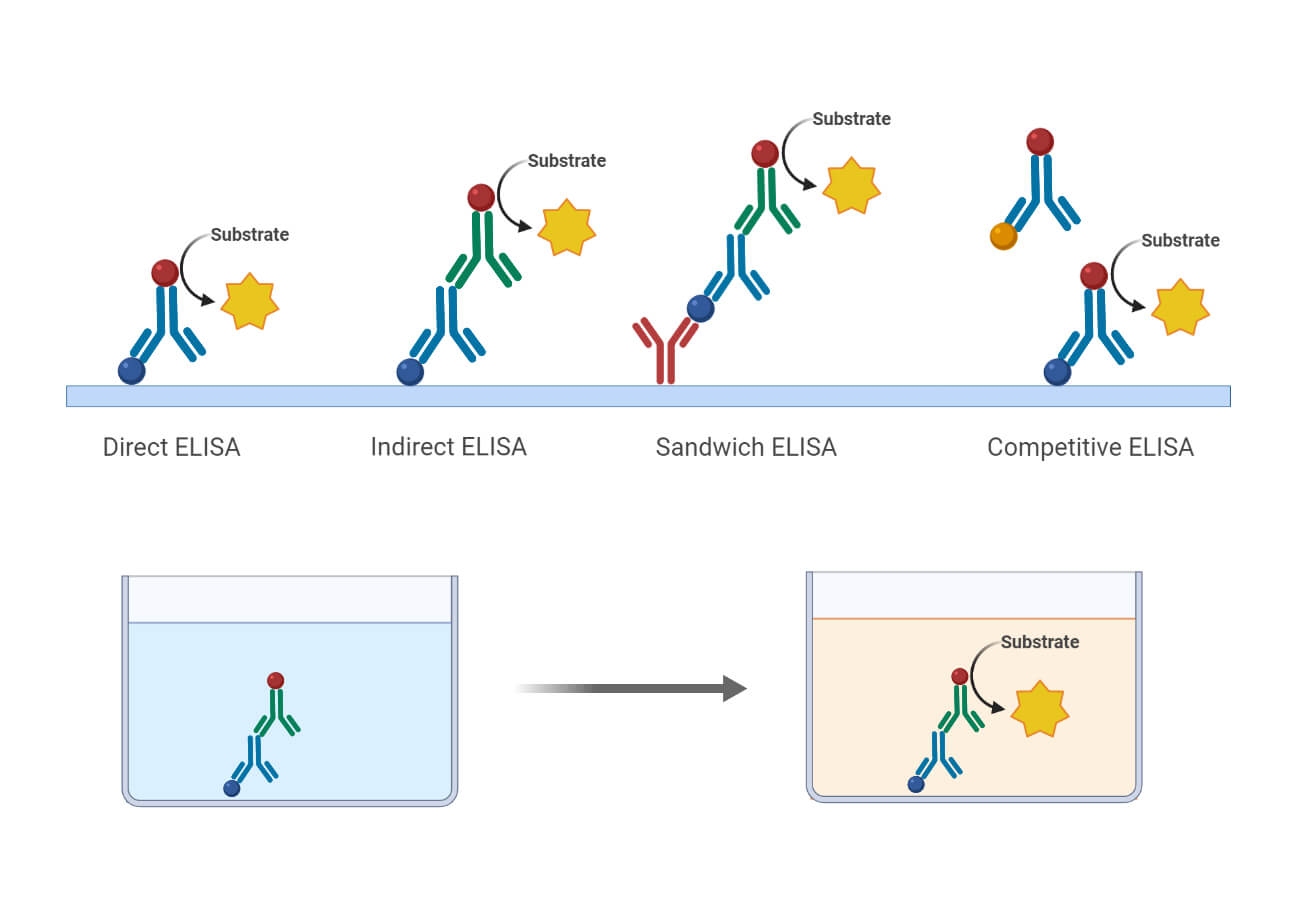
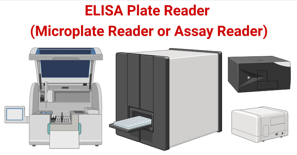
The test is conducted in an ELISA plate, also called a 96-well polystyrene plate or microplate, in one of two configurations: either the antigen or the capture antibody is bonded to the bottom of each well. The antigen is affixed to the bottom of each well when the assay’s objective is to determine whether an antibody is present in a clinical sample. The capture antibody is connected to the bottom of each well if the objective is to identify the antigen in a clinical sample (also known as a sandwich ELISA). The introduction and incubation of the target sample is only the beginning of the ELISA process. To determine whether binding between the capture and target antibody and antigen took place, the addition of a tagged detection antibody is the next step. A fluorescent, chemiluminescent, or chromogenic substance is used to label the detection antibody so that an ELISA reader can measure its concentration. ELISA plate reader is also termed as microplate reader or assay reader.

The spectrophotometry performed by ELISA readers measures the amount of light absorbed and reflected by a sample, such as a protein. Fluorescence and luminescence can also be measured with ELISA plate readers. When exposed to light, chemical dyes fluoresce—emitting a particular color or wavelength. A substance is identified and measured by the quantity of reflection, absorption, and color. Most ELISA readers also include software that transforms the raw intensity measurements into quantitative curves with dilution data and error. Calibration curves are included in the software to guarantee that the instrument is used to its fullest potential while maintaining the highest levels of quality and reproducibility.
Scientists use them for testing proteins and enzymes. They are also employed in the detection of HIV and the measurement of nucleic acids. It verifies the existence of antibodies or antigens of an infectious agent in an organism, antibodies from a vaccine, or auto-antibodies, for instance, in rheumatoid arthritis.
Interesting Science Videos
Principle of ELISA Plate Reader
The primary operation of the microplate reader is to measure the energy difference in light before and after passing through the test sample using a photoelectric colorimeter or spectrophotometer. The energy difference in light induced by the test substance’s absorption is typically linearly related to the test substance’s concentration.
The wavelength range of the microplate reader is typically 400 to 750 nm and is restricted by filters or diffraction gratings (nanometres). Some readers do their analyses between 340 and 700 nm while operating in the ultraviolet spectrum. Optic fibers are used in the optical system to provide light to the sample-containing microplate wells. The diameter of the light beam that travels through the sample ranges from 1 to 3 mm. The light emitted by the sample is detected by a detection device, which amplifies the signal and calculates the sample’s absorbance. It is transformed into data via a reading system, enabling the interpretation of the test results. Several microplate readers use double-beam light systems.
Types of ELISA Plate Reader
Based on the detection technologies, there are two main types of microplate readers, namely:
a. Dedicated Microplate Readers
One technology- absorbance, luminescence, or fluorescence- can be measured by specialized microplate readers. Although they lack the versatility of multimode microplate readers, they frequently provide greater sensitivity and dependability and are frequently more affordable.
b. Multimode Microplate Readers
Microplate readers with many detection modes can employ multiple technologies. Including all detection modalities into a single instrument is more complicated than specialized microplate readers. Some design concessions must be made, but they offer much flexibility. Multimode microplate readers have historically been expensive. However, some models offer a high degree of modularity, allowing the instrument to be built with only the required features.
Instrumentation of ELISA Plate Reader
- Source of light
Generally, xenon flash lamps are used as a source of light.
- Filters and monochromators
These are two competing wavelength selection technologies integral to microplate reader design. Monochromators change polychromatic light into monochromatic light. The light paths for excitation and detection include optical filters with particular wavelengths and bandwidths. Filters are less expensive compared to monochromators. These are placed between the source of light and the sample.
- Microplate
Microplates are designed in such a way that they have wells with a limited volume for the sample to be subjected. 96, 384, and 1536 wells per plate are the most popular well densities for screening.
- Microplate Reader
Different technologies such as absorbance, fluorescence, and luminescence are used to detect and quantify chemical, biological or physical events within the well of a microplate.
- Software for results
Different software can be installed and employed to simplify the process of data analysis.
Parts of ELISA Plate Reader
Some of the major parts of ELISA reader are listed and briefly explained. They are:
- Printer: ’PRINT’ key, when pressed, helps to get a printout of the current screen displayed with the help of the printer.
- Touch Screen: It displays the menus and the text box of the parameter, which together form the touch zone.
- Keypad: Several keys such as ‘YES’, ‘NO’,’PRINT’, ’ENTER’, ’ESC’, ’SHAKE’, and navigation keys are present with their respective functions.
- Plate Carrier/Holder: It holds the microtiter plate and moves it inside. A Stepper motor and timing belt is used to power it. It precisely positions each plate considerably below each channel’s optical path.
It also comprises an ON/ OFF switch panel, USB cable connector, SMPS, Cooling Fan, and Serial RS232 output.
Detection Methods Used
Different detection methods used by microplate readers are:
- Absorbance
The biological specimen is illuminated by a light source employing a certain wavelength throughout this detection process. Optical filters or monochromators are used to choose the wavelength in this case. The amount of initial light that passes through the biological sample is determined using a light detector that is situated on the opposite side of the plate from the well. Usually, the concentration of the target molecule present inside the wells is correlated with the amount of transmitted light. Additional conventional colorimetric analyses have been incorporated into the wells of an ELISA microplate reader to properly examine the specimens. They are employed in a number of tests, including ELISA, protein, and nucleic acid assays.
- Fluorescence Intensity Detection
Typically, the equipment for fluorescence detection is more expensive. The instrumentation for this kind of microplate reader consists of:
First optical system: The biological sample is illuminated by this system utilizing a certain wavelength of light. A monochromator or optical filter typically selects it.
The illumination system: As the light passes through the specimen, fluorescence (a type of light) is released.
Second optical system: The microplate reader’s emission system, which gathers the emitted light and distinguishes it from the excitation light. A monochromator system is utilized for this separation. A light detector called a photomultiplier tube is also used to measure the signal (PMT).
- Time-Resolved Fluorescence
The measurement of fluorescence intensity by the multi-well reader and the time-resolved fluorescence are somewhat comparable. The time of the excitation and measurement processes is the only difference between the two. The processes of photon excitation and photon emission are inherently synchronous when determining the sample’s fluorescence intensity. While the excitation process in the plate reader is going on, the biological sample’s light output is being monitored.
Use of certain fluorescent chemicals known as lanthanides is essential to the fluorescence optical system. These molecules have the odd characteristic of emitting in the reader for milliseconds after the excitation has ended. The fluorescein fluorescent dye, which emits after being stimulated for a few nanoseconds, is a common fluorescent dye used in fluorescence intensity measurements. In the microplate reader, a pulsed light source is used to excite lanthanides. Measurements are made following the excitation procedure. It is mostly used in the pharmaceutical industry for drug screening purposes.
- Luminescence
A chemical and biochemical process takes place inside the microplate reader’s wells to produce the luminescence detecting technique. In comparison to the methods mentioned above, luminescence detection is a simpler optical phenomenon. It is not necessary to use certain excitation wavelengths or a light source for the excitation of light beams.
The components of a typical luminescent optical system are:
- A light-tight reading chamber and
- A PMT detector
ELISA Plate Reader Operating Procedure
The mechanisms of operation for sandwich ELISA assay with the use of absorbance ELISA microplate reader are described below:
- The capture antibody binds ELISA microplate wells.
- The antigen in the sample binds to the capture antibody after adding it to the well.
- The unbound material is removed from the microplate, leaving only the target antigen.
- Add a detection antibody. An enzyme-conjugated detection antibody binds to a second location on the target antigen.
- Wash the microplate – Unbound antibodies are removed, leaving just those specific for the desired target.
- Add substrate. The substrate is transformed by the enzyme on the detecting antibody, resulting in a color change.
- Read the plate. The microplate reader picks up the colored reaction result and produces optical density (OD) measurements.
Operation mechanism of ELISA reader
Before starting
- To change the temperature in real-time, click on “instrument” on the left, then “temperature,” and then check “temperature control.”
- In the “Action” bar in the left column, drag it to the process list beneath the 96-well plate, drag it up and down to sort, then click the trash can to remove the choice. You can create procedures that are tailored to your requirements.
During starting
- Power up and let it warm up for ten minutes.
- To start a new process, launch the program and choose “New.”
- In the left column, choose “Absorbance.” In the center of the screen is a schematic of a 96-well plate.
- The hole that has been chosen will turn blue and can be used in accordance with the spotting hole.
- Insert the 96-well plate, configure the settings, and then select “start” to measure the absorbance.
- Wait a moment; an Excel file with the results and other parameters will soon be produced. Process the data by saving and copying the file to the hard drive.
- Compute the results – The amount of antigen in each sample is estimated and examined.
Applications of ELISA Plate Reader
- It facilitates the reader to diagnose various diseases based on the reaction. AIDS, Ebola, Lyme disease, rotavirus, cancer, anemia, and rotavirus are a few of these.
- It helps to detect any food allergens present in the food industry.
- It is used to perform antibody tests and protein and enzyme assays.
- It is also employed in the quantitation of nucleic acids.
Advantages of ELISA Plate Reader
- An ELISA plate reader requires about two to 100 microliters to obtain a response; this is a significantly smaller sample size.
- ELISA plate readers can analyze more samples (typically 96 wells) in less time.
- It facilitates both quantitative and qualitative analysis.
- A good microplate reader will perform tests with excellent accuracy, sensitivity, and speed.
Limitations of ELISA Plate Reader
- These are not portable, delicate, and expensive.
- Because of the prolonged exposure to air throughout this lengthy measuring process, biological and chemical constituents in the microplates may experience significant changes in their optical characteristics (for example, color and absorbance). As a result, the microplate reader’s full-spectrum measurement cannot accurately measure the test substance.
- The microplate reader only offers numerical data regarding the absorbance of specific wavelengths throughout the measuring process; it cannot obtain a real-time spectrogram.
Precautions
- The placement of the instrument should be away from high magnetic fields and interference voltages.
- The instrument needs to be in a location with noise levels that are no more than 40 dB.
- Direct sunlight should be avoided to prevent the aging of optical components.
- Operating ambient temperatures and humidity levels should range from 15 to 40 degrees Celsius and 15 to 85%, respectively.
- Be careful not to drop samples or reagents within the instrument or on its interior, and thoroughly clean the area after use.
- Don’t turn the power off while you’re measuring.
ELISA Plate Reader Examples
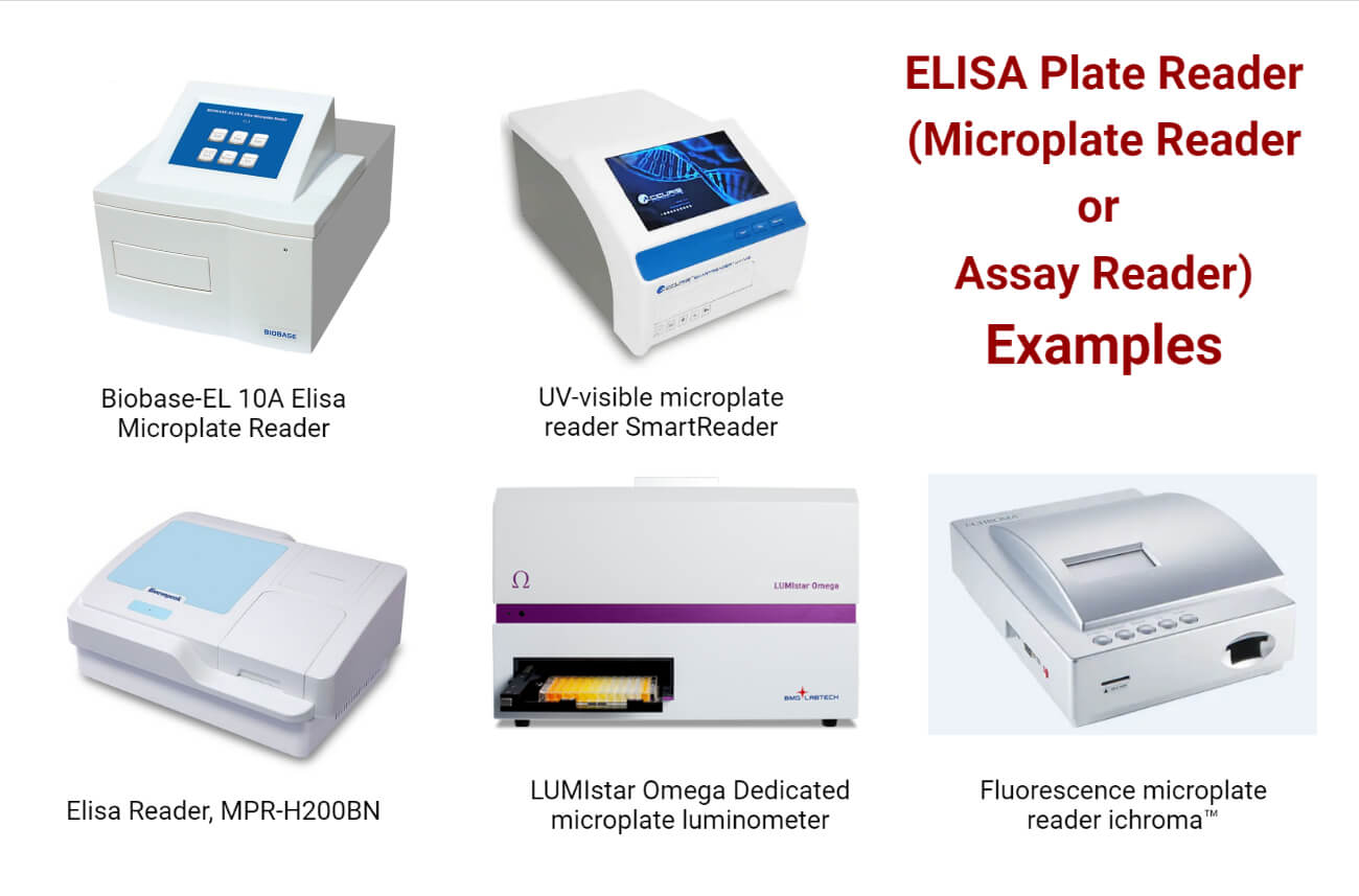
Absorbance microplate reader BIOBASE-EL 10A (Biobase)
- There are three distinct linear vibration plate functions.
- Dependable and accurate center positioning function.
- Optional eight-channel fiber measurement system.
- Set the food safety-specific inhibition rate measurement module to your desired settings.

UV-visible microplate reader SmartReader (BENCHMARK SCIENTIFIC)
- The monochromator-based optical system eliminates the need for filters and enables the exact selection of wavelengths from 200 nm to 1000 nm.
- The huge touchscreen command center’s user-friendly interface enables quick setup of new protocols and the changing and operating of already-established protocols.
- The experiment’s findings can be improved by controllable plate shaking and incubation.
- Enzyme kinetics and bacterial growth experiments can be processed at temperatures ranging from ambient +5°C to 45°C.
ELISA microplate reader MPR-H200BN (Bioevopeak)
- Single- or dual-wavelength measurements, thorough qualitative and quantitative data-evaluation functions with cut-offs, curve-fits, and transformation formulas.
- A reliable optical system with 8-channel optical fiber scanning and an automatic plate-centering mechanism precisely sets the cell’s center.
- Performing 12 tests total on a single plate.
Luminescence microplate reader LUMIstar Omega (BMG LABTECH)
- It offers incredibly accurate readings for all luminescence applications in kinetic and end-point assays.
- Additionally, multiple wavelength detection and BRET applications are made possible by an integrated filter wheel and the optional Simultaneous Dual Emission detection.
Fluorescence microplate reader ichroma™ (Boditech Med Inc.)
- Fast patient sample treatment and saved data search are made possible by the intuitive and user-friendly interface.
- Due to its solidity and moderate weight, it is simple to carry and install.
- There is storage for 100 patient test results.
- An optimized optical module for the immunofluorescence test.
- Both single and multi-measurement modes.
References
- http://cepclab.org.in/?p=436
- https://www.sepmag.eu/blog/elisa-reader
- https://bmet.ewh.org/bitstream/handle/20.500.12091/259/Microplate%20Reader.pdf?sequence=1&isAllowed=y
- https://www.protocols.io/view/microplate-reader-operating-procedure-6qpvrox62vmk/v2
- https://hudsonrobotics.com/elisa-microplate-reader-principle-and-uses/
- https://www.apexscientific.co.za/internal/producttraining/elisa-readers-and-washers/elisa-reader/
- https://www.diasource-diagnostics.com/var/ftp_diasource/IFO/DIA2000.pdf
- https://www.smacgigworld.com/blog/working-parameters-and-detection-methods-of-microplate-reader.php
- https://www.moleculardevices.com/applications/enzyme-linked-immunosorbent-assay-elisa
- https://www.diasource-diagnostics.com/var/ftp_diasource/IFO/DIA2000.pdf
- https://www.mdpi.com/2079-6374/12/5/284/htm
- https://www.rollmed.net/news/precautions-for-using-the-microplate-reader-61561251.html
- https://www.medicalexpo.com/prod/biobase/product-84845-651627.html
- https://www.medicalexpo.com/prod/bioevopeak/product-301335-1058984.html
- https://www.medicalexpo.com/prod/bmg-labtech/product-80666-663128.html
- https://www.medicalexpo.com/prod/boditech-med-inc/product-67878-444624.html
