Interesting Science Videos
What is Dengue and Dengue Virus?
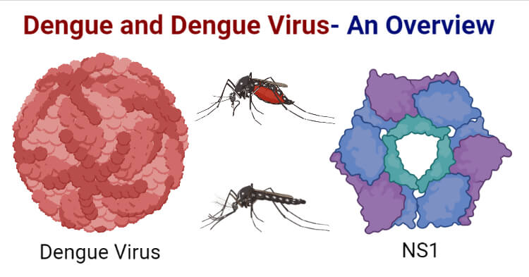
- Dengue is a mosquito-borne infection caused by a single-stranded RNA virus belonging to the Flaviviridae family known as the Dengue virus (DENV).
- It is transmitted by different species of female Aedes mosquito, primarily Aedes aegypti followed by Aedes albopictus and other species of Aedes.
- The dengue virus has 4 antigenically distinct serotypes; DENV-1, DENV-2, DENV-3, and DENV-4. A fifth serotype (DENV-5) has been detected in Malaysia in October 2013.
- The disease mainly occurs in tropical and subtropical regions with approximately 390 million dengue virus infections per year of which 96 million are symptomatic.
- With the dramatic increase in the global incidence of dengue in recent decades, about half of the world’s population is now at risk of the disease.
Structure and Genome of Dengue Virus
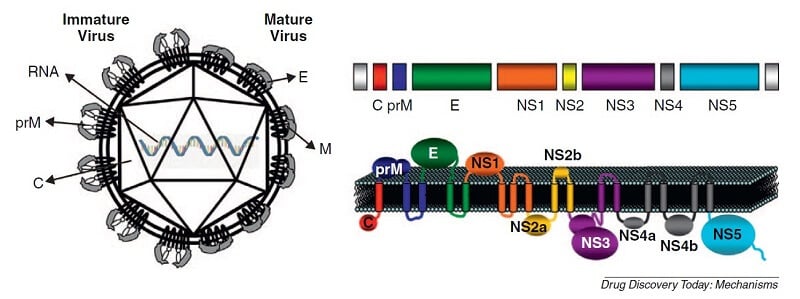
Figure: The dengue virus structure and genome. (a) Schematic representation of structural proteins in immature and mature dengue viral particles. (b) Schematic representation of the flavivirus genome organization and expression: structural and nonstructural protein-coding regions and topology of the polyprotein in the membrane of the endoplasmic reticulum. Image Source: https://doi.org/10.1016/j.ddmec.2011.09.001.
- The dengue virus genome consists of a single-stranded positive-sense RNA.
- The genome consists of ten genes that are translated into a single polyprotein that encodes for three structural proteins, namely, capsid (C), pre membrane (prM/M), envelope(E), and 7 nonstructural proteins (NS1, NS2A, NS2B, NS3, NS4A, NS4B, and NS5).
- The C protein is essential for specific encapsidation of its RNA genome, M protein plays an important role in the arrangement and maturation of the DENV particle and the E protein present on the viral surface is essential for the initial attachment of the virus to the host cell. The nonstructural proteins help in viral replication and assembly.
- The dengue virus is roughly spherical in its symmetry with approximately 50nm diameter. The nucleocapsid consists of the viral RNA and C proteins that form the core of the virus.
- The nucleocapsid is surrounded by a lipid bilayer of the viral envelope that is embedded with 180 E and M proteins across the layer.
- The dengue virus exhibits different conformations and surface structures during its maturation and infection, made possible by the flexibility of the envelope protein.
- The E protein is the major exposed antigen of the dengue virus against which the antibodies develop during the infection.
- The E protein of the four different serotypes of DENV shares 60-70% similarity in their amino acid sequence.
Epidemiology of Dengue
- Dengue has become one of the most widespread mosquito-borne diseases across the world with incidence increasing 30-fold in the last five decades.
- It is endemic to 128 countries currently, mostly targeting the developing countries. The most prevalent regions include America, South-East Asia, and Western Pacific, while Asia itself harboring almost 70% of the disease incidences.
- The highest number of dengue cases globally was reported in 2019.
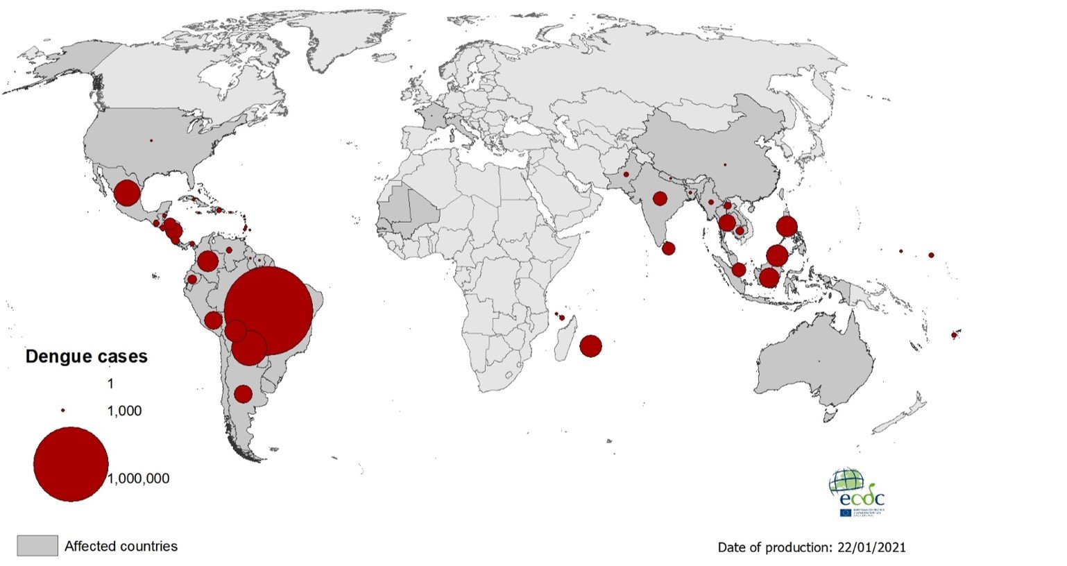
Figure: Geographical distribution of dengue cases reported worldwide, 2020. Image Source: European Centre for Disease Prevention and Control.
Transmission of Dengue Virus
- The transmission of the dengue virus occurs through the bite of female mosquitoes of the genus Aedes, primarily Aedes aegypti followed by Aedes albopictus and Aedes polynesiensis depending on the geographical region.
- After feeding on a DENV-infected individual, the virus replicates in the midgut of the mosquito, reaches the hemocoel and hemolymph, and further disseminates to other secondary tissues like the salivary glands.
- Studies have shown the presence of the viral particles within the nervous system, salivary glands, foregut, midgut, fat body, epidermal cells, ovary, and internal body wall lining cells of the mosquito whereas their absence from muscle, hindgut, and Malpighian tubules.
- The extrinsic incubation period (EIP), which is the time taken from entry of the virus to actual transmission to the new host, takes about 8-12 days with temperatures between 25-28°C.
- Once a mosquito is infected, it is capable of transmitting the virus throughout its lifetime.
- While the primary mode of transmission of DENV involves mosquito vectors, there is evidence of the possibility of maternal transmission (from pregnant mother to her child) that seems to be linked to the timing of the infection during the pregnancy. The babies conceived from mothers with DENV infection during pregnancy may suffer from pre-term birth, low birth weight, and fetal distress.
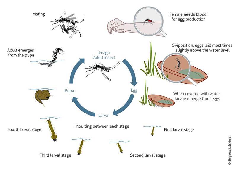
Figure: Life Cycle of Aedes albopictus. Image Source: Biogents Inc.
Dengue Virus Replication
The various stages in the viral replication include:
- Virus entry: The virus enters into the host cell through receptor-mediated endocytosis although the exact cell-surface receptors have not yet been identified. Some possible cellular receptors include various glycoproteins (i.e heparin sulfates), dendritic cell-specific intercellular adhesion molecule-3-grabbing non-integrin, or a mannose receptor. The viral E glycoprotein interacts with the receptors and mediates attachment and entry through the formation of endosomes.
- Membrane fusion and release of nucleocapsid: Once internalized, the low pH of the endosome induces a conformational change in the E protein and mediates fusion of the viral and endosomal membrane of the host cells, and releases the nucleocapsid into the cytoplasm.
- Translation replication of genome vRNA: The nucleocapsid opens to uncoat the viral genome which then uses the host cell’s machinery to replicate itself. It uses the ribosomes on the host’s ER and translates the positive-sense viral RNA into a single polyprotein. The polyprotein is further cleaved into individual structural and nonstructural proteins by NS2B or NS3 viral protease and host proteases.
- Virus assembly: The newly synthesized viral RNA gets enclosed in the C protein forming the nucleocapsid and entering the rough ER where it gets enveloped and surrounded by the M and E proteins.
- Transport of immature viral particles: The immature virus particles travel through the secretory pathway-Golgi apparatus. The low pH of the Golgi network causes rearrangement of the prM by furin protease and finally forms the infectious mature form.
- Release of mature virus: The mature dengue viruses are then finally released from the cell and become capable of infecting other host cells.
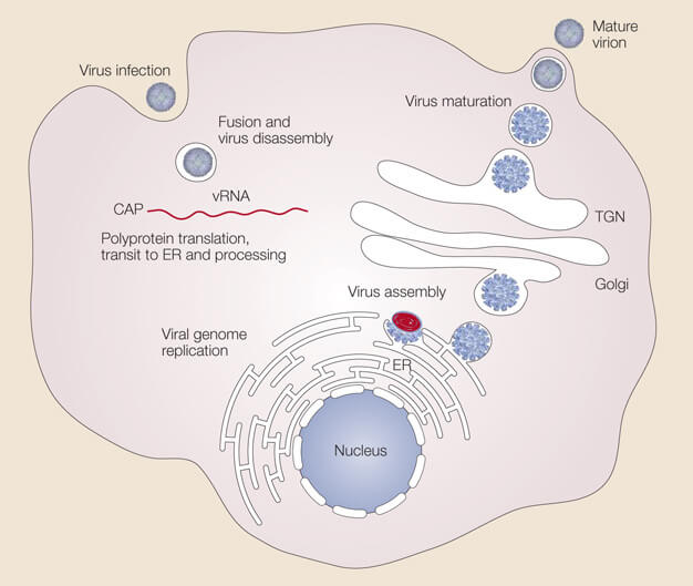
Figure: Dengue Virus Replication. Image Source: https://doi.org/10.1038/nrmicro1067.
Pathogenesis of Dengue
- Most primary infections of dengue with a certain serotype are asymptomatic and only lead to mild febrile illness, although some individuals can develop hemorrhagic fever.
- Secondary infection with a different serotype may however lead to severe clinical manifestations like dengue hemorrhagic fever (DHF) or dengue shock syndrome (DSS).
- An individual develops immunity after infection from a serotype against it, however, they are still prone to future infections from the other serotypes of the virus as the heterologous immunity declines gradually in 1-2 years.
- Many viral, as well as host factors, contribute to the pathogenesis of DENV such as the non-structural protein 1 (NS1) viral antigen, dengue virus genome variations, subgenomic RNA, antibody-dependent enhancement (ADE), T-cells, anti-DENV NSI antibodies, and autoimmunity.
- After the virus enters the bloodstream of an individual through the mosquito bite, it infects the nearby skin cells as well as specialized immune cells known as Langerhans cells.
- The viral antigen is then presented on the surface of Langerhans cells that travel to the lymph nodes and help activate the innate immune response.
- The dengue virus further infects the macrophages and monocytes and further disseminate throughout the body which results in viremia in the individual.
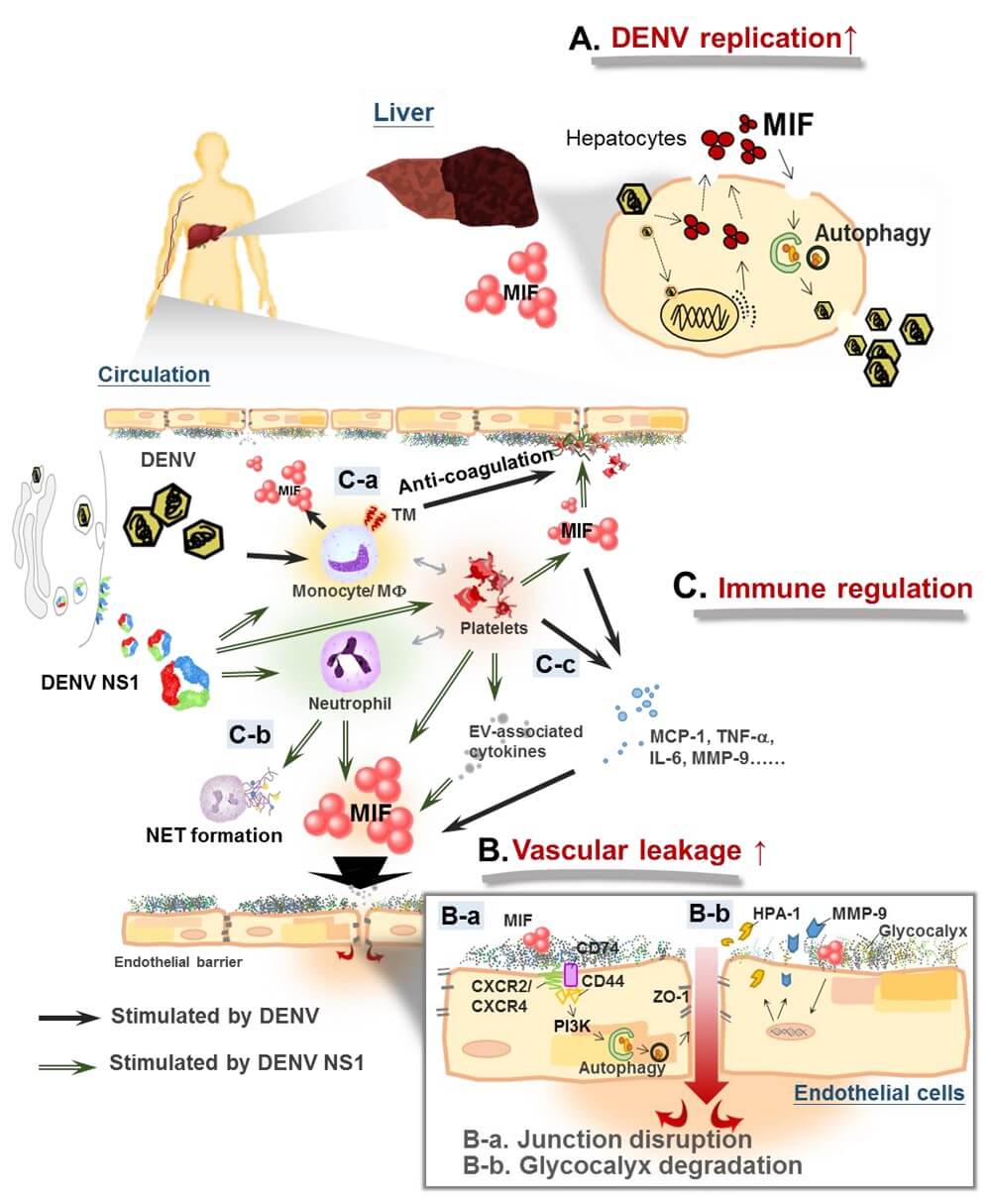
Figure: Macrophage migration inhibitory factor (MIF) mediates immune response crosstalk in dengue pathogenesis. Image Source: https://doi.org/10.3390/microorganisms8060891.
Immune response
- To fight against the virus, the infected cells produce a type of cytokines known as the interferons that affect the viral replication and also activate the innate and adaptive immune responses.
- Once the adaptive immune response activates, B cells produce IgM and IgG antibodies that are released in the blood and lymph fluid.
- These cells recognize the viral particles and help neutralize them.
- The cytotoxic T cells also help in the adaptive immune response.
- The complement system is also activated by the innate immune response.
Clinical Manifestations of Dengue
Dengue infections may be asymptomatic or symptomatic causing undifferentiated fever, dengue fever, dengue hemorrhagic fever, or dengue shock syndrome.
Undifferentiated fever
It usually follows primary infections but can also occur during secondary infections and is often not distinguishable from other viral infections.
Dengue fever
It may occur during primary or secondary infections. It is usually self-limiting and lasts for 5-7 days. Some of the common symptoms include:
- High fever
- Severe headache (especially in the retro-orbital area)
- Arthralgia
- Myalgia
- Anorexia
- Abdominal discomfort
- Vomiting
- Macular popular rash (in some cases)
The clinical features also vary according to the age of the patients. Leukopenia and thrombocytopenia are usually observed in patients of all ages. Some cases of DF may also lead to bleeding complications like epistaxis, gastrointestinal bleeding, gingival bleeding, haematuria, and menorrhagia (in women).
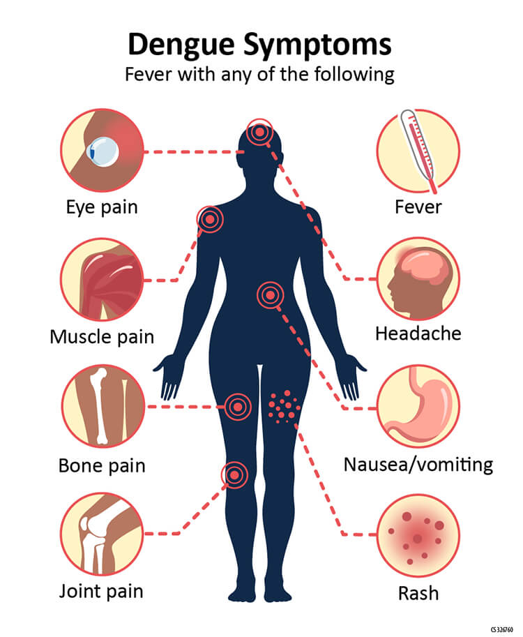
Figure: Dengue Symptoms. Image Source: CDC.
Dengue hemorrhagic fever
It usually occurs after secondary infection but may sometimes occur after primary infection as well. It causes an acute onset of fever for 2-7 days and the patient shows hemorrhagic manifestations with a positive tourniquet test.
General symptoms include:
- High intermittent fever
- Severe headache
- Flushing
- Myalgia and arthralgia
- Vomiting
- Anorexia
- Acute abdominal pain
Other than these, it may also cause bleeding manifestations like bleeding from gums, petechiae, haematemesis, menorrhagia in females, and circulatory disturbances like low blood pressure, tachycardia, etc. Further complications may also include encephalitis, liver failure, myocarditis, and disseminated intravascular coagulation that may lead to massive bleeding.
Dengue Shock Syndrome
Dengue shock syndrome is characterized by DHF accompanied by unstable pulse pressure, narrow pulse pressure, restlessness, cold blotchy skin, circumoral cyanosis, and circulatory disturbances. DSS is generally associated with high mortality rates in patients.
Diagnosis of Dengue Virus
- Diagnosis of dengue infection based only on clinical symptoms are unreliable as it produces a wide range of symptoms that are often non-specific.
- Early detection and treatment of the infection can prevent the development of severe manifestations as well as the death of the patients.
- The dengue infections can be diagnosed by virus isolation techniques in suitable culture media, detection by RNA amplification tests, detection of viral antigens by ELISA, or through rapid kit tests.
- NS1 antigen detections are also commercially available for its diagnosis that give results within a few hours. Rapid antigen kits can provide results in less than an hour. However, these tests are not type-specific and cannot differentiate the serotypes of the virus causing infection.
- The virus isolation, serotype identification, and nucleic acid detection are usually performed for diagnosis till the 5th day of infection.
- The virus isolation and serotype identification techniques use whole blood, serum, and tissues as their specimen and provide results in about 1-2 weeks while the nucleic acid procedure gives results in 1-2 days and uses tissue, whole blood, serum, and plasma as its specimen. ELISA and rapid tests give results in 1-2 days and 30 minutes respectively with serum, plasma, whole blood as the specimen and are performed after 5 days of infection.
- IgM antibodies are detectable within a week after infection and remain so for about 3 months and are indicative of recent DENV infection while IgG antibodies take longer to develop in the body but remain in the body for years and hence are indicative of a past DENV infection.
Treatment of Dengue
- Specific treatment for dengue fever is not yet available.
- Fluid replacement and antipyretic therapy with paracetamol or acetaminophen can be used during the febrile phase.
- Non-steroid anti-inflammatory drugs like ibuprofen and aspirin should be avoided in case of dengue infections. Intravenous fluid administration is considered mandatory in cases of shock, severe vomiting, and prostration.
- Crystalloids make up the first-line treatment choice of intravenous fluid.
- A major concern of bleeding in patients with low platelet count must be managed by transfusion of fresh whole blood.
Vaccines of Dengue
- A tetravalent vaccine is preferred to protect individuals from all four dengue virus types.
- Although the various types of vaccines (live attenuated, chimeric, DNA, and subunits vaccine) have shown positive results, none of them entirely suffice for routine use.
- Dengvaxia has been licensed and made available in some countries for people between the ages of 9 to 45 years.
- However, the WHO has recommended the vaccine to people who have had previous dengue infection only as it imposes an increased risk of disease severity in people who get infected with the virus for the first time after their vaccination.
Prevention and control of Dengue
- Control of mosquito breeding should be done by preventing egg-laying in or near water through environmental modification and management.
- Proper covering and cleaning of domestic use stored water must be done regularly.
- Mosquito repellents, insecticides, coils, vaporizers, oils, window screens, etc. must be used to prevent mosquito bites.
- Clothes that do not expose too much skin must be worn in areas with a higher risk of dengue infections.
- Education and awareness programs should be conducted for people and communities that provide information to them about the cause, clinical symptoms, treatments, and management of the disease.
References
- (2009). Dengue guidelines for diagnosis, treatment, prevention and control : new edition. World Health Organization. https://apps.who.int/iris/handle/10665/44188
- Bäck AT, Lundkvist A. Dengue viruses – an overview. Infect Ecol Epidemiol. 2013;3:10.3402/iee.v3i0.19839. Published 2013 Aug 30. doi:10.3402/iee.v3i0.19839
- Hasan S, Jamdar SF, Alalowi M, Al Ageel Al Beaiji SM. Dengue virus: A global human threat: Review of literature. J Int Soc Prev Community Dent. 2016;6(1):1-6. doi:10.4103/2231-0762.175416
- Henchal EA, Putnak JR. The dengue viruses. Clin Microbiol Rev. 1990;3(4):376-396. doi:10.1128/CMR.3.4.376
- https://www.nature.com/scitable/topicpage/host-response-to-the-dengue-virus-22402106/
- Bhatt, P., Sabeena, S.P., Varma, M. et al. Current Understanding of the Pathogenesis of Dengue Virus Infection. Curr Microbiol 78, 17–32 (2021). https://doi.org/10.1007/s00284-020-02284-w
- Martina BE, Koraka P, Osterhaus AD. Dengue virus pathogenesis: an integrated view. Clin Microbiol Rev. 2009;22(4):564-581. doi:10.1128/CMR.00035-09
- Niyati Khetarpal, Ira Khanna, “Dengue Fever: Causes, Complications, and Vaccine Strategies“, Journal of Immunology Research, vol. 2016, Article ID 6803098, 14 pages, 2016. https://doi.org/10.1155/2016/6803098.
- https://www.who.int/news-room/fact-sheets/detail/dengue-and-severe-dengue
- https://www.nature.com/scitable/topicpage/dengue-viruses-22400925/
