Bacteria are pleomorphic i.e. they show different morphological characters. Based on their shape, bacteria are mainly classified into four main groups viz. coccus (spherical shaped), bacillus (rod-shaped), spirilla or spirochete (spiral-shaped), and vibrio (comma shaped). However, some species show distinguishing morphological figures such as elliptical, oval, V- or J- shaped filamentous, branched, coccobacillus, etc.

Interesting Science Videos
Coccobacilli Characteristics
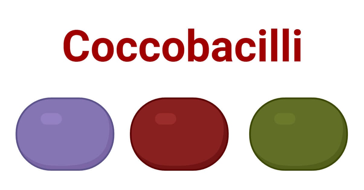
Coccobacilli are bacteria halfway between bacillus (rod) and coccus (spherical) in shape. They are very short bacillus, with their breadth nearly equal to their cell length. Under the microscope, they appear as slightly compressed spheres or ovals.
- The sizes of coccobacilli are different among different species, but they are generally smaller and stouter than bacillus.
- Both Gram-positive and Gram-negative coccobacilli have been identified. Despite their unique shape, they resemble their characters to other respective types of bacteria – like cell wall and membrane compositions, metabolic characters, genome, cytoplasmic contents, etc.
Some Examples of Coccobacilli
There are different pathogenic coccobacillus bacteria causing a wide range of infections including respiratory tract infections (RTIs), meningitis, STDs (sexually transmitted diseases), etc. Some common pathogens with associated diseases are listed below:
Haemophilus influenzae
Haemophilus influenzae are small Gram-negative pleomorphic, facultative anaerobic coccobacilli in the family Pasteurellaceae. They measure from 0.3 to 1 μm in diameter. They are associated with a wide range of infections like meningitis, bloodstream infections, otitis media, conjunctivitis, bacterial influenza, RTIs, etc.
Haemophilus ducreyi
Haemophilus ducreyi are Gram-negative, facultative anaerobic, coccobacilli causing genital ulceration/chancroid. Under the microscope, they appear as coccobacilli arranged in short chains or clumps, generally in a ‘rail-track’ arrangement.
Coxiella burnetii
Coxiella burnetii is a small Gram-negative, obligate intracellular coccobacilli bacterium causing Q fever. They are found in two morphological forms; the dormant small cell variant (SCV) measuring about 0.3 μm in diameter and the metabolically active large cell variant (LCV) measuring about 2.0 μm in diameter.
Brucella spp.
Brucella spp. are small, nonmotile, facultatively intracellular Gram-negative coccobacilli measuring about 0.6 to 1.5 μm by 0.5-0.7 μm in diameter. They are responsible for brucellosis in humans and cattle and other domestic animals.
Bordetella pertussis
Bordetella pertussis is a small strict aerobic, Gram-negative coccobacilli measuring about 0.8 by 0.4 μm in size and causing pertussis (whooping cough).
Chlamydia trachomatis
Chlamydia trachomatis is a Gram-negative obligate parasitic coccobacilli bacterium causing chlamydia (a major STD) and other diseases like cervicitis, urethritis, salpingitis, lymphogranuloma venereum, etc.
Gardnerella vaginalis
Gardnerella vaginalis is small anaerobic or facultative anaerobic Gram-variable coccobacilli causing bacterial vaginosis.
Yersinia pestis
Yersinia pestis is Gram-negative, facultative anaerobic coccobacilli measuring about 0.5 μm by 1 to 2 μm in size and causing plague diseases.
Francisella tularensis
Francisella tularensis is a very small capsulated, Gram-negative coccobacilli measuring just about 0.3 to 0.7 μm by 0.2 μm in size. F. tularensis is responsible for tularemia, a zoonotic disease manifesting fever, skin ulcers, lymphadenitis, pneumonia, upper RTI, etc.
Kingella kingae
Kingella kingae is a Gram-negative beta-hemolytic coccobacilli measuring about 0.6 to 1 μm by 1 to 3 μm in size and causing a wide range of diseases like bacteremia, osteoarthritis, endocarditis, etc.
Acinetobacter baumannii (during stationary growth phase)
A. baumannii is a Gram-negative opportunistic pathogenic coccobacilli bacterium responsible for different nosocomial infections. They typically measure about 0.9 to 1.6 m by 1.5 to 2.5 μm in size.
References
- Khattak ZE, Anjum F. Haemophilus influenzae Infection. [Updated 2023 Apr 27]. In: StatPearls [Internet]. Treasure Island (FL): StatPearls Publishing; 2023 Jan-. Available from: https://www.ncbi.nlm.nih.gov/books/NBK562176/
- Humphreys, T. L., & Janowicz, D. M. (2015). Haemophilus ducreyi: Chancroid. Molecular Medical Microbiology (Second Edition), 1437-1447. https://doi.org/10.1016/B978-0-12-397169-2.00080-9
- lton GG, Forsyth JRL. Brucella. In: Baron S, editor. Medical Microbiology. 4th edition. Galveston (TX): University of Texas Medical Branch at Galveston; 1996. Chapter 28. Available from: https://www.ncbi.nlm.nih.gov/books/NBK8572/
- Mohseni M, Sung S, Takov V. Chlamydia. [Updated 2023 Aug 8]. In: StatPearls [Internet]. Treasure Island (FL): StatPearls Publishing; 2023 Jan-. Available from: https://www.ncbi.nlm.nih.gov/books/NBK537286/
- Yagupsky, P. (2015). Kingella kingae: Carriage, Transmission, and Disease. Clinical Microbiology Reviews, 28(1), 54-79. https://doi.org/10.1128/CMR.00028-14
- https://www.onlinebiologynotes.com/francisella-tularensis-morphology-culture-characteristics-pathogenesis-diagnosis-and-treatment/
- Howard, A., Feeney, A., & Sleator, R. D. (2012). Acinetobacter baumannii: An emerging opportunistic pathogen. Virulence, 3(3), 243-250. https://doi.org/10.4161/viru.19700
- https://www.osmosis.org/answers/coccobacilli
- Yersinia pestis. (2023, August 19). In Wikipedia. https://en.wikipedia.org/wiki/Yersinia_pestis
- https://www.dictionary.com/browse/coccobacilli
- https://byjus.com/biology/bacteria/#classification
- https://microbiologyinfo.com/different-size-shape-and-arrangement-of-bacterial-cells/
- https://microbeonline.com/size-of-bacteria/
- https://www.vetbact.org/?artid=181
- https://microbeonline.com/gram-negative-cocci-coccobacilli-medical-significance-list-bacteria-diseases/#Gram_Negative_Coccobacilli
- https://www.osmosis.org/learn/Haemophilus_ducreyi_(Chancroid)
