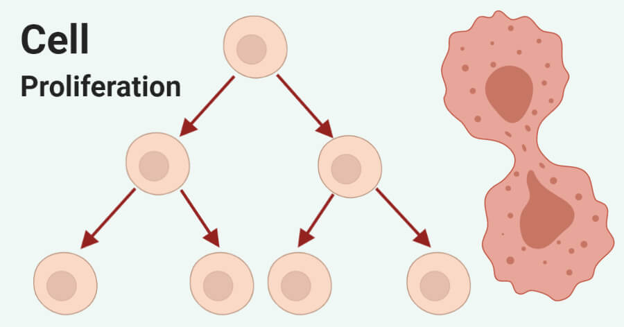
Cell proliferation is the process of increase in the number of cells which occurs as a result of regulated cell growth and cell division.
- Cell proliferation is responsible for the exponential increase in the cell number, resulting in rapid tissue growth.
- The process is balanced by cell division and cell differentiation or cell death, which maintains an appropriate number of cells in the body.
- Cell proliferation is an important process that is essential for fundamental living processes like embryonic development, organ growth, and other physiological processes.
- Even though growth is a consequence of both an increase in cell number as well as the cell size, the term ‘proliferation’ indicates the increase in cell number as a function of time.
- The increase in cell number results via a sequence of steps involving cell growth and cell division.
- Cell proliferation, however, if disturbed or affected by unwanted factors, might result in abnormal cell division leading to dreadful diseases like cancer.
- The progression through the cell cycle or cell proliferation is regulated by closely related factors in the body.

Interesting Science Videos
Normal cell proliferation
Normal cell proliferation is indicated by a balance between cell growth, cell division, cell differentiation, and cell death.
- All of these processes are equally important during normal cell proliferation, and any changes in these processes might result in abnormal cell proliferation leading to diseases.
- During the process of the normal cell cycle, cell proliferation and apoptosis play important roles.
- The process of normal cell proliferation is highly regulated to ensure that the number of cells in the body remains virtually unchanged with the number of new cells formed being equal to the number of cell deaths.
- In the case of normal cell proliferation, when an appropriate number of cells are produced, the inhibitory factors trigger a negative feedback mechanism to reduce and eventually stop the rate of cell growth. This ensures that the number of cells doesn’t exceed the required number.
- Most of the cells found in living beings (except cells of bone marrow and epidermis) remain in a non-proliferative state unless they are stimulated to divide for repair, which is considered the normal stage.
- There are different genes in the body that ensure that cell proliferation occurs in a normal fashion. Two of the important genes include proto-oncogenes and tumor suppressor genes.
- Normal cell proliferation requires both normal cell division and cell differentiation. In normal cell division, the cell cycle occurs via tightly regulated steps that together ensure the division of a cell.
- In the case of normal cell differentiation, the new cells formed either differentiate to become different cells with different functions (stem cells) or help in tissue repair.
- In order to ensure normal cell proliferation, it is important that each step in the cell cycle occurs in the designated way to ensure normal cell growth, division, and differentiation.
Abnormal cell proliferation
Abnormal cell proliferation is indicated by the over-proliferation of cells and accumulation of such cells in an abnormal fashion.
- Abnormal cell proliferation can occur either due to abnormal cell division or by abnormal cell differentiation.
- The biology of division, differentiation, and apoptosis is mostly similar in both normal and abnormal cell proliferation, with differences in the process of the relation of these processes.
- In the case of abnormal cell proliferation, these processes are not regulated, which causes abnormalities.
- Abnormal cell proliferation results in the formation of neoplasm which is an abnormal mass of tissue where the growth and division of the cells are uncoordinated and continue in the same excessive manner even after the cessation of the stimuli that caused it.
- In such neoplasms, four distinct cellular functions are inappropriately regulated.
- At first, the negative feedback mechanism of normal cell proliferation is ineffective, followed by distortion in the differentiation process.
- The cells might either be blocked at a particular stage of differentiation or might be differentiated into inappropriate abnormal cell types.
- Abnormal differentiation then results in destabilization of the chromosomal and genetic organization of the cells. Finally, the process of apoptosis or regulated cell death is affected.
- All of these processes, individually or together, result in abnormal cell proliferation.
- Abnormal cell proliferation doesn’t always result in cancerous cells. Abnormal cell proliferation of non-cancerous cells results in hyperplasia where the uncontrolled dividing of cells leads to the formation of tissue with an unusually large number of structurally normal cells.
- However, the process of abnormal cell proliferation in cancerous cells results in the formation of tumors that can be either benign or malignant.
- The process of abnormal cell proliferation is initially stimulated by genetic alteration, caused by different factors, like mutations and radiation that affect the cell proliferation process.
Cell proliferation assay
- The measurement of the rate of cell proliferation provides insightful information about the process of cell proliferation as well as cell growth.
- The rate of cell proliferation might differ in different cells depending on the cell type, stage of development, and the presence of various growth factors.
- The measurement of the rate of cell proliferation in in-vitro studies is helpful in different cytotoxic and apoptosis studies, where that in in-vivo studies helps in understanding the cell proliferation steps and the developmental stages of different animals.
- These studies also help in understanding the pathology of cancerous tissues.
- The basic principle of all cell proliferation is the detection of cell viability and the measurement of the number of cells or the changes in the rate of division of cells.
- Depending on the factors that are considered during the measurement of cell proliferation, these assays can be divided into four classes;
1. Rate of deoxyribonucleic acid (DNA) synthesis-based assays
- During the process of cell proliferation, DNA replication takes place before cell division; thus, the determination of the rate of DNA replication has a direct relationship with the rate of cell proliferation.
- The amount of DNA synthesized can be measured by adding labeled nucleotides or synthetic nucleoside analogs in the growth medium.
- These nucleosides are then incorporated into the DNA which can then be measured.
- For the determination of DNA synthesis, radioactive-labeled 3H-thymine and BrdU (5-bromo-2’deoxiuridine) are used.
- The amount of DNA is then measured by measuring the radioactivity produced by these nucleotides via immunoassays like ELISA.
2. Metabolic activity-based assays
- These assays are based on the measurement of levels of essential metabolites like ATP or the reduction potential of cells for salts like tetrazolium or resazurin.
- The concentration of ATP as well as the ratios of NADPH/NADP, FADH/FAD, FMNH/FMN, and NADH/NAD all increase during cell proliferation.
- In the presence of these metabolite intermediates, the tetrazolium salts are reduced to a formazan produced via cellular dehydrogenases or reductases that can then be detected by the resulting colorimetric change.
- In the case of resazurin dye, the nonfluorescent blue redox dye gets reduced into resorufin, which produces red fluorescence.
- The measurements in this assay are based on the absorption of media containing dye solution using a spectrophotometer or microplate reader.
3. Antigens associated cell proliferation assay
- The rate of cell proliferation can also be determined by detecting the antigens present on the proliferating cells, which are not present in non-proliferating cells by using antigen-specific antibodies.
- For this, different antibodies are produced that are targeted at different antigens found in proliferative cells in different stages.
- One classic example of this is the anti-K-676 antibodies that are used in the detection of protein expressed during the S< G2 and M phases of the cell cycle in humans.
- These antibodies, however, do not detect cells in the G0 and G1 phases of the cell cycle as S, G2, and M phases of the cell cycle are proliferative phases, but G0 and G1 are resting phases.
- The antigen-antibody association can then be detected by immunological assays by either observing under a fluorescence microscope or by measuring with a flow cytometer.
4. Variations in adenosine triphosphate (ATP) concentration
- The detection of the amount of ATP present inside the cells is directly proportional to the rate of proliferation of cells as there is a tight regulation of intracellular ATP within the cells.
- The ability to synthesize ATP is lost as the cells lose membrane integrity or cell viability. Any remaining ATP in the cell is also removed by ATPases.
- The concentration of ATP in cells can be measured by using a bioluminescence-based assay involving the enzyme luciferase and the substrate luciferin.
- In the presence of ATP, luciferase produced light, and the intensity of the light is directly proportional to the concentration of ATP.
- This assay has an advantage as it is rapid and doesn’t involve any incubation step for the conversion of substrate into a colored compound.
- Besides, the assay is also highly sensitive as it can be used with less than 10 cells per well.
- As the measurement of ATP is a kit-based method, the protocol involved in the assay might differ from the manufacturer.
Cell proliferation and differentiation
- The growth of multicellular organisms is characterized by the rapid proliferation of embryonic cells, followed by their differentiation to produce different specialized cells that together make up different tissues and organs.
- The proliferation of embryonic cells results in the formation of a mass of cells which then undergo differentiation, which makes up an essential step of cell growth.
- With the differentiation of such cells, the rate of proliferation usually decreases as the well-differentiated cells usually don’t proliferate.
- Most of the cells in adult living beings are arrested in the G0 stage of the cell cycle.
- Very few types of differentiated cells never divide again, but others can resume proliferation as required in order to replace the lost cells during injury or cell death.
- The rate of proliferation also depends on the extent of differentiation with poorly differentiated cells being highly proliferative and well-differentiated cells being either unable to proliferate or proliferating at a prolonged rate.
- In the case of abnormal proliferation of cancerous cells, the neoplasms are either poorly differentiated or moderately differentiated.
- Thus, the degree of differentiation affects the nature of cancers. Aggressive cancers are generally poorly differentiated, whereas less aggressive cancers are moderately or well-differentiated.
- Cell proliferation and differentiation are closely related and have an inverse relationship.
- Precursor cells continue to divide until the fully differentiated state, whereas terminal differentiation of cells usually coincides with proliferation arrest and permanent exit from the division cycle.
- Besides, the decision between proliferation and differentiation is made during the G1 phase depending on the cell’s response to external signals.
- Thus, the process of cell proliferation and differentiation might occur one after the other depending on the external signals or the cellular mechanisms.
Cell proliferation and cancer cells

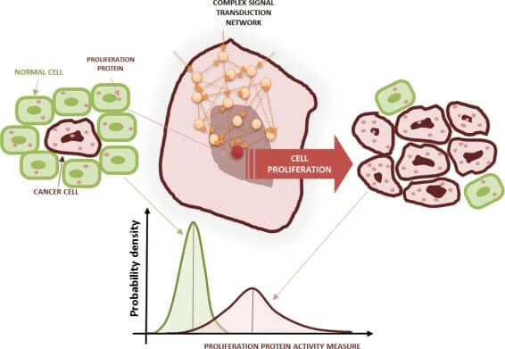
Figure: Cancer cell proliferation. Green cells are normal cells and red cells are tumor cells. The proliferation activity of normal and tumor cells can be measured by looking at the activation of a proliferation protein, which is driven by a complex network based on protein interactions. In a population of cells the proliferation activity can be described by means of the probability density for the proliferation protein; e.g., phosphorylated form of the extracellular signal-regulated kinase (ERK). The plots at the bottom show an example of the probability density of a proliferation indicator in the tumor (red line) and normal cells (green line), respectively. Image Source: https://doi.org/10.1186/s12918-015-0216-5
- Cancer, on the fundamental level, is the abnormal proliferation of cells, resulting in an increase in tumor cell number, which ultimately results in adverse effects on the host.
- Based on multiple studies, it has been speculated that cell proliferation plays a critical role in different steps of cancer development.
- Cell proliferation in either a limited cycle or in multiple cycles affects initiation, promotion or selection, and progression during cancer development.
- Even though cell proliferation is considered a significant risk factor for cancer, there have not been studies that show that cell proliferation acts as a carcinogen.
- As cell proliferation is the central and key phenotypic expression in all types of malignant tumors, its involvement in cancer is undisputed, but the extent of the effect is not yet understood.
- Cell proliferation in cancer is accompanied by not only the disturbance in the balance between cell division and cell death but also by other associated changes like invasion and metastasis.
- It has been observed that in both in vivo and in vitro cancer development, different carcinogenic agents require at least a single round o cell proliferation to initiate the process.
- Different carcinogenic agents like chemicals, radiation, and viruses interact with the genome of the target cells and result in different forms of mutations.
- The fixation of the genomic changes in order to generate mutation often requires a round of proliferation.
- Thus, in the cases of cancers where mutations play an important role, the round of cell proliferation is essential.
- It has been assumed that the rate-limiting step of the carcinogenic process of cell proliferation and not the exposure to an adequate level of a carcinogen.
- However, there are some criticisms to this belief supported by evidence like the lack of association between cell proliferation and cancer occurrence in some organs and tissues.
- Therefore, abnormal cell proliferation is an important factor that affects the initiation and progression of cancer of various organs but cannot be considered the driving factor in all forms of cancers.
Cell proliferation and stem cells
- The self-renewing tissues of the living beings like the hematopoietic system and the skin are capable of renewing themselves because they contain a small population of precursor cells, called the stem cells.
- Stem cells have an unaltered proliferative self-renewal capacity extending to at least a single lifespan of an organism.
- In the case of embryonic stem cells, the mechanism of the cell cycle is quite different. The first cell cycles of these cells lack gap phases but consist of alternating S and M phases.
- The stem cells either in the embryonic stage or later, divide to form daughter cells that either differentiate or remain as stem cells.
- The process of stem cell proliferation can be distinctly observed in the case of blood cell differentiation.
- Most blood cells have limited life spans, ranging from less than a day to a few months. These cells are then continually replaced by other cells that are produced from a common stem cell.
- The daughter cells from the stem cells then differentiate into different types of cells that function distinct functions.
- Among all stem cells, the embryonic stem cells have the broadest differentiative capacity as they can give rise to all of the differentiated cell types in adult organisms.
- The proliferation of stem cells is an important process that has therapeutic applications as the stem cells isolated from adult tissues give rise to not only to blood cells but also many other cell types like neurons and connective tissues.
Cell proliferation diseases
Based on the understanding of the biochemical mechanisms of pathogenesis of several diseases, it has been observed that excessive proliferation and turnover of cellular matrix contribute to the pathogenesis. Cell proliferation plays a vital role in degenerative diseases where the cells do not replicate enough as well as in cancers, where the cells proliferate excessively. Some of the common diseases that result from excessive proliferation include;
1. Cancer
- Cancer is a disease that results from abnormal proliferation of different kinds of cells in the cells; thus, there hundred of different types of cancer that might vary in behavior and response to treatment.
- The most important feature of cancer is tumor clonality, where large tumors are developed from single cells that proliferative excessively.
- Abnormal proliferation is influenced by various factors like mutations, radiation, chemical agents, or even viruses.
- At the molecular level, cancer is a multistep process beginning with mutations followed by the selection of cells with the progressively increasing capacity of these cells for proliferation, survival, and invasion.
2. Pulmonary fibrosis
- Pulmonary fibrosis is a chronic lung disease caused by the excessive accumulation of extracellular matrix resulting in remodeling of the lung architecture.
- Idiopathic pulmonary fibrosis is the most common form of this disease with no known effective therapy.
- Based on the pathological findings of the diseases, it has been assumed that the disease is caused as a result of an imbalance between proliferation and apoptosis of fibroblasts and the accumulation of the matrix substances.
- During the disease, fibroblast proliferation surpasses apoptosis resulting in the accumulation of the extracellular matrix.
- It has been speculated that factors like inflammation, oxidative stress, and coagulation disturbances aid in the progression of the disease.
3. Rheumatoid arthritis
- Rheumatoid arthritis is a chronic inflammatory disease caused by the inflammation of synovial joints.
- Rheumatoid arthritis is stimulated by the dysregulated proliferation of T-cell. During the diseases, the balance between apoptosis and cell proliferation is disturbed, which skews the balance towards cell survival.
- This phenomenon is often termed as ‘apoptosis resistance’ where the fibroblast-like synoviocyte cells proliferate excessively.
Factors affecting cell proliferation
The overall process of cell proliferation occurs via different phases of the cell cycle. The cell cycle and, in turn, cell proliferation is influenced by a combination of cell-intrinsic and cell-extrinsic interactions or factors. Some of the common factors that influence cell proliferation include;
1. Growth factors
- Growth factors are large and complex proteins that are mostly present on the plasma membrane.
- These factors, after binding to the receptors, can induce transmission of a signal to the cytoplasm through the activation of the kinase.
- The signal is then transduced to the cell nucleus via secondary messengers.
2. Enzymes
- Different proteins and enzymes are also involved in the regulation of cell proliferation where they act to provide energy and help in rapid energy synthesis.
- Other proteins might have housekeeping functions that help in the growth of cells to maintain a metabolic balance.
- Besides, complex multi-enzyme complexes are also known to regulate the process of DNA synthesis and cell replication.
- Cyclin-dependent kinases are an important group of enzymes that arrest the cells in the G1 phase as a response to stimuli like growth factors, DNA damage, cellular stress, and differentiation.
3. Genes
- There are different specific genes that are involved in the regulation of cell proliferation.
- Some of the common ones include proto-oncogenes that are involved in normal cell growth but might become an oncogene after being overactive and causing the growth of cancer cells.
- Tumor suppressor genes are the other types of genes that are responsible for the production of tumor suppressor proteins that helps in cell growth.
- In addition to these, there are several other genes like H2AFZ and EXO1 that directly affect several steps of the cell proliferation process.
References and Further Readings
- Dang C, Gilewski TA, Surbone A, et al. Cell Proliferation. In: Kufe DW, Pollock RE, Weichselbaum RR, et al., editors. Holland-Frei Cancer Medicine. 6th edition. Hamilton (ON): BC Decker; 2003. Available from: https://www.ncbi.nlm.nih.gov/books/NBK12640/
- Andreeff M, Goodrich DW, Pardee AB. Cell Proliferation and Differentiation. In: Kufe DW, Pollock RE, Weichselbaum RR, et al., editors. Holland-Frei Cancer Medicine. 6th edition. Hamilton (ON): BC Decker; 2003. Chapter 3.Available from: https://www.ncbi.nlm.nih.gov/books/NBK13866/
- Andreeff M, Goodrich DW, Pardee AB. Proliferation. In: Kufe DW, Pollock RE, Weichselbaum RR, et al., editors. Holland-Frei Cancer Medicine. 6th edition. Hamilton (ON): BC Decker; 2003. Available from: https://www.ncbi.nlm.nih.gov/books/NBK13035/
- Feitelson, Mark A et al. “Sustained proliferation in cancer: Mechanisms and novel therapeutic targets.” Seminars in cancer biology 35 Suppl,Suppl (2015): S25-S54. doi:10.1016/j.semcancer.2015.02.006
- Cooper GM. The Cell: A Molecular Approach. 2nd edition. Sunderland (MA): Sinauer Associates; 2000. Cell Proliferation in Development and Differentiation.Available from: https://www.ncbi.nlm.nih.gov/books/NBK9906/
- Chao, Dennis L et al. “Cell proliferation, cell cycle abnormalities, and cancer outcome in patients with Barrett’s esophagus: a long-term prospective study.” Clinical cancer research : an official journal of the American Association for Cancer Research 14,21 (2008): 6988-95. doi:10.1158/1078-0432.CCR-07-5063
- Cooper GM. The Cell: A Molecular Approach. 2nd edition. Sunderland (MA): Sinauer Associates; 2000. The Development and Causes of Cancer.Available from: https://www.ncbi.nlm.nih.gov/books/NBK9963/
- Debta P. (2016) Abnormal Cells Proliferation. Adv Cancer Prev. 1:109. doi: 10.4172/2472-0429.1000109
- Yadav, Kamalendra & Singhal, Nitin & Rishi, Vikas & Yadav, Hariom. (2001). Cell Proliferation Assays. eLS.
- Adan A, Kiraz Y, Baran Y. Cell Proliferation and Cytotoxicity Assays. Curr Pharm Biotechnol. 2016;17(14):1213-1221. doi: 10.2174/1389201017666160808160513. PMID: 27604355.
- Koyanagi M, Kawakabe S, Arimura Y. A comparative study of colorimetric cell proliferation assays in immune cells. Cytotechnology. 2016 Aug;68(4):1489-98. doi: 10.1007/s10616-015-9909-2. Epub 2015 Aug 18. PMID: 26280992; PMCID: PMC4960196.
- Ruijtenberg, Suzan, and Sander van den Heuvel. “Coordinating cell proliferation and differentiation: Antagonism between cell cycle regulators and cell type-specific gene expression.” Cell cycle (Georgetown, Tex.) 15,2 (2016): 196-212. doi:10.1080/15384101.2015.1120925
- Farber E. (1995) Cell Proliferation as a Major Risk Factor for Cancer: A Concept of Doubtful Validity. Cancer Research 55. 3759-3762.
- Liu, L., Michowski, W., Kolodziejczyk, A. et al.The cell cycle in stem cell proliferation, pluripotency and differentiation. Nat Cell Biol 21, 1060–1067 (2019). https://doi.org/10.1038/s41556-019-0384-4
- Malemud, Charles J. “Defective T-Cell Apoptosis and T-Regulatory Cell Dysfunction in Rheumatoid Arthritis.” Cells 7,12 223. 22 Nov. 2018, doi:10.3390/cells7120223
- Todd, N.W., Luzina, I.G. & Atamas, S.P. Molecular and cellular mechanisms of pulmonary fibrosis. Fibrogenesis Tissue Repair5, 11 (2012). https://doi.org/10.1186/1755-1536-5-11
- Schonrock, Kuhlmann, Adler, Bitsch, & Bruck. (1998). Identification of glial cell proliferation in early multiple sclerosis lesions. Neuropathology and Applied Neurobiology, 24(4), 320–330.doi:10.1046/j.1365-2990.1998.00131.x
- Sporn MB, Harris ED Jr. Proliferative diseases. Am J Med. 1981 Jun;70(6):1231-5. doi: 10.1016/0002-9343(81)90832-9. PMID: 6263092.
- Loeb MJ. Factors affecting proliferation and differentiation of Lepidopteran midgut stem cells. Arch Insect Biochem Physiol. 2010 May;74(1):1-16. doi: 10.1002/arch.20349. PMID: 20422716.
