X-rays make up X-radiation, a form of electromagnetic radiation.
Most X-rays have a wavelength ranging from 0.01 to 10 nanometers, corresponding to frequencies in the range 30 petahertz to 30 exahertz (3×10¹⁶ Hz to 3×10¹⁹ Hz) and energies in the range 100 eV to 100 keV, produced by the deceleration of high-energy electrons.
Interesting Science Videos
What is X-Ray Spectroscopy?
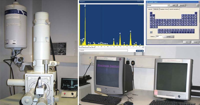
X-ray spectroscopy is a general term for several spectroscopic techniques for the characterization of materials by using x-ray excitation.

Source: University College Cork
Principle of X-Ray Spectroscopy
- XRF works on methods involving interactions between electron beams and x-rays with samples.
- It is made possible by the behavior of atoms when they interact with radiation.
- When materials are excited with high-energy, short wavelength radiation (e.g., X-rays), they can become ionized.
- When an electron from the inner shell of an atom is excited by the energy of a photon, it moves to a higher energy level.
- When it returns to the low energy level, the energy which it previously gained by the excitation is emitted as a photon which has a wavelength that is characteristic for the element (there could be several characteristic wavelengths per element).
- Thus atomic X-rays emitted during electronic transitions to the inner shell states in atoms of modest atomic number.
- These X-rays since have characteristic energies related to the atomic number, and each element therefore has a characteristic X-ray spectrum which can be used to identify the element.
Working of X-Ray Spectroscopy
- An XRF spectrometer works because if a sample is illuminated by an intense X-ray beam, known as the incident beam, some of the energy is scattered, but some is also absorbed within the sample in a manner that depends on its chemistry.
- The incident X-ray beam is typically produced from a Rh target, although W, Mo, Cr and others can also be used, depending on the application.
- When x-ray hits sample, the sample emits x-rays along a spectrum of wavelengths characteristic of the type of atoms present.
- If a sample has many elements present, the use of a Wavelength Dispersive Spectrometer allows the separation of a complex emitted X-ray spectrum into characteristic wavelengths for each element present.
- Various types of detectors used to measure intensity of emitted radiation.
- The intensity of the energy measured by these detectors is proportional to the abundance of the element in the sample.
- The exact value for each element is derived from standards from prior analyses from other techniques.
Instrumentation of X-Ray Spectroscopy
Components for X-ray spectroscopy are:
- X-ray generating equipment (X-ray tube)
- Collimator
- Monochromators
- Detectors
A. X-ray generating equipment (X-ray tube)
- X-rays can be generated by an X-ray tube.
- X-rays tube is a vacuum tube that uses a high voltage to accelerate the electrons released by a hot cathode to a high velocity.
- The high velocity electrons collide with a metal target, the anode, creating the X-rays.
B. Collimators
- A collimator is a device that narrows a beam of particles or waves.
- Narrow mean to cause the directions of motion to become more aligned in a specific direction (i.e., collimated or parallel).
- Collimation is achieved by using a series of closely spaced ,parallel metal plates or by a bundle of tubes ,0.5 or less in diameter.
C. Monochromator
- Monochromator crystals partially polarize an unpolarized X-ray beam.
- The main goal of a monochromator is to separate and transmit a narrow portion of the optical signal chosen from a wider range of wavelengths available at the input.
Types of Monochromator
- Metallic Filter Type
- Diffraction grating type
D. X-ray Detectors
The most commonly employed detectors include:
- Solid State Detectors
- Scintillation Detectors
Solid-State Detectors
- The charge carriers in semiconductor are electrons and holes.
- Radiation incident upon the semiconducting junction produces electron-hole pairs as it passes through it.
- Electrons and holes are swept away under the influence of the electric field, and the proper electronics can collect the charge in a pulse.
Scintillation detectors
Scintillation detectors consist of a scintillator and a device, such as a PMT (Photomultiplier tubes), that converts the light into an electrical signal.
- It consists of an evacuated glass tube containing a photocathode, typically 10 to 12 electrodes called dynodes, and an anode.
- Electrons emitted by the photocathode are attracted to the first dynode and are accelerated to kinetic energies equal to the potential difference between the photocathode and the first dynode.
- When these electrons strike the first dynode, about 5 electrons are ejected from the dynode for each electron hitting it.
- These electrons are attracted to the second dynode, and so on, finally reaching the anode.
- Total amplification of the PMT is the product of the individual amplifications at each dynode.
- Amplification can be adjusted by changing the voltage applied to the PMT.
Applications of X-Ray Spectroscopy
X-ray spectrometry is used in a wide range of applications, including
- Research in igneous, sedimentary, and metamorphic petrology
- Soil surveys
- Mining (e.g., measuring the grade of ore)
- Cement production
- Ceramic and glass manufacturing
- Metallurgy (e.g., quality control)
- Environmental studies (e.g., analyses of particulate matter on air filters)
- Petroleum industry (e.g., sulfur content of crude oils and petroleum products)
- Field analysis in geological and environmental studies (using portable, hand-held XRF spectrometers)
Advantages of X-Ray Spectroscopy
- X-ray spectroscopy is an excellent method to determine the structure of a compound.
- In the event when other spectral methods fail to reveal a compound’s identity, X-ray spectroscopy is the method of choice for structural determination where the other parameters such as bond lengths and bond angles are also determined.
Limitations of X-Ray Spectroscopy
- The technique requires the availability of a compound as a single crystal.
- Most chemists find this process very tedious, time consuming and it requires a skillful hand.
References
- https://slideplayer.com/slide/5770713/
- http://instructor.physics.lsa.umich.edu/adv-labs/X-Ray_Spectroscopy/x_ray_spectroscopy_v2.pdf
- https://www.iucr.org/__data/assets/pdf_file/0013/733/chap16.pdf
- http://www.issp.ac.ru/ebooks/books/open/X-Ray_Spectroscopy.pdf
- https://en.wikipedia.org/wiki/X-ray_spectroscopy
- https://www.britannica.com/science/X-ray-spectroscopy
- http://umich.edu/~jphgroup/XAS_Course/Harbin/Lecture1.pdf
- https://www.ixasportal.net/ixas/images/ixas_mat/Giuliana_Aquilante.pdf
- http://www.spectroscopyonline.com/x-ray-spectroscopy
- https://www.slideshare.net/nanatwum20/xrf-xray-fluorescence
