- Hypersensitivity refers to increased reactivity or exaggerated immune response of a host to an antigen to which it has been previously exposed.
- According to the most widely accepted classification of Gell and Coombs, there are four main types of hypersensitivity reactions as type I, II, III and IV.
- Type I, II and III are immunoglobulin-mediated (immediate) hypersensitivity reactions while type IV reaction is lymphoid cell-mediated or simply cell mediated hypersensitivity (delayed-type).

- Type IV hypersensitivity reaction also known as cell mediated hypersensitivity or delayed type of hypersensitivity is the T lymphocytes mediated destruction of cells along with dendritic cells, macrophages and cytokines playing major roles.
- The reaction is mediated by specific subsets of CD4+ helper T cells (Th-1 and Th-17 cells) or by CD8+ cytotoxic T cells.
- Type IV hypersensitivity occurs 24 hours after contact with an antigen, usually starting at 2 or 3 days and often last for many days.
- For this reason, type IV hypersensitivity reaction is termed as “delayed hypersensitivity”.
- Type IV hypersensitivity is unique in that, unlike the first three types of hypersensitivity which are antibody mediated, type IV hypersensitivity is cell mediated and also a delayed reaction.
Interesting Science Videos
Mechanism of Type IV (Cell Mediated) Hypersensitivity
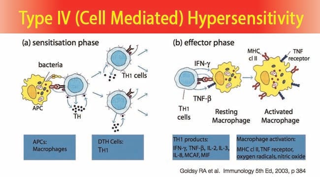
- In type IV hypersensitivity reaction, when a sub-population of CD4 Th1 cells encounter certain type of antigens, they produce cytokines which induce a localized inflammatory reaction mediated by non-specific inflammatory cells most prominently marcrophages.
- The antigens involved may be either intracellular pathogens such as Mycobacterium tuberculosis, Listeria monocytogens, Histoplasma capsulatum, Herpes Simplex virus etc. or contact antigens such as Nickel salts, Poison ivy etc.
- The reaction is accomplished in two phases: the initial sensitization phase and the later effector phase.
- In the sensitization phase, the primary contact with the antigen is established. During this period, specific Th cells are sensitized and are clonally expanded.
- In the effector phase, a subsequent exposure to the same antigen induces the delayed type hypersensitivity response.
- Cytokines such as IL-2 and interferon gamma is released inducing the further release of other Th1 cytokines, which mediates the immune response activating macrophages and other non-specific inflammatory cells.
- Activated CD8+ T cells on the other hand, destroy target cells on contact, whereas activated macrophages produce hydrolytic enzymes and on presentation with certain intracellular pathogens, transform into multinucleated giant cells.
Variants of Type IV (Cell Mediated) Hypersensitivity
-
Contact Hypersensitivity
- Occurs after sensitization with simple chemicals (eg, nickel, formaldehyde), plant materials (poison ivy), some cosmetics, soaps, and other substances.
- Small molecules enter the skin and acting as haptens attach to body proteins which induce cell-mediated hypersensitivity particularly in the skin.
- On subsequent exposure to the agent, the sensitized person develops erythema, itching, vesication, eczema, or necrosis of skin within 12-48 hours.
-
Tuberculin -type Hypersensitivity
- Occurs due to sensitization of soluble antigens of microorganisms during many infectious diseases.
- It is exemplified by tuberculin reaction in which when a small amount of tuberculin is injected into the skin of a person previously exposed to Mycobacterium tuberculosis, mononuclear cells accumulate in the subcutaneous tissue along with abundance of CD4 Th1 cells leading to induration and redness developing at its peak in 24–72 hours.
Examples of Type IV (Cell Mediated) Hypersensitivity
- The tuberculin reaction (Mantoux test): This is a ‘recall’ response to purified mycobacterial antigens and is used as the basis of a diagnostic skin test for an immune response to tuberculosis.
- Granuloma formation: The inability to kill intracellular pathogens in macrophages often results in a chronic stimulation of the pathogen specific T cells. The cytokines produced are responsible for ‘walling off’ the macrophages containing the persistent antigens and thus the production of granulomas.
- Allergic contact dermatitis: Environmental chemicals, metals or topical medications causing epidermal necrosis, inflammation, skin rash and blisters.
- Type-1 diabetes: The killing of the pancreatic islet cells by cytotoxic T cells resulting in insulin deficiency.
References
- Brooks, G. F., Jawetz, E., Melnick, J. L., & Adelberg, E. A. (2010). Jawetz, Melnick, & Adelberg’s medical microbiology. New York: McGraw Hill Medical.
- Kristine, K. (2009, September 23). Real examples of hypersensitivity reactions. Retrieved from https://www.pathologystudent.com/real-examples-of-hypersensitivity-reactions.
- Owen, J. A., Punt, J., & Stranford, S. A. (2013). Kuby Immunology (7 ed.). New York: W.H. Freeman and Company.
- Playfair, J., & Chain, B. (2001). Immunology at a Glance. London: Blackwell Publishing.
