Trichophyton is a dermatophyte fungus classified along with Epidermophyton and Microsporum spp. Dermatophytic fungi are known to cause of dermatophytosis, a cutaneous infection of the skin, hair, and nails, in humans and animals. Trichophyton spp specifically causes tinea infections such as tines pedis (Athletes foot), tinea capitis (ringworms), tinea crusi (jock itch), and infections of the nails, beards, skin, and scalp. It has a global distribution, with varients of its species spread in various parts of the globe. There are several species of Trichophyton each with prevalent occurrences and effects depending on transmission and the host of infection.
Interesting Science Videos
Habitat of Trichophyton spp
- Trichophyton spp is distributed globally with prevalence in the tropical and Mediterranean regions.
- Based on habitat prevalence, there are two types of trichophyton groups i.e anthropophilic and zoophilic.
- Anthrophilic Trichophytons are those transmitted from person to person and cause human infections, while zoophilic Trichophytons have an animal-human transmission route causing either, human or animal infections.
- Another group is the geophilic species that exist in the soil developing on soil decomposing keratinous materials such as hair, feathers, hooves, and horns.
- Geophilic species may cause both human and animal infections.
Morphology of Trichophyton spp
- Trichophyton morphologies vary depending on the growth temperature, growth medium, and the type of species.
- Most of these species produce either macroconidia or microconidia or both.
- The Macroconida are small, cigar-shaped (cylindrical), with thin to the thick-walled cell walls, and they tend to occur in clusters.
- Their occurrence shapes and sizes depending on the medium of growth.
- Microconidia, are formed in small numbers or they may be produced numerously.
- The microconidia, appear spherical in shape, they may occur singly or in clusters and they are produced on the terminal branches on the hyphal structures.
- Trichophytons produces spiral-shaped hyphal structures, from which the conidial spores originate.
- They do not form conidiophores.

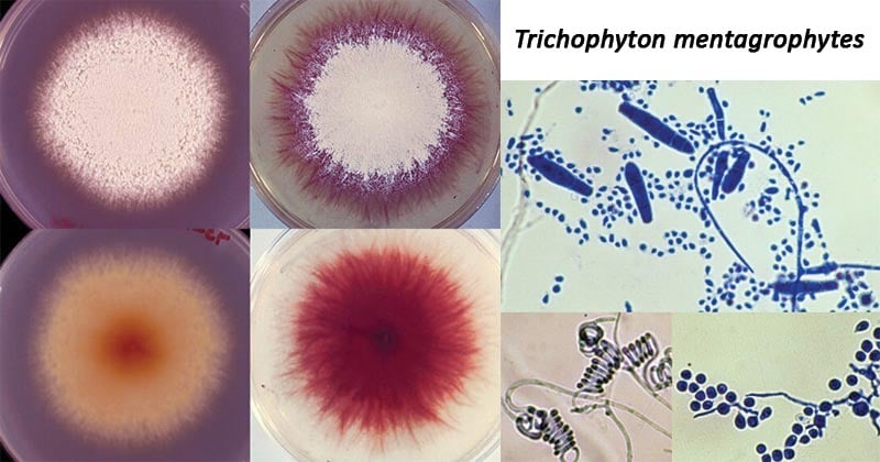
Figure: Cultures of Trichophyton mentagrophytes; Microconidia, macroconidia, chlamydoconidia, and spiral hyphae in T. mentagrophytes. Image Source: The University of Adelaide.
Cultural Characteristics of Trichophyton spp
- Trichophytons grow well in Sabouraud Dextrose agar within 2 weeks at room temperature producing cylindrical, smooth-walled macroconidia and characteristics microconidia.
- Different growth textures can be seen depending on the type of species, such as Trichophyton mentagrophyte produces a cottony to granular growth, with grape clusters of spherical microconidia on terminal branches. They also form coiled or spiral hyphae
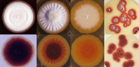
- Trichophyton rubrum develops white, cottony surface and deep red nondiffusible pigment in reverse growth. They produce small pear-shaped microconidia
- Trichophyton tonsurans produce a flat, powdery to a velvety colony on the obverse surface that becomes reddish-brown on reverse; with elongated microconidia.

Figure: Trichophyton rubrum produces both red and yellow pigments. Culture colors may range from none to dark red to dark yellow, with all combinations in between. The images above show the same strain grown on lactritmel agar that promotes red pigmentation and on mycobiotic agar that shows an underlying yellow pigmentation. Image Source: The University of Adelaide.
Selected Species of Trichophyton
Trichophyton verrucosum
- This species has a global distribution.
- It causes ringworms in cattle, donkeys, dogs, goats, sheep, and horses.
- In culture, at room temperature, it produces characteristic growth colonies which are flat, white/cream color, with an occasional dome, with a glabrous texture.
- Formation of macroconidia are rare, and when formed, they have a rat-tail or string bean shape, while microconidia are tear-shaped.
- Human infection is associated with inflammation of hair shafts (ectothrix), tinea capitis, and tinea barbae.
Trichophyton rubrum
- It is the most common cause of dermatophytosis.
- Its occurrence is worldwide.
- It is anthropophilic species of dermatophytes.
- It is a keratinophilic filamentous fungus.
- It affects human feet, skin, and fingernails, causing chronic infections on these body parts.
- Its infections may be characterized by granulomatous lesions.
- Microscopically, endothrix and ectothrix invasion may be observed on infected hair.
- The cultural appearances include pigmented shades of white to cream to yellow to brown, and to deep red, with a waxy, cotton, or smooth texture.
- Reverse pigmentation may be characterized by colorless to yellowish to yellow-brown to wine red.
- They form both macroconidia and microconidia.
- Macroconidia may or may not occur, and if they occur, they are in large numbers, with terminal projections.
- Microconidia may or may not occur, with varying shapes and sizes. They may be slender clavate to pyriform.
- The formation of the macroconidia and microconidia depends majorly on the type of infection caused by the fungus, for example, tinea pedis and onychomycosis produces slender clavate microconidia with no macroconidia, while in tinea corporis, microconidia are produced which occur in large numbers with clavate to pyriform shapes and moderate numbers of macroconidia which are thin-walled, cigar-shaped.
Trichophyton mentagrophyte
- This is a zoophilic species with a cosmopolitan distribution.
- It commonly affects animals such as mice, guinea pigs, cats, horses, sheep, and rabbits.
- It is known to cause skin and scalp infections, known as ringworms, in humans, majorly in rural areas.
- It grows in SDA media at room temperature producing a cottony-granular growth with clustered occurring macroconidia.
- Few or no microconidia are produced.
- At 37°C, they produce 2-5 celled thin-walled, club-shaped to cigar-shaped macroconidia and numerous microconidia which also over in clusters, or may occur singly.
Pathogenesis of Trichophyton spp
Transmission of Trichophyton spp
- The transmission of Trichophyton is via direct or indirect contact with an infected individual or through spores.
- Transmission can be anthropophilic or zoophilic or geophilic.
- Anthrophilic transmission is by direct contact or through fomites, such as contaminated towels, clothing, shared shower stalls.
- Zoophilic transmission is from animals to the human host.
- Geophilic transmission occurs from soil or infected animals into the human host.
- Dermatophytosis caused by Trichophyton can be contracted through contact with infected humans or animals or with contaminated objects, such as towels, bedding, manicure appliances, and hairbrushes.
- Furthermore, Trichophyton related skin conditions are frequently spread in areas such as pools, spas, locker rooms, and shared shower facilities.
Clinical Manifestations of Trichophyton spp infections
Trichophytons as dermatophytes cause dermatophytosis infection in humans. These infections include:
Tinea corporis (ringworm)
- It is caused by Trichophyton mentagrophytes.
- It affects the nonhairy, smooth skin
- It is characterized by circular patches with advancing red, vesiculated border and central scaling.
- The infection is pruritic
Tinea pedis (athlete’s foot)
- It is caused by Trichophyton rubrum and Trichophyton mentagrophytes
- It occurs in the interdigital spaces on feet of persons wearing shoes
- It is characterized by acute infection associated with itching, and red vesicular and chronic infection also characterized by itching, scaling, fissures.
Tinea cruris (jock itch)
- It is caused by Trichophyton rubrum and Trichophyton mentagrophytes
- It is associated with groin Erythematous scaling lesions in the intertriginous area., and the lesions are pruritic.
Tinea capitis
- It is caused by Trichophyton tonsurans, Trichophyton mentagogrophytes.
- It affects the scalp hair, the endothrix (fungus inside hair shaft), ectothrix (fungus on the surface of hair).
- It is characterized by circular bald patches with short hair stubs or broken hair within hair follicles.
- Kerion hypersensitivity reaction is rare.
Tinea barbae
- It affects the beard hair
- It is characterized by Edematous, erythematous lesion
- It is caused by Trichophyton mentagrophytes, Trichophyton rubrum, Trichophyton verrucosum
Tinea unguium (onychomycosis)
- It affects the nail
- The nails thickened or crumbling distally; discolored; lusterless.
- It is usually associated with tinea pedis
- It is caused by Trichophyton rubrum, Trichophyton mentagrophytes
Trichophytid Reaction
- During dermatophytosis, some patients may develop hypersensitivity to the elements produced by the fungus, developing allergic reactions known as dermatophytids commonly on the hands. The diagnosis includes a skin test.
Laboratory Diagnosis of Trichophyton spp infections
Specimen: Skin scrapings, nail scrapings, hair plucks
Microscopic Examination
- 10–20% potassium hydroxide, with or without calcofluor white, and the specimens skin or nails or hair.
- Generally, Trichophyton shows branching hyphae or chains of arthroconidia (arthrospores) in skin or nail samples.
- In hairs, Trichophyton tonsurans and Trichophyton violaceum are observed by the production of arthroconidia inside the hair shaft (endothrix)
Cultural Examination
- Using inhibitory mold agar or SDA medium containing cycloheximide and chloramphenicol which suppresses mold and bacterial growth. Growth is observed within 1-3 weeks at room temperature.
- On SDA medium, Trichophytons produce a cottony growth, or velvety, depending on the species.
- Macroconidia and microconidia develop ranging in size, shape, and occurrence depending on the species of Trichophyton.
Treatment of Trichophyton spp infections
- Treatment is by use of topical antifungals or antibiotics.
- Tinea capitis (scalp infection) are treated for several weeks with oral administration of griseofulvin or terbinafine. Shampoos and miconazole creams are also helpful. ketoconazole and itraconazole have also shown effectiveness.
- Itraconazole and terbinafine are used for the treatment of tinea corporis, tinea pedis.
- Tinea unguium (onychomycosis) or Nail infections are treated with oral itraconazole or terbinafine or surgical removal of the nail.
Prevention and Control of Trichophyton spp infections
- Maintenance of proper hygiene limits the spread of Trichophyton.
- Proper and good ventilation can reduce contracting dermatophytes infection such as Athletes foot, which requires wearing cotton socks and well-ventilated footwear.
References
- Medical Microbiology by Jawertz, 23rd Edition
- Microbiology by Prescott, 5th Edition
- https://mycology.adelaide.edu.au/descriptions/dermatophytes/trichophyton/
- https://www.frontiersin.org/articles/10.3389/fmicb.2015.00801/full
- https://microbewiki.kenyon.edu/index.php/Trichophyton_rubrum
- https://en.wikipedia.org/wiki/Trichophyton
Sources
- 4% – https://www.bustmold.com/resources/mold-library/trichophyton/
- 1% – https://www.slideshare.net/rubaiyakabir/dermatophytes-69013431
- 1% – https://www.researchgate.net/publication/15578547_The_Dermatophytes
- 1% – https://www.ncbi.nlm.nih.gov/pmc/articles/PMC5885633/
- 1% – https://medical-dictionary.thefreedictionary.com/dermatophytid
- 1% – https://basicmedicalkey.com/medical-mycology/
- <1% – https://www.sciencedirect.com/topics/agricultural-and-biological-sciences/trichophyton-mentagrophytes
- <1% – https://www.sciencedirect.com/science/article/pii/B9780121617752500335
- <1% – https://www.researchgate.net/publication/6356667_Tinea_capitis_Ringworm_of_the_scalp
- <1% – https://www.researchgate.net/publication/5439502_Tinea_pedis_in_athletes
- <1% – https://www.ncbi.nlm.nih.gov/pmc/articles/PMC4804599/
- <1% – https://www.ncbi.nlm.nih.gov/pmc/articles/PMC1521225/
- <1% – https://www.msdmanuals.com/professional/dermatologic-disorders/fungal-skin-infections/tinea-cruris-jock-itch
- <1% – https://www.microscopemaster.com/bacteria-size-shape-arrangement.html
- <1% – https://www.healthline.com/health/disease-transmission
- <1% – https://www.canada.ca/en/public-health/services/laboratory-biosafety-biosecurity/pathogen-safety-data-sheets-risk-assessment/epidermophyton-floccosum-microsporum-trichophyton.html
- <1% – https://dermis.net/dermisroot/en/15678/diagnose.htm
- <1% – https://catalog.hardydiagnostics.com/cp_prod/Content/hugo/TrichophytonAgar.htm
- <1% – https://archive.org/stream/Chapter01ClinicalChemistry/SUCCESS%20IN%20CLINICAL%20LABORATORY%20SCIENCE/Chapter%2007%20-%20Mycology_djvu.txt
