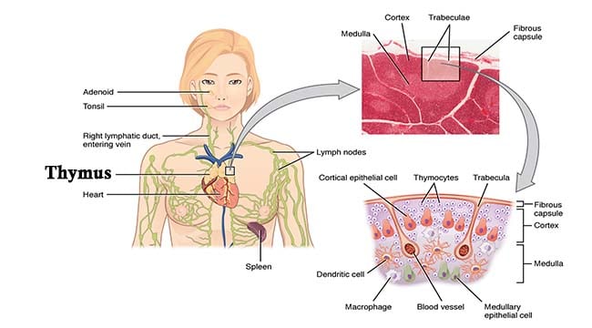
- The thymus is a lymphocyte-rich, bilobed, encapsulated organ located behind the sternum, above and in front of the heart.
- The activity of the thymus is maximal in the fetus and in early childhood and then undergoes atrophy at puberty although never totally disappearing.
- The thymus is derived from the third and fourth pharyngeal pouches during embryonic life and attracts (with chemoattractive molecules) circulating T cell precursors derived from hemopoietic stem cells (HSC) in the bone marrow.

- It is essential for the maturation of T cells and the development of cell-mediated immunity thus referred to as primary lymphoid organ.
- In fact, the term ‘T cell’ means thymus-derived cell and is used to describe mature T cells.
- It is composed of cortical and medullary epithelial cells, stromal cells, interdigitating cells and macrophages.
- These cells are important in the differentiation of the immigrating T cell precursors and their ‘education’ (positive and negative selection) prior to their migration into the secondary lymphoid tissues.
Interesting Science Videos
Structure of Thymus
- It is a pink, flattened, asymmetrical structure lying between sternum and pericardium in anterior mediastinum.
- It is large in infants weighing upto 70 g while atrophied in adult to about 3g.
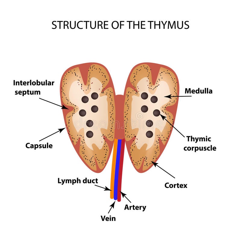
- The thymus consists of two lobes joined by aerolar tissues.
- The two thymic lobes are surrounded by a thin connective tissue capsule.
- Fibrous extensions of capsule around the thymus called trabeculae or septa divide thymus into lobules.

- Each lobule has:
-
An Outer Cortex
- Dark-staining outer part packed with lymphocytes, compartmentalized by elongated epithelial cells.
- It consists of immature T -cells.
- The process of proliferation and selection occurs mainly here.
-
An Inner Medulla
- Lighter central zone with fewer lymphocytes but more epithelial reticular cells.
- It consists of mature T-cells.
- It is predominantly the epithelial part, to which cortical lymphocytes migrate before export via venules and lymphatics.
- Also, the final stages of selection may occur at the cortico-medullary junction.
- Medulla also consists of thymic corpuscles alternatively called Hassall’s corpuscles which are oval structures consisting of round whorls of flattened epithelial cells. They are believed to be aged and degenerated cells.
Functions of Thymus
-
T cell maturation and development
Immature T cell precursors travel from the bone marrow to the thymus (to become thymocytes) where they generate antigen specificity, undergo thymic education, and then migrate to the peripheral lymphoid tissues as mature T cells.
The main functions of the thymus as a primary lymphoid organ are to:
(a) To produce sufficient numbers (millions) of different T cells each expressing unique T cell receptors (generate diversity) such that in every individual there are at least some cells potentially specific for each foreign antigen in the environment.
(b) To select T cells for survival in such a way that the chance for an auto-immune response is minimized.
-
Production of Hormones
The thymus has an interactive role with the endocrine system. Thymic epithelial cells produce the hormones thymosin and thymopoietin and in concert with cytokines (such as IL-7) are probably important for the development and maturation of thymocytes into mature T cells.
Reference
- Brooks, G. F., Jawetz, E., Melnick, J. L., & Adelberg, E. A. (2010). Jawetz, Melnick, & Adelberg’s medical microbiology. New York: McGraw Hill Medical.
- Lydyard, P.M., Whelan,A.,& Fanger,M.W. (2005).Immunology (2 ed.).London: BIOS Scientific Publishers.
- Owen, J. A., Punt, J., & Stranford, S. A. (2013). Kuby Immunology (7 ed.). New York: W.H. Freeman and Company.
- Playfair, J., & Chain, B. (2001). Immunology at a Glance. London: Blackwell Publishing.


This is awesome.
Please keep sending me notes on microbiology or other areas of Laboratory studies.